. 2010 Mar 19;285(12):8646-55.
doi: 10.1074/jbc.M109.034926. Epub 2010 Jan 15.

A lectin isolated from bananas is a potent inhibitor of HIV replication

Affiliations

A lectin isolated from bananas is a potent inhibitor of HIV replication

Michael D Swanson et al. J Biol Chem. .

Abstract

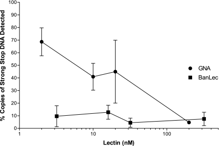
BanLec is a jacalin-related lectin isolated from the fruit of bananas, Musa acuminata. This lectin binds to high mannose carbohydrate structures, including those found on viruses containing glycosylated envelope proteins such as human immunodeficiency virus type-1 (HIV-1). Therefore, we hypothesized that BanLec might inhibit HIV-1 through binding of the glycosylated HIV-1 envelope protein, gp120. We determined that BanLec inhibits primary and laboratory-adapted HIV-1 isolates of different tropisms and subtypes. BanLec possesses potent anti-HIV activity, with IC(50) values in the low nanomolar to picomolar range. The mechanism for BanLec-mediated antiviral activity was investigated by determining if this lectin can directly bind the HIV-1 envelope protein and block entry of the virus into the cell. An enzyme-linked immunosorbent assay confirmed direct binding of BanLec to gp120 and indicated that BanLec can recognize the high mannose structures that are recognized by the monoclonal antibody 2G12. Furthermore, BanLec is able to block HIV-1 cellular entry as indicated by temperature-sensitive viral entry studies and by the decreased levels of the strong-stop product of early reverse transcription seen in the presence of BanLec. Thus, our data indicate that BanLec inhibits HIV-1 infection by binding to the glycosylated viral envelope and blocking cellular entry. The relative anti-HIV activity of BanLec compared favorably to other anti-HIV lectins, such as snowdrop lectin and Griffithsin, and to T-20 and maraviroc, two anti-HIV drugs currently in clinical use. Based on these results, BanLec is a potential component for an anti-viral microbicide that could be used to prevent the sexual transmission of HIV-1.


Figures

FIGURE 1.
FIGURE 1.
BanLec has antiviral activity against multiple HIV-1 isolates with different tropisms. A, TZM-bl cells were pretreated with different concentrations of BanLec before infection with the R5 tropic isolates NL(AD8) and 81A-4, dual tropic 89.6, and X4 tropic NL4-3. Forty-eight hours after exposure to virus, luciferase activity was determined by measuring relative luminescent units (RLU). The averages from three separate experiments were used for the calculation of IC50 values, which were determined by nonlinear regression. The IC50 for viral inhibition were as follows: NL(AD8) = 2.06 nm, 81A-4 = 0.69 nm, 89.6 = 0.48 nm, NL4-3 = 0.49 nm. B, Magi-CCR5 indicator cells were used to determine anti-viral activity of BanLec against multiple strains of HIV-1. Forty hours after exposure to virus, infected cells were quantified by staining for β-galactosidase activity. Infectivity of BanLec-treated virus is presented as a percent of positively infected cells as compared with the PBS control. Error bars represent S.D. from three separate experiments.
FIGURE 2.
FIGURE 2.
BanLec inhibits infection of HIV-1 pseudotyped with envelopes from multiple primary isolates. TZM-bl cells were infected with HIV-1 pseudotyped with primary HIV-1 envelope proteins from subtype B (A) and subtype C (B) in the presence of different concentrations of BanLec. Forty-eight hours later, luciferase activity was assessed. The IC50 values were determined as in Fig. 1 and are shown in Table 1. Results shown are the average of three independent experiments, and error bars represent the S.D. RLU, relative luminescent units.
FIGURE 3.
FIGURE 3.
BanLec inhibits HIV-1 infection of MDM. A, MDM were pretreated with BanLec for 30 min before the addition of 100 TCID50 of HIV-1 NL(AD8). Twenty-four hours later, the media was removed, and the cells were washed with PBS to eliminate remaining virus. Fresh media containing BanLec or PBS was added to the cells. A sample of culture supernatant was taken every 3 days for p24 quantification by ELISA and replaced with new media containing lectin in PBS or PBS alone as a control. On day 15, viability was assessed by an MTT assay, which indicated no cellular toxicity (data not shown). B, MDM were pretreated and infected with HIV-1 as described above. 24 h post-infection the cells were washed with PBS to remove residual virus and cultured in media containing BanLec or PBS. Seven days post-infection, supernatants were removed for determination of p24 antigen as detected by ELISA. The concentration for a 50% reduction in p24 production was calculated to be 9.72 nm. Cellular viability was assessed by an MTT assay, and no toxicity was observed (data not shown). Results shown in panels A and B are representative of three and two separate experiments, respectively.
FIGURE 4.
FIGURE 4.
BanLec inhibits production of early HIV-1 reverse transcription products in peripheral blood lymphocytes. Peripheral blood lymphocytes were treated with different lectin concentrations 30 min before infection with HIV-1 Bru. Three hours post-infection, cellular DNA of the infected cells was harvested, and strong-stop DNA was quantified by real-time PCR. The number of copies was normalized to a PBS-treated control (100%). The known anti-HIV lectin GNA (circles) was used as a positive control and to assess the relative molar potency of BanLec (squares).
FIGURE 5.
FIGURE 5.
BanLec binds to glycosylated gp120. A, shown is dose-dependent binding of BanLec to glycosylated gp120. BanLec was used to coat a 96-well ELISA plate. Serial dilutions of gp120 were added in duplicate to the wells. gp120 was detected with an anti-gp120 antibody. Results are representative of four independent experiments. B, methyl α-d-mannopyranoside inhibits interaction of BanLec with gp120. ELISA plates were coated with BanLec as in panel A. Serial dilutions of methyl α-d-mannopyranoside were added to wells along with a constant amount of gp120. The amount of gp120 bound was determined using the standard curve produced in panel A. Abs, absorbance.
FIGURE 6.
FIGURE 6.
Binding of gp120 by BanLec blocks access to the anti-HIV monoclonal antibody 2G12. Recombinant gp120-coated ELISA plates were treated with different concentrations of BanLec before incubation with the 2G12 antibody, which recognizes the high mannose structures found at positions Asn-295, -332, and -392 of the gp120 protein. Unbound antibody was removed, and the amount of antibody remaining was determined by comparison to a standard curve. The results shown represent the average from three separate experiments. Error bars represent S.E. The effect of increasing amounts of BanLec was determined to be significant by 1-way analysis of variance testing (p < 0.01).
FIGURE 7.
FIGURE 7.
BanLec primarily inhibits binding of HIV to the cellular membrane. TZM-bl cells were spin-infected at 16 °C, a temperature that allows for attachment of virus but does not allow fusion events to occur. The unbound virus was removed, and the cells were incubated with media containing inhibitors (CD4-IgG2, T-20, maraviroc, or BanLec) on ice for 30 min, and then the plates were shifted to 37 °C to allow for fusion and infection to be completed (○). The results were compared with a standard infection procedure (pre-attachment) in which the virus and inhibitors were incubated together on ice for 30 min and then added to TZM-bl cells and incubated at 37 °C (●). The results shown are the averages from three separate experiments. Nonlinear regression analysis was used for curve fitting and calculation of IC50 values (Table 2). RLU, relative luminescent units.
FIGURE 8.
FIGURE 8.
Comparison of the anti-HIV activity of BanLec to the anti-HIV lectins GNA and GRFT. TZM-bl cells were pretreated with BanLec, GRFT, or GNA diluted in PBS or PBS alone, as a control, for 30 min before infection by the R5 tropic HIV-1 virus 81-A. Forty-eight hours later, luciferase activity was measured. The results are normalized to infected cells treated with PBS alone. The average of three separate experiments is shown and was used to calculate IC50 values by nonlinear regression. The calculated IC50 values are the following: GNA = 34.3 nm, BanLec 3.18 nm, and GRFT 0.42 nm. RLU, relative luminescent units.

References

    1. Executive Summary United Nations Programme on HIV/AIDS (UNAIDS) and World Health Organization (WHO) (2008) Report on the Global AIDS Epidemic
    1. Underhill K., Montgomery P., Operario D. (2007) BMJ 335, 248. - PMC - PubMed
    1. Randolph M. E., Pinkerton S. D., Bogart L. M., Cecil H., Abramson P. R. (2007) Arch. Sex. Behav. 36, 844–848 - PMC - PubMed
    1. Crosby R., Yarber W. L., Sanders S. A., Graham C. A. (2005) J. Am. Coll. Health 54, 143–147 - PubMed
    1. Gupta G. R. (2002) BMJ 324, 183–184 - PMC - PubMed

Publication types