. 2010 Mar 19;285(12):9221-32.
doi: 10.1074/jbc.M109.073650. Epub 2010 Jan 15.

Orphan nuclear receptor DAX-1 acts as a novel corepressor of liver X receptor alpha and inhibits hepatic lipogenesis

Affiliations

Orphan nuclear receptor DAX-1 acts as a novel corepressor of liver X receptor alpha and inhibits hepatic lipogenesis

Balachandar Nedumaran et al. J Biol Chem. .

Abstract

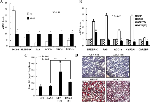
DAX-1 (dosage-sensitive sex reversal adrenal hypoplasia congenital critical region on X chromosome, gene 1) is a member of the nuclear receptor superfamily that can repress diverse nuclear receptors and has a key role in adreno-gonadal development. Our previous report has demonstrated that DAX-1 can inhibit hepatocyte nuclear factor 4alpha transactivity and negatively regulate gluconeogenic gene expression (Nedumaran, B., Hong, S., Xie, Y. B., Kim, Y. H., Seo, W. Y., Lee, M. W., Lee, C. H., Koo, S. H., and Choi, H. S. (2009) J. Biol. Chem. 284, 27511-27523). Here, we further expand the role of DAX-1 in hepatic energy metabolism. Transfection assays have demonstrated that DAX-1 can inhibit the transcriptional activity of nuclear receptor liver X receptor alpha (LXRalpha). Physical interaction between DAX-1 and LXRalpha was confirmed Immunofluorescent staining in mouse liver shows that LXRalpha and DAX-1 are colocalized in the nucleus. Domain mapping analysis shows that the entire region of DAX-1 is involved in the interaction with the ligand binding domain region of LXRalpha. Competition analyses demonstrate that DAX-1 competes with the coactivator SRC-1 for repressing LXRalpha transactivity. Chromatin immunoprecipitation assay showed that endogenous DAX-1 recruitment on the SREBP-1c gene promoter was decreased in the presence of LXRalpha agonist. Overexpression of DAX-1 inhibits T7-induced LXRalpha target gene expression, whereas knockdown of endogenous DAX-1 significantly increases T7-induced LXRalpha target gene expression in HepG2 cells. Finally, overexpression of DAX-1 in mouse liver decreases T7-induced LXRalpha target gene expression, liver triglyceride level, and lipid accumulation. Overall, this study suggests that DAX-1, a novel corepressor of LXRalpha, functions as a negative regulator of lipogenic enzyme gene expression in liver.


Figures

FIGURE 1.
FIGURE 1.
DAX-1 represses the transcriptional activity of LXRα. A, Western blot analysis. Western blot analysis was performed using the protein extracts from mouse tissues (upper panel) and cell lines (lower panel). B and C, HepG2 (B) and 293T (C) cells were transfected with pcDNA3-HA-LXRα (200 ng), HA-DAX-1(50, 100, and 200 ng), and LXRE-luc (200 ng). As positive control, we transfected HA-SHP and HA-LXRα and LXRE-luc in the presence of ligand. Effect of DAX-1 alone with basal reporter activity was also shown. D, 293T cells were transfected with HA-LXRα (10 μg) and FLAG-hDAX-1 (5 and 10 μg). E, HepG2 cells were transfected with sihDAX-1-1 (200 pmol) or sihDAX-1-2 (50 and 200 pmol), and 24 h later HA-LXRα (200 ng) and LXRE-Luc (200 ng) were transfected. After 24 h, the cells were harvested, and luciferase and β-galactosidase assays were performed. F, HepG2 cells were transfected with sihDAX-1-1 (200 pmol) and sihDAX-1-2 (50, 100, and 200 pmol). After 48 h transfection cells (D and E) were harvested for Western blot analysis with the indicated antibodies.
FIGURE 2.
FIGURE 2.
Interaction between DAX-1 and LXRα both in vitro and in vivo. A, in vivo interaction between DAX-1 and LXRα. 293T cells were cotransfected with expression vectors for HA-LXRα together with pEBG-DAX-1 (GST-DAX-1) or GST alone (pEBG) as a control. The complex formation (upper panel, GST purification.) and the amount of HA-LXRα used for the in vivo binding assay (lower panel, Lysate) were determined by anti-HA antibody. The same blot was stripped and re-probed with an anti-GST antibody (middle panel) to confirm the expression levels of the GST fusion protein (GST-hDAX-1) and the GST control (GST). WB, Western blot. B, endogenous interaction between LXRα and DAX-1 in HepG2 cells. Protein extracts from HepG2 cells treated with vehicle (DMSO) or T7 were coimmunoprecipitated (IP) using DAX-1 antibody or secondary antibody alone (negative control) and Western blotted with LXRα antibody. Inputs (10%) for DAX-1 and LXRα are shown in the bottom panels. C, coimmunoprecipitation assays with liver extracts (n = 4) demonstrate the functional association between LXRα and DAX-1. Protein extracts from livers were immunoprecipitated using DAX-1 antibody or IgG alone (negative control) and Western-blotted with LXRα (upper two panels) antibody. Expression of LXRα and DAX-1 (lower two panels) from 10% of lysate were analyzed by Western blotting with specific antibodies. D, subcellular localization of DAX-1 and LXRα. HeLa cells were transiently transfected with pEGFP-DAX-1 or pEGFP with pCDNA3/HA-LXRα. The yellow stain in the merged image indicates the colocalization of DAX-1 and LXRα. Data shown are representative cells from one of three independent experiments. DAPI, 4′,6-diamidino-2-phenylindole. E, in vivo immunofluorescent staining. Paraffin sections of normal (ad libitum) mouse liver samples were used for immunofluorescent staining. The hepatic DAX-1 and LXRα proteins were detected with anti-DAX-1 and LXRα antibodies and visualized with red fluorescence for DAX-1 and green fluorescence for LXRα. Pictures are shown at ×400 magnification with a confocal microscope.
FIGURE 3.
FIGURE 3.
Mapping of interaction domain between DAX-1 and LXRα. A, schematic representation of LXRα deletion constructs (upper panel). In vitro MBP pulldown assay was performed using bacterially expressed various MBP-LXRα deletion constructs and 35S-labeled HA-DAX-1 WT (lower panel). B, schematic representation of DAX-1 deletion constructs (upper panel). In vitro MBP pulldown assay was performed using bacterially expressed MBP-LXRα WT and in vitro translated 35S-labeled different HA-DAX-1 deletion constructs. Then cell lysates were immunoprecipitated with amylose beads and detected using phosphorimager (lower panel). NT, N terminus; CT, C terminus. C, schematic representation of DAX-1 WT and DAX-1 mL1 (mutant of first LXXLL motif) constructs (upper panel). MBP pulldown assay was performed using bacterially expressed MBP-LXRα WT and in vitro translated 35S-labeled HA-DAX-1 WT and HA-DAX-1 mL1 proteins. The cell lysates were then immunoprecipitated with amylose beads and detected using phosphorimager (lower panel).
FIGURE 4.
FIGURE 4.
DAX-1 competes with SRC-1 for LXRα transactivation. A, HepG2 cells were transfected using 200 ng of LXRE-Luc with indicated amounts of LXRα, SRC-1, and DAX-1 expression vectors. Cells were harvested 40 h after transfection, and lysates were utilized for luciferase and β-galactosidase assay. The results shown are the mean of β-galactosidase values from three independent experiments. Effects of DAX-1 and SRC-1 alone on the basal reporter activity are also shown. B, in vitro MBP competition assay. MBP-fused full-length LXRα (upper panel) or MBP-LXRαC (lower panel) bound to amylose beads was incubated with 35S-labeled full-length HA-hDAX-1, in the presence of increasing amounts of cold methionine-labeled SRC-1 (1, 2, 4, or 8 μl). After washing, bound proteins were subjected to SDS-PAGE, and the amount of MBP-bound HA-DAX-1 was visualized via autoradiography.
FIGURE 5.
FIGURE 5.
DAX-1 decreases LXRα agonist-mediated target gene promoter activity and expression. A, HepG2 cells were transfected with SREBP-1c-Luc and with LXRα and DAX-1, and cell lysates were utilized for luciferase and β-galactosidase assays. The results shown are means of β-galactosidase values from three independent experiments. B, chromatin immunoprecipitation analysis using antibodies for LXRα, DAX-1, SRC-1, and acetyl-H3. PCR amplification of immunoprecipitated chromatin fragments was conducted using primer pairs specific for the proximal, regulatory region (1) and a distal, nonregulatory region (2) of the SREBP-1C gene promoter (left panel). Cell extracts from primary hepatocytes treated with vehicle (DMSO) or LXRα agonist (T7) with or without infection of Ad-DAX-1 were immunoprecipitated with LXRα, DAX-1, SRC-1, and acetyl-H3 antibodies (right panel). After reverse cross-linking, DNA was extracted, and PCR was performed using primers for LXR-RE containing proximal and nonspecific distal region of SREBP-1c promoter. As a negative control, cell extracts were incubated with IgG without any preincubation of primary antibody. Inputs (5%) for both proximal and distal SREBP promoter are shown. C and D, quantitative PCR analysis was performed using total RNA extracted from HepG2 (C), and RT-PCR analysis was performed using total RNA from rat primary hepatocytes (D) after the treatment of LXRα agonist with or without adenovirus DAX-1 infection. DAX-1, SREBP-1C, FAS, and β-actin genes amplified using specific primers for DAX-1, SREBP-1C, FAS, and β-actin were used for PCR. E, Student's t test. Western blot analysis showing protein level of DAX-1 from HepG2 cells infected with mock and Ad-shDAX-1 (right panel).
FIGURE 6.
FIGURE 6.
DAX-1 lowers serum triglyceride and lipid accumulation in liver. Comparison of DAX-1 and lipogenic gene expressions in normal and db/db mice. A, quantitative PCR analysis of hepatic mRNA levels of DAX-1, SRC-1, PGC-1a, SREBP-1C, FAS and acetyl-coenzyme A carboxylase in normal and db/db mice (*, p < 0.01; n = 4). B, DAX-1 decreases T7-mediated lipogenic gene expression in mice. Male 8-week-old C57BL6 mice were provided with the meal form of a standard rodent diet. T0901317 (LXR agonist, 50 mg/kg body weight) or vehicle (1% methylcellulose and 1% Tween 80) were administered by oral gavage each day for 1 week. Recombinant adenovirus (0.5 × 10⁹ plaque-forming unit) GFP (n = 5) or DAX-1 (n = 5) were delivered by tail vein injection on the 4th day of oral gavage. Three days after adenovirus GFP (n = 3) or DAX-1 (n = 3) injection, mice were sacrificed, and the expressions of SREBP-1c, FAS, and ACC1α were analyzed by real time quantitative RT-PCR. All data were normalized to ribosomal L32 expression. C, liver triglyceride level is decreased by DAX-1. Hepatic TAG levels were analyzed from mouse liver tissue infected with adenoviruses as in B. D, DAX-1 decreases the lipid accumulation in liver. Oil Red O staining was performed from the liver samples as in B. Data in A–C are represented as mean ± S.D.

Similar articles

Cited by

References

    1. Nedumaran B., Hong S., Xie Y. B., Kim Y. H., Seo W. Y., Lee M. W., Lee C. H., Koo S. H., Choi H. S. (2009) J. Biol. Chem. 284, 27511–27523 - PMC - PubMed
    1. Giguère V. (1999) Endocr. Rev. 20, 689–725 - PubMed
    1. Aranda A., Pascual A. (2001) Physiol. Rev. 81, 1269–1304 - PubMed
    1. Iyer A. K., McCabe E. R. (2004) Mol. Genet. Metab. 83, 60–73 - PubMed
    1. Zhang H., Thomsen J. S., Johansson L., Gustafsson J. A., Treuter E. (2000) J. Biol. Chem. 275, 39855–39859 - PubMed

Publication types

LinkOut - more resources