

An FMRI study of frontostriatal circuits during the inhibition of eye blinking in persons with Tourette syndrome
- PMID: 20080981
- PMCID: PMC4295823
- DOI: 10.1176/appi.ajp.2009.08121831
An FMRI study of frontostriatal circuits during the inhibition of eye blinking in persons with Tourette syndrome
Abstract
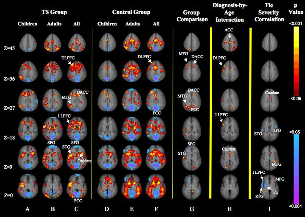
Objective: The authors sought to study activity in neural circuits that subserve the inhibition of a semi-involuntary motor behavior, eye blinking, in children and adults with Tourette syndrome and in healthy comparison subjects.
Method: Functional magnetic resonance imaging was used to scan 120 participants (51 with Tourette syndrome and 69 comparison subjects) as they either blinked normally or successfully inhibited eye blinking. The authors compared the blood-oxygen-level dependent signal during these two conditions across the Tourette and comparison groups.
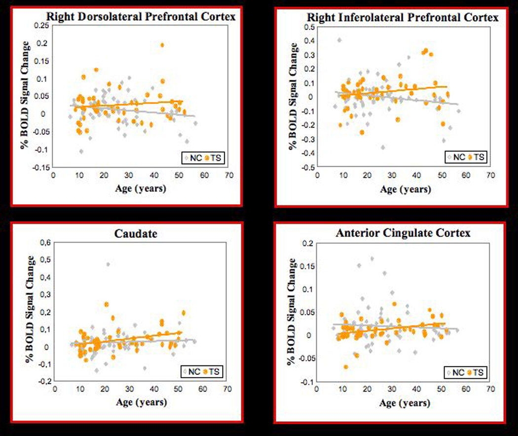
Results: Relative to comparison subjects, patients with Tourette syndrome activated more strongly the frontal cortex and striatum during eye blink inhibition. Activation increased more with age in the dorsolateral and inferolateral prefrontal cortex and caudate nucleus in the Tourette group relative to comparison subjects. In addition, the Tourette group more strongly activated the middle frontal gyrus, dorsal anterior cingulate, and temporal cortices. The severity of tic symptoms in the Tourette group correlated inversely with activation in the putamen and inferolateral prefrontal cortex.
Conclusions: Frontostriatal activity is increased in persons with Tourette syndrome during the inhibition of eye blinks. Activation of frontostriatal circuits in this population may help to maintain regulatory control over semi-involuntary behaviors, whether these are tics or eye blinks.
Figures
Similar articles
-
A developmental fMRI study of self-regulatory control in Tourette's syndrome.Am J Psychiatry. 2007 Jun;164(6):955-66. doi: 10.1176/ajp.2007.164.6.955. Am J Psychiatry. 2007. PMID: 17541057 Free PMC article.
-
Deficient activity in the neural systems that mediate self-regulatory control in bulimia nervosa.Arch Gen Psychiatry. 2009 Jan;66(1):51-63. doi: 10.1001/archgenpsychiatry.2008.504. Arch Gen Psychiatry. 2009. PMID: 19124688 Free PMC article.
-
Neural substrates of self-regulatory control in children and adults with Tourette syndrome.Can J Psychiatry. 2009 Sep;54(9):579-88. doi: 10.1177/070674370905400902. Can J Psychiatry. 2009. PMID: 19751546 Free PMC article.
-
Functional disturbances within frontostriatal circuits across multiple childhood psychopathologies.Am J Psychiatry. 2009 Jun;166(6):664-74. doi: 10.1176/appi.ajp.2009.08091354. Epub 2009 May 15. Am J Psychiatry. 2009. PMID: 19448188 Free PMC article. Review.
-
[Structural and functional neuroanatomy of attention-deficit hyperactivity disorder (ADHD)].Encephale. 2009 Apr;35(2):107-14. doi: 10.1016/j.encep.2008.01.005. Epub 2008 Jul 7. Encephale. 2009. PMID: 19393378 Review. French.
Cited by
-
Impaired automatic but intact volitional inhibition in primary tic disorders.Brain. 2020 Mar 1;143(3):906-919. doi: 10.1093/brain/awaa024. Brain. 2020. PMID: 32125364 Free PMC article.
-
Striatal magnetic resonance spectroscopy abnormalities in young adult SAPAP3 knockout mice.Biol Psychiatry Cogn Neurosci Neuroimaging. 2016 Jan 1;1(1):39-48. doi: 10.1016/j.bpsc.2015.10.001. Biol Psychiatry Cogn Neurosci Neuroimaging. 2016. PMID: 26858992 Free PMC article.
-
Impaired Motor Timing in Tourette Syndrome: Results From a Case-Control Study in Children.Front Neurol. 2020 Oct 29;11:552701. doi: 10.3389/fneur.2020.552701. eCollection 2020. Front Neurol. 2020. PMID: 33192986 Free PMC article.
-
The buildup of an urge in obsessive-compulsive disorder: Behavioral and neuroimaging correlates.Hum Brain Mapp. 2020 Apr 15;41(6):1611-1625. doi: 10.1002/hbm.24898. Epub 2020 Jan 9. Hum Brain Mapp. 2020. PMID: 31916668 Free PMC article.
-
Multispectral brain morphometry in Tourette syndrome persisting into adulthood.Brain. 2010 Dec;133(Pt 12):3661-75. doi: 10.1093/brain/awq300. Epub 2010 Nov 10. Brain. 2010. PMID: 21071387 Free PMC article.
References
-
- Spessot AL, Peterson BS. In: Tourette’s syndrome: a multifactorial, developmental psychopathology, in Developmental Psychopathology. 2nd ed. Cicchetti D, Cohen DJ, editors. Vol. 3. Hoboken, NJ: John Wiley & Sons; 2006. pp. 436–469.
-
- Leckman JF, Walker DE, Cohen DJ. Premonitory urges in Tourette’s syndrome. Am J Psychiatry. 1993;150:98–102. - PubMed
-
- Peterson BS, Thomas P, Kane MJ, Scahill L, Zhang H, Bronen R, King RA, Leckman JF, Staib L. Basal ganglia volumes in patients with Gilles de la Tourette syndrome. Arch Gen Psychiatry. 2003;60:415–424. - PubMed
Publication types
MeSH terms
Grants and funding
LinkOut - more resources
Full Text Sources
Medical
