. 2010 Feb;137(3):405-15.
doi: 10.1242/dev.045369.

Pax6-dependent Shroom3 expression regulates apical constriction during lens placode invagination

Affiliations

Pax6-dependent Shroom3 expression regulates apical constriction during lens placode invagination

Timothy F Plageman Jr et al. Development. 2010 Feb.

Abstract

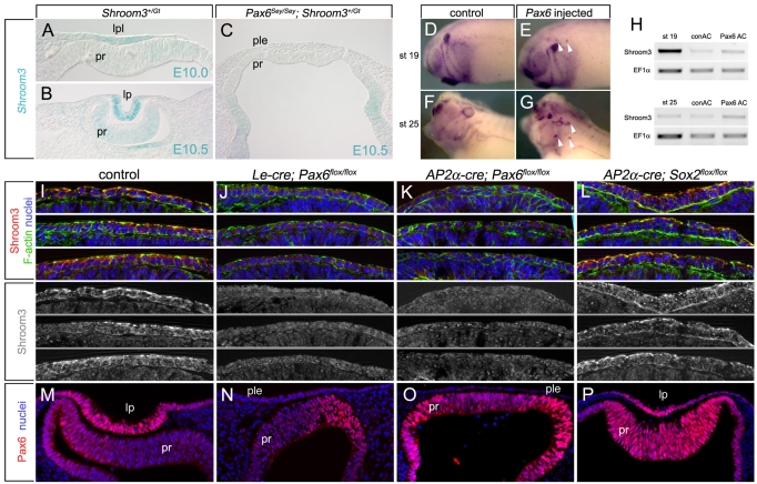
Embryonic development requires a complex series of relative cellular movements and shape changes that are generally referred to as morphogenesis. Although some of the mechanisms underlying morphogenesis have been identified, the process is still poorly understood. Here, we address mechanisms of epithelial morphogenesis using the vertebrate lens as a model system. We show that the apical constriction of lens epithelial cells that accompanies invagination of the lens placode is dependent on Shroom3, a molecule previously associated with apical constriction during morphogenesis of the neural plate. We show that Shroom3 is required for the apical localization of F-actin and myosin II, both crucial components of the contractile complexes required for apical constriction, and for the apical localization of Vasp, a Mena family protein with F-actin anti-capping function that is also required for morphogenesis. Finally, we show that the expression of Shroom3 is dependent on the crucial lens-induction transcription factor Pax6. This provides a previously missing link between lens-induction pathways and the morphogenesis machinery and partly explains the absence of lens morphogenesis in Pax6-deficient mutants.


Figures

Fig. 1.
Fig. 1.
Shroom3 is expressed in the invaginating lens pit and is required for apical constriction. (A,B) X-gal staining of cryosections through the lens pit of Shroom3+/Gt mouse embryos indicating expression of Shroom3 mRNA. (C,D) Immunofluorescent staining of cryosections of wild-type embryos indicating Shroom3 protein localization. lp, lens pit; pr, presumptive retina. (E,F) Whole-mount view of the eye region of control (E) and Shroom3 homozygous mutant (F) embryos. The lens pit is outlined (dashed line). (G-J) Surface view of 28-somite (~E10.0) control (G,I) or Shroom3 homozygous mutant (H,J) lens placode immunofluorescently stained with a ZO1 antibody (red) and phalloidin (green) to identify apical junctions between cells. Lens pit invagination is first detectable at 28 somites. I and J are magnifications of the bracketed area in G and H, respectively. (K) Quantification of the apical area per cell for lens pit cells from control (gray) and Shroom3Gt/Gt (green) embryos that were somite-staged as indicated. One-way ANOVA analysis was performed to generate P-values and indicate statistical significance. NS, not significant.
Fig. 2.
Fig. 2.
F-actin and myosin II are mislocalized in the lens pits of Shroom3 mutant embryos. (A-F,H-M) Cryosectioned E10.5 lens pits from control or Shroom3Gt/Gt mutant mice labeled with phalloidin to visualize F-actin (A-F) or with an antibody specific to myosin II (H-M). D-F and K-M are magnifications of the indicated regions of A-C and H-J, respectively. The blue and green lines in I refer to regions of distinct curvature in control and Shroom3Gt/Gt lens pit as quantified in O. lp, lens pit; pr, presumptive retina. (G,N) Quantification of F-actin (G) and myosin II (N) labeling intensity along the apical and basal surfaces of the lens pit in E10.5 control and Shroom3Gt/Gt embryos. *, P<0.05 by Student's t-test. (O) Quantification of rate of change of curvature along the basal surface of control (gray line) and Shroom3Gt/Gt (red line) lens pits. Regions where the mutant shows lower than normal curvature (blue lines) or higher than normal curvature (green line) are indicated.
Fig. 3.
Fig. 3.
Shroom3-deficient embryos have altered lens pit cell shapes. (A-D) Representative images of β-catenin (red) and phalloidin (green) labeled lens pit cryosections from control and Shroom3Gt/Gt mouse embryos. The boxed areas in A and C are magnified in B and D, respectively. (E-I) Outlines of individual lens pit cells were traced (dashed white lines) and cell profiles placed on a grid (E,F) to permit quantification of cell width at seven evenly spaced positions (G). Cell height was also quantified (H) and an average cell shape calculated (I). In I, the average cell dimensions are indicated relative to the basal dimension of the control cell. This shows that the Shroom3Gt/Gt lens pit cell is shorter and less apically constricted than in the control. (G,H) Error bars are s.e.m.; *, P<0.05. (J,K) Chicken lens pits were electroporated with a GFP- or V5-tagged dominant-negative chicken Shroom3-expressing plasmid. Cryosections were immunofluorescently labeled with phalloidin to visualize F-actin and with GFP or V5 antibodies. Positive cells are outlined (dashed white line). Arrowheads indicate ectopic basal actin. Gray lines between panels indicate that the panels are separated color channels of the same image. (L) Quantification of cell width along the apical/basal axis of chicken lens pit cells expressing GFP (gray, control), Shroom3-Flag (blue), DN-cShroom3-V5 (red) or Mena-dn (green). The error bars are s.e.m. The green asterisks indicate a significant difference between Mena-dn and GFP and the red asterisks a significant difference between DN-cShroom-V5 and GFP (P<0.05). (M) The average shape of cells expressing the indicated transgene is depicted with the relative change in apical and basal cell width. NS, not significant.
Fig. 4.
Fig. 4.
The FPn domain of Shroom3 is required for apical constriction and apical Vasp localization. (A) The wild-type and mutant mouse Shroom3 proteins used. (B) Alignment of the amino acid sequences of human (h), mouse (m) and chicken (c) Shroom proteins in the proline-rich (FPn) motif region. The consensus binding site for the Mena/Vasp EVH1 domain is indicated (shaded orange, below the alignment: x, any amino acid; f, hydrophobic amino acid). The red box indicates the amino acids that are deleted in the Shroom3ΔFPn mutant. (C) The domain structure of mouse wild-type Mena, dominant-negative Mena (Mena-dn), Vasp and Evl. (D) MDCK cells were transiently transfected with the indicated plasmid(s) and immunofluorescently labeled to detect protein expression. Cells were co-labeled with ZO1 to detect apical junctions. Apical structures are detected in the xy plane, whereas the apical-basal axis is visualized in the xz plane. (E) Quantification of the average apical area for cells expressing the indicated proteins. Error bars are s.e.m. (F) MDCK cells were transiently transfected with Shroom3-Flag or Shroom3-ΔFPn-Flag and immunofluorescently stained to visualize ectopic Shroom3 and endogenous Vasp. As in D, apical structures are detected in the xy plane, whereas the apical-basal axis is visualized in the xz plane. Hoechst 33258 labeling was used to visualize nuclei.
Fig. 5.
Fig. 5.
Apical Vasp localization is reduced in the Shroom3 mutant lens pit. (A-F) Cryosections of control (A-D) and Shroom3 mutant (E,F) mouse lens pits labeled with phalloidin to visualize F-actin, Hoechst to mark nuclei, and with antibodies specific to Shroom3 or Vasp. The indicated regions of A, C and E are magnified in B, D and F, respectively. Single-channel and merged images are shown in each row. (G-I) Quantification of labeling intensity for Shroom3 (G), F-actin (H) and Vasp (I) in control (gray lines) and Shroom3Gt/Gt (green lines) lens pit epithelial cells along a basal-to-apical line interval. Statistical significance between the control and mutant was determined for each value along the axis using Student's t-test and subtracted from 1. The pixels with values above 0.95 (P<0.05) are indicated by the blue line.
Fig. 6.
Fig. 6.
Pax6 is necessary and sufficient for Shroom3 expression in the lens. (A-C) Shroom3 expression from the lacZ promoter trap allele was assessed in control (A,B) or Pax6Sey/Sey (C) mouse embryos by X-gal staining. (D-G) Control or Pax6 mRNA-injected Xenopus embryos were assayed for Shroom3 expression by in situ hybridization. Arrowheads indicate ectopic Shroom3 expression. (H) RNA was isolated from animal caps derived from control or Pax6 mRNA-injected Xenopus embryos and analyzed by RT-PCR using primers specific to Xenopus Shroom3 or the housekeeping gene Ef1α. (I-L) Control mouse embryos (I) or those conditionally deficient for Pax6 (Le-Cre; Pax6flox/flox; AP2α-Cre; Pax6flox/flox) (J,K) or Sox2 (AP2α-Cre; Sox2flox/flox) (L) in the surface ectoderm were cryosectioned and labeled for Shroom3. (M-P) Pax6 expression in E10.5 wild-type or conditional mutant mouse lens pits as assessed by immunofluorescence. Hoechst 33258 labeling was used to visualize nuclei. lpl, lens placode; ple, presumptive lens ectoderm; lp, lens pit; pr, presumptive retina.
Fig. 7.
Fig. 7.
A model: Pax6-dependent expression of Shroom3 in the lens pit induces apical constriction and facilitates placodal invagination. A model illustrating Shroom3 activity is an important component of the morphogenesis mechanisms required for invagination of the lens pit. (A) The activity of Shroom3 becomes important at ~E9.5, just prior to lens pit invagination. (B) At this time, inductive signaling that involves Fgf receptor signaling via the Frs2α adapter (blue), Bmp7 signaling (orange) and the Meis transcription factors (gray), results in the transcriptional upregulation of Pax6 via the EE and SIMO control elements. In turn, Pax6 is required, directly or indirectly, for the expression of Shroom3 and engagement of the machinery that results in apical constriction in lens pit epithelial cells. (C) It is likely that Shroom3-induced apical constriction is one of several mechanisms of morphogenesis that combine to achieve placodal invagination and the formation of a lens pit at E10.5.

Similar articles

Cited by

References

    1. Altmann C. R., Chow, R. L., Lang, R. A. and Hemmati-Brivanlou, A. (1997). Lens induction by Pax-6 in Xenopus laevis. Dev. Biol. 185, 119-123. - PubMed
    1. Ashery-Padan R., Marquardt, T., Zhou, X. and Gruss, P. (2000). Pax6 activity in the lens primordium is required for lens formation and for correct placement of a single retina in the eye. Genes Dev. 14, 2701-2711. - PMC - PubMed
    1. Ball L. J., Kuhne, R., Hoffmann, B., Hafner, A., Schmieder, P., Volkmer-Engert, R., Hof, M., Wahl, M., Schneider-Mergener, J., Walter, U., et al. (2000). Dual epitope recognition by the VASP EVH1 domain modulates polyproline ligand specificity and binding affinity. EMBO J. 19, 4903-4914. - PMC - PubMed
    1. Ball L. J., Jarchau, T., Oschkinat, H. and Walter, U. (2002). EVH1 domains: structure, function and interactions. FEBS Lett. 513, 45-52. - PubMed
    1. Barrett K., Leptin, M. and Settleman, J. (1997). The Rho GTPase and a putative RhoGEF mediate a signaling pathway for the cell shape changes in Drosophila gastrulation. Cell 91, 905-915. - PubMed

Publication types

MeSH terms