. 2010 Feb;137(3):467-76.
doi: 10.1242/dev.041335.

The EVERSHED receptor-like kinase modulates floral organ shedding in Arabidopsis

Affiliations

The EVERSHED receptor-like kinase modulates floral organ shedding in Arabidopsis

Michelle E Leslie et al. Development. 2010 Feb.

Abstract

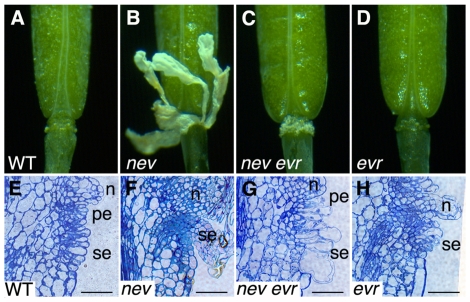
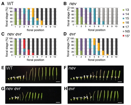
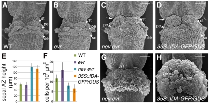
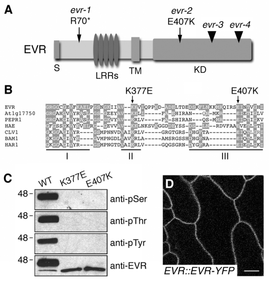
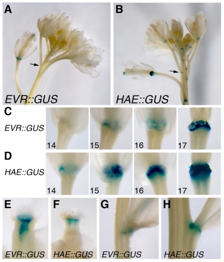
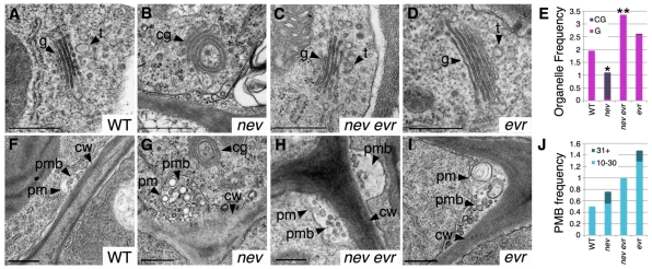
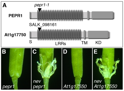
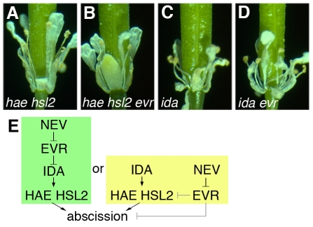
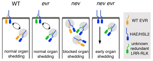
Plant cell signaling triggers the abscission of entire organs, such as fruit, leaves and flowers. Previously, we characterized an ADP-ribosylation factor GTPase-activating protein, NEVERSHED (NEV), that regulates membrane trafficking and is essential for floral organ shedding in Arabidopsis. Through a screen for mutations that restore organ separation in nev flowers, we have identified a leucine-rich repeat receptor-like kinase, EVERSHED (EVR), that functions as an inhibitor of abscission. Defects in the Golgi structure and location of the trans-Golgi network in nev abscission zone cells are rescued by a mutation in EVR, suggesting that EVR might regulate membrane trafficking during abscission. In addition to shedding their floral organs prematurely, nev evr flowers show enlarged abscission zones. A similar phenotype was reported for plants ectopically expressing INFLORESCENCE DEFICIENT IN ABSCISSION, a predicted signaling ligand for the HAESA/HAESA-LIKE2 receptor-like kinases, indicating that this signaling pathway may be constitutively active in nev evr flowers. We present a model in which EVR modulates the timing and region of abscission by promoting the internalization of other receptor-like kinases from the plasma membrane.


Figures

Fig. 1.
Fig. 1.
Mutations in EVR rescue organ separation in nev flowers. (A) Wild-type flower after abscission (stage 17). (B) nev mutant flowers (stage 17) retain their floral organs indefinitely. (C) Organ shedding is rescued in nev evr flowers (stage 17). (D) evr flower after abscission (stage 17). (E-H) Longitudinal sections of wild-type (E), nev (F), nev evr (G) and evr (H) flowers (stage 16) stained with Toluidine Blue. In nev evr flowers (G), the remaining abscission zone (AZ) cells expand to a greater extent than do those of wild type (E). The sepal (se) and petal (pe) AZ regions are indicated, as are the nectaries (n). Scale bars: 50 μm.
Fig. 2.
Fig. 2.
Abscission occurs prematurely in nev evr flowers. Progression of flower development in wild-type, nev, nev evr and evr plants from the first open flower to maturing fruit. (A-D) From the first open flower (position 1), stages were assessed for up to 10 flowers per inflorescence (n=15 inflorescences per genotype). For each position, the percentage of flowers at each stage is shown. In wild-type flowers (A), organ separation (stage 16) is first observed at position 5.7±1.0. By position 8, all flowers have shed their organs (stage 17). nev flowers (B) retain their floral organs and are labeled as NS (non-shedding, stage 16 on). In nev evr flowers (C), organ separation (stage 15*) is first observed at position 3.4±0.5 and is complete by position 6. In evr flowers (D), organ separation (stage 16) is first observed at position 5.9±0.8 and is complete by position 9. (E-H) evr-2 fruit (stage 17; 8.7±0.3 mm; n=11) are 79% the length of wild type (11.0±0.5 mm; n=11). nev-3 evr-2 fruit (6.4±0.3 mm; n=13) are 88% the length of nev-3 fruit (7.3±0.5 mm; n=15). Scale bars: 5 mm.
Fig. 3.
Fig. 3.
Ectopic AZ cell expansion occurs in nev evr flowers. (A-D) Scanning electron micrographs (SEMs) of flowers immediately after organ separation (first stage 17 flower). Owing to increased cell expansion and the presence of additional cells, nev evr flowers (C), like flowers constitutively expressing IDA (D), develop larger AZs than do wild-type (A) and evr (B) flowers, which cover the stem-like gynophore of the fruit (arrows). (E,F) Quantification of AZ size (E) and cell expansion (F) in wild-type and mutant flowers (n≥4, second stage 17 flower). In nev evr and 35S::IDA-GFP/GUS flowers, the average heights of the sepal AZs are 2-fold greater than those of wild-type or evr flowers (E). A decrease in the number of sepal AZ cells in a defined area was observed for nev evr and 35S::IDA-GFP/GUS flowers compared with wild-type and evr flowers, suggesting that cell expansion contributes in part to the increase in AZ size (F). (G,H) The expanding AZs of older nev evr (G) and 35S::IDA-GFP/GUS (Col) (H) flowers (stage 17) envelop the nectaries and form visible collars of tissue at the fruit bases. The sepal (se), petal (pe) and stamen (st) AZs cannot be distinguished. Scale bars: 100 μm.
Fig. 4.
Fig. 4.
EVR encodes a membrane-localized LRR-RLK. (A) Diagram of the EVR protein, with the sites of the identified evr mutations indicated. The regions corresponding to the signal peptide (S), leucine-rich repeats (LRRs), transmembrane domain (TM) and kinase domain (KD) are indicated. Point mutations are marked by arrows, and T-DNA insertions by arrowheads. (B) Sequence alignment of kinase subdomains I-III from EVR and related LRR-RLKs from Arabidopsis and Lotus japonicus. Amino acids conserved between EVR and other proteins are shaded. The sites of the conserved glutamic acid in subdomain III affected by the evr-2 mutation and an invariant lysine in subdomain II required for kinase activity are marked by arrows above the alignment. (C) The recombinant EVR KD autophosphorylates at serine, threonine and tyrosine residues in vitro. Mutations in subdomains II (K377E) and III (E407K) of the kinase domain block the kinase activity of EVR. EVR antiserum recognizes ~46 and ~40 kDa phosphorylated and unphosphorylated proteins, respectively. (D) EVR localizes to the plasma membrane. Fluorescent localization of the EVR-YFP marker in epidermal cells of wild-type leaf petioles (stems). Scale bar: 10 μm.
Fig. 5.
Fig. 5.
EVR is expressed in organ AZs. (A-H) The regulatory regions of EVR and HAE direct expression of β-glucuronidase (GUS) in AZs (A-D), in internal tissues of the floral style (E,F), and at the junction between the cauline leaves and the inflorescence stem (G,H). The HAE::GUS marker is also expressed at the bases of the floral pedicels (B, arrow). Within floral AZs (C), the EVR promoter directs GUS expression prior to organ separation (stage 15), during abscission (stage 16), and as the remaining cells form protective scar tissue (stage 17). GUS expression directed by the HAE promoter shows a similar, yet stronger profile in AZs, with an earlier stage of initiation (Jinn et al., 2000).
Fig. 6.
Fig. 6.
Mutations in EVR restore Golgi structure and location of the TGN in nev flowers. Transmission electron micrographs and analysis of cells in sepal AZ regions at the time of organ separation for wild-type (stage 16), nev (stage 16 non-shedding), nev evr (stage 15*) and evr (stage 16) flowers. (A-D) Instead of the flat stacks of Golgi cisternae characteristic of wild type (A), circularized multilamellar structures are observed in nev cells (B). Golgi with a wild-type appearance are found in nev evr (C) and evr cells (D). We frequently observed vesicular-tubular structures characteristic of the trans-Golgi network (TGN) (81%, n=16) closely associated with the Golgi cisternae (34±21 nm, n=13) in wild-type cells (A), whereas the TGN (15%, n=13) was less often observed near the circularized multilamellar structures (40±25 nm, n=2) of nev cells (B). The location of the TGN was restored in nev evr cells (83% associated with Golgi, n=24; 37±12 nm, n=20) and was unaffected by loss of EVR alone (76% associated with Golgi, n=21; 38±25 nm, n=15). (E) Frequency of flat Golgi cisternae (G, pink) and circularized multilamellar structures (CG, purple) per cell in sections of wild-type and mutant sepal AZ regions. For each genotype, n (cells)≥11. Statistical differences between nev and wild type, and between nev evr and nev tissues are indicated by single and double asterisks, respectively (Fisher's exact test, P<0.0001). A statistical difference was not detected between evr and wild-type tissues. (F-I) Paramural bodies (PMBs) were observed in the cells of wild-type (F), nev (G), nev evr (H) and evr (I) flowers. Whereas PMBs were observed in cells from each genotype, PMBs with greater than 30 vesicles were only observed in nev and evr cells. (J) Frequency of PMBs (10-30 and 31+ vesicles) per cell in sections of wild-type and mutant sepal AZ regions. For each genotype, n (cells)≥11. Statistical differences in PMB accumulation were not detected. cg, circularized multilamellar structures; cw, cell wall; g, Golgi cisternae; pm, plasma membrane; pmb, paramural body; t, trans-Golgi network. Scale bars: 0.5 μm.
Fig. 7.
Fig. 7.
Mutations in PEPR1 or At1g17750 do not rescue shedding in nev flowers. (A) Diagrams of the predicted PEPR1 and At1g17750 proteins, with the sites of the T-DNA mutations indicated by arrowheads. The regions corresponding to the signal peptide (S), leucine-rich repeats (LRRs), transmembrane domain (TM, and kinase domain (KD) are indicated. (B-E) Floral organ shedding occurs normally in pepr1 (B) and At1g17750 (D) mutant flowers (stage 17), but is blocked in nev pepr1 (C) and nev At1g17750 (E) flowers (stage 17).
Fig. 8.
Fig. 8.
Mutations in EVR do not restore abscission in ida or hae hsl2 mutant flowers. (A-D) Organ separation is blocked in ida (C) and hae hsl2 (A) mutant flowers (stage 17), and is not rescued in ida evr (D) or hae hsl2 evr (B) flowers (stage 17). (E) Alternative pathways for the relationships between NEV, EVR, IDA and HAE/HSL2. In both, EVR is predicted to act as a negative regulator of abscission and to function downstream of NEV. In the simplest model, NEV and EVR act upstream of both IDA and HAE/HSL2. Alternatively, NEV and EVR could function in a converging pathway, in which EVR acts upon HAE/HSL2, or further downstream.
Fig. 9.
Fig. 9.
A model for EVR function during the transition to floral organ separation. In wild-type AZ cells before organ shedding, EVR might inhibit cell separation by interacting with ligand-binding LRR-RLKs, such as HAE/HSL2, that trigger cell wall loosening and separation. This interaction could promote the internalization and recycling of inactive receptor complexes through the endosomal system. Continued signaling from a ligand-activated HAE/HSL2 complex would lead to organ shedding in older flowers. The EVR RLK may act redundantly with another LRR-RLK(s), such that loss of EVR alone would not alter the timing of abscission. Disrupting NEV activity might alter the trafficking of receptor complexes containing EVR or EVR-like RLKs, thereby blocking a signal required for cell separation. Mutations in EVR could bypass the requirement for NEV in floral organ shedding, resulting in constitutive signaling of the HAE/HSL2 LRR-RLKs, premature activation of organ separation, the enlargement of AZ regions, and deregulated AZ cell expansion.

References

    1. Alexandersson E., Saalbach, G., Larsson, C. and Kjellbom, P. (2004). Arabidopsis plasma membrane proteomics identifies components of transport, signal transduction and membrane trafficking. Plant Cell Physiol. 45, 1543-1556. - PubMed
    1. Alonso J. M., Stepanova, A. N., Leisse, T. J., Kim, C. J., Chen, H., Shinn, P., Stevenson, D. K., Zimmerman, J., Barajas, P., Cheuk, R., et al. (2003). Genome-wide insertional mutagenesis of Arabidopsis thaliana. Science 301, 653-657. - PubMed
    1. Ascencio-Ibáñez J. T., Sozzani, R., Lee, T.-J., Chu, T.-M., Wolfinger, R. D., Cella, R. and Hanley-Bowdoin, L. (2008). Global analysis of Arabidopsis gene expression uncovers a complex array of changes impacting pathogen response and cell cycle during geminivirus infection. Plant Physiol. 148, 436-454. - PMC - PubMed
    1. Blázquez M. A., Soowal, L. N., Lee, I. and Weigel, D. (1997). LEAFY expression and flower initiation in Arabidopsis. Development 124, 3835-3844. - PubMed
    1. Boller T. and Felix, G. (2009). A renaissance of elicitors: perception of microbe-associated molecular patterns and danger signals by pattern-recognition receptors. Annu. Rev. Plant Biol. 60, 379-406. - PubMed

Publication types

MeSH terms