. 2010 Feb;137(3):507-18.
doi: 10.1242/dev.043257.

Insights into the organization of dorsal spinal cord pathways from an evolutionarily conserved raldh2 intronic enhancer

Affiliations

Insights into the organization of dorsal spinal cord pathways from an evolutionarily conserved raldh2 intronic enhancer

Hozana A Castillo et al. Development. 2010 Feb.

Abstract

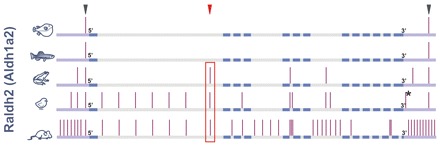
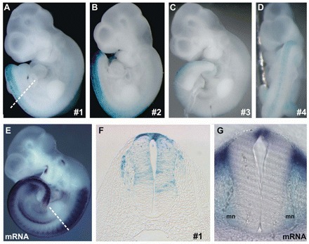
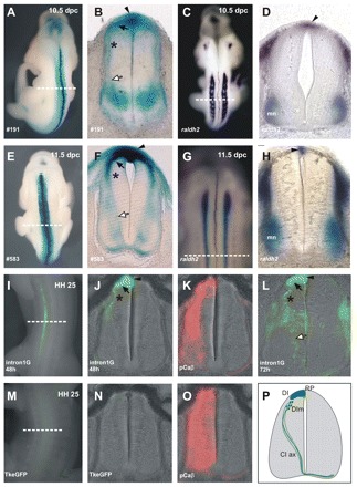
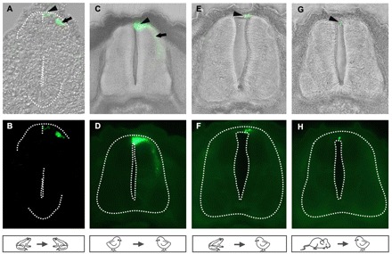
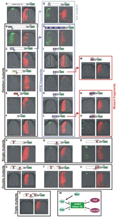
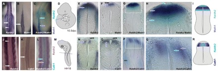
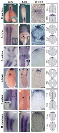
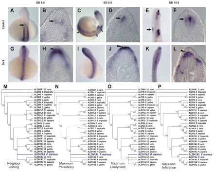
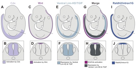
Comparative studies of the tetrapod raldh2 (aldh1a2) gene, which encodes a retinoic acid (RA) synthesis enzyme, have led to the identification of a dorsal spinal cord enhancer. Enhancer activity is directed dorsally to the roof plate and dorsal-most (dI1) interneurons through predicted Tcf- and Cdx-homeodomain binding sites and is repressed ventrally via predicted Tgif homeobox and ventral Lim-homeodomain binding sites. Raldh2 and Math1/Cath1 expression in mouse and chicken highlights a novel, transient, endogenous Raldh2 expression domain in dI1 interneurons, which give rise to ascending circuits and intraspinal commissural interneurons, suggesting roles for RA in the ontogeny of spinocerebellar and intraspinal proprioceptive circuits. Consistent with expression of raldh2 in the dorsal interneurons of tetrapods, we also found that raldh2 is expressed in dorsal interneurons throughout the agnathan spinal cord, suggesting ancestral roles for RA signaling in the ontogenesis of intraspinal proprioception.


Figures

Fig. 1.
Fig. 1.
Evolutionary conservation of raldh2. Five raldh2 orthologs (fugu, zebrafish, frog, chick and mouse) were aligned using Homo sapiens as baseline. Vertical bars, conserved non-coding elements (CNEs). The red box highlights raldh2 intron 1G conservation. Arrowheads indicate the three CNEs chosen for transient transgenesis. Exons, blue bars. Introns, gray bars. 5′ and 3′ regions, violet bars. The asterisk indicates the CNE conserved between chicken and human, but lost from mouse.
Fig. 2.
Fig. 2.
Mouse Raldh2 intron 1G CNE activity in transient transgenic mice. (A-D) The CNE drives lacZ expression (blue) at the posterior dorsum in all (4/4) transient transgenic mice harvested at 10.5 dpc. (E) This β-galactosidase field is a subset of the endogenous Raldh2 expression domain, as indicated by Raldh2 in situ hybridization (ISH). (F) Thoracic transverse section depicts dorsal spinal cord (SC) and dorsal root ganglia enhancer activation. (G) Brachial transverse section shows Raldh2 expression in the roof plate (RP) and motoneurons (mn). Dashed lines in A and E indicate the plane of the sections in F and G, respectively.
Fig. 3.
Fig. 3.
The Raldh2 intron 1G enhancer is a roof plate and dorsal interneuron enhancer in amniotes. (A,E) Dorsal views of stable transgenic mice. (B,F) Transverse sections at the brachial SC of the stable transgenic mice shown in A and E, respectively. The enhancer is active in the RP (arrowhead), in dorsal interneurons (DI) (black arrow) and in ventrally migrating dorsal interneurons (DIm) (asterisk). β-galactosidase expression is also observed in axons of commissural interneurons (CI ax) (white arrow). (C,D,G,H) ISH shows Raldh2 expression in the 10.5-11.5 dpc mouse brachial RP. (C,G) Dorsal views. (D,H) Transverse sections. (I-O) Enhancer activity is conserved in chicken. (I) Chicken thoracic SC 48 hours after Raldh2 intron 1G electroporation. (J) Section of the embryo in I showing RP (arrowhead), dorsal interneuron (black arrow) and migrating dorsal interneuron expression (asterisk). (L) Chicken thoracic SC 72 hours after Raldh2 intron 1G electroporation showing enhancer activation in the RP (arrowhead), dorsal interneuron (black arrow) and migrating dorsal interneurons (asterisk), as well as reporter expression in CI ax (white arrow). (K,O) RFP expression (red) driven by the chicken beta-actin promoter (positive control). (M,N) eGFP expression driven by the minimal Tk promoter. Dashed lines indicate plane of section in adjacent panels. (P) Scheme of enhancer-driven expression in mouse and chicken embryonic SC. mn, motoneurons.
Fig. 4.
Fig. 4.
The raldh2 intron 1G enhancer is a tetrapod dorsal spinal cord enhancer. (A,B) Injection of the Xenopus raldh2 intron 1G CNE into Xenopus laevis blastomeres activates eGFP expression in the RP (arrowhead) and in dIs (arrow) at NF25. (C-H) Electroporation of chicken embryos. Frog (E,F) and mouse (G,H) enhancers retain RP, but not interneuron, activity in the electroporated chicken thoracic SC at HH25, contrasting with robust activity of the chicken enhancer in the chicken RP (arrowhead) and dIs (arrow) (C,D). The neural tube boundary is outlined.
Fig. 5.
Fig. 5.
Dissection of the chicken Raldh2 intron 1G enhancer. (A) The wild-type chicken enhancer drives RP and dorsal-most interneuron expression. (B) Removal of the 5′ 144 bp expands expression throughout the SC, highlighting the presence of strong inhibitors of ventral interneuron expression. (C) Further removal of 279 bp abrogates RP and interneuron expression, indicating that this fragment contains a RP and interneuron activator element (RPIE). (D,E) Further removal of 95 bp (D) and 207 bp (E) does not reveal relevant cis regulators. (F) A minimal Tk promoter does not drive significant eGFP expression. (G) Mutation of a Lim-homeodomain site within the 5′ 144 bp repressor releases interneuron expression. (H) The isolated RPIE drives RP and interneuron expression. (I-L) RPIE dissection into four overlapping fragments (A-D) indicates that RP and interneuron activities reside in fragments B and D (J,L). (M) Mutation of a double Tcf/Cdx site in fragment B abolishes RP and interneuron expression. (N) Mutation of a double Tcf/Cdx site in D1 abrogates RP and interneuron expression. (O) Mutation of a double Tcf/Tgif site (D2) does not change RP expression, but expands expression into more-ventral interneurons (arrows). (P-R) Single mutations in double Tcf/homeodomain sites in the context of the full chicken Raldh2 intron 1G enhancer. (P) 5′ Tcf/Cdx mutation limits RP expression and restricts interneuron expression. (Q) 3′ Tcf/Cdx mutation limits RP expression and eliminates interneuron expression. (R) Tcf/Tgif mutation derepresses ventral interneuron expression. (S-U) Double mutants B+D1, B+D2 and D1+D2 do not eliminate RP expression. (V) Dorsal SC expression is eliminated in the triple mutant. (W) Model of Raldh2 intron 1G regulation in the SC. HD, homeodomain.
Fig. 6.
Fig. 6.
A novel spinal cord Raldh2 domain in dorsal interneurons. (A,J) Bilateral Raldh2 expression in dIs of 10.5 dpc mouse (A) and chicken (J) embryos. Arrows and arrowheads point to interneuron and RP domains, respectively. Raldh2 expression in the chicken dorsal SC shifts from bilateral interneuron fields at brachial levels to a single midline RP domain at cervical levels. (B,K) Mouse Math1/chicken Cath1 expression marks dorsal-most (dI1) interneurons. (C,L) Double ISH for Raldh2 and Math1/Cath1 indicates overlapping Raldh2 and Math1/Cath1 domains in dI1. (D,M) Diagrams depicting embryo position in A-C and J-L and section levels (dashed lines) in E-H and N-Q. (E) Raldh2 expression in the 10.5 dpc mouse lumbar SC from the RP (arrowhead) to adjacent dIs (arrow). (F) Math1 expression labels mouse dI1 (arrow). Note the lack of staining in the RP (open arrowhead). (G) Double ISH for Raldh2 (light blue) and Math1 (dark blue). (H) Enlargement of the boxed region in G. Raldh2 expression spreads from the RP (arrowhead) through the Math1 domain (dark-blue arrow), emerging ventral to dI1 (light-blue arrow). (I) Scheme of Raldh2 and Math1 expression in the mouse dorsal lumbar SC. (N) Raldh2 expression in the HH18 chicken brachial SC. Raldh2 is expressed in the ventricular zone of dIs, but not in the RP (open arrowhead). (O) Cath1 expression labels dI1 (arrow). Note the lack of staining in the RP (open arrowhead). (P) Double ISH for Raldh2 (light blue) and Cath1 (dark blue). Note the dorsal expression of Raldh2 and Cath1 and ventral expression of Raldh2 in motoneurons. (Q) Enlargement of the boxed region in P. Raldh2 is expressed in a ventricular (medial) subset of the Cath1 interneuron (dI1) domain. (R) Scheme of Raldh2 and Cath1 expression.
Fig. 7.
Fig. 7.
Ontogeny of raldh2 expression in the vertebrate dorsal spinal cord. (A-C) In the lamprey (Petromyzon marinus) embryo, raldh2 is expressed in dIs (C, arrow), but not in the RP. (D-F) raldh2 expression in zebrafish embryos. (D) No raldh2 expression is detected in the zebrafish neural tube 24 hours post-fertilization (hpf), but at 31 hpf raldh2 expression is detected in a small domain in the hindbrain/SC transition at the pectoral fin bud level (E, asterisk; E,F, arrowheads). (G-I) raldh2 ISH in stage-matched medaka (Oryzias latipes) embryos indicates onset of expression in the same hindbrain/SC domain as in zebrafish (H,I). (J-R) Frog (J-L), chicken (M-O) and mouse (P-R) embryos express raldh2 in the RP (arrowheads) and in dIs (arrows). (M-O) SC Raldh2 expression starts in chicken interneurons at HH18, at the forelimb bud level (M, arrows). Raldh2 expression is restricted to the RP in HH32 chicken SC (M-O). In frogs (J-L) and mice (P-R), raldh2 is initially expressed in RP and in dIs, but is subsequently restricted to the RP. Insets show whole embryos, with plane of section indicated (dashed line). Inset in M shows Raldh2 staining (arrows) in chicken brachial sections. Expression patterns are represented schematically on the right.
Fig. 8.
Fig. 8.
raldh2 expression in the lamprey spinal cord. (A-F) Time course of raldh2 SC expression in the lamprey embryo. SC raldh2 expression starts at embryonic day (ED) 6.5 in dIs (arrow in B), in a small anterior domain (arrow in A). At ED 8.5, raldh2 expression extends to encompass the whole AP axis of the embryonic SC (C, arrows), where it is restricted to dIs (D, arrow). Later (ED 10.5), raldh2 expression is restricted to dIs of the anterior SC domain (E,F, arrows). (G-L) Expression of the dorsal marker zic-1 labels RP and dIs throughout the lamprey neural tube, indicating that a RP is present in lamprey (arrowhead) and highlighting the restriction of lamprey raldh2 expression to dIs. (M-P) Phylogenetic trees constructed from the alignment of vertebrate Aldh protein sequences. Nodes with bootstraps inferior to 80%, 80%, 80% and 90% were collapsed in neighbor-joining (NJ), maximum parsimony (MP), maximum likelihood (ML) and Bayesian inference (BI) trees, respectively.
Fig. 9.
Fig. 9.
Model for dorsal spinal cord raldh2 regulation. (A,B) Cdx genes are expressed throughout the posterior SC. (C,D) Wnt genes are expressed in the RP throughout the anterior neural tube and the SC. (E,F) Combined expression of Lim-homeodomain genes, such as Lhx1-3,9 and Tgif, define a SC domain ventral to the RP and dorsal-most interneurons. (G,H) Intersection of positive (Cdx and Wnt) and negative Lim-homeodomain and Tgif regulators of the raldh2 intron 1G enhancer in the embryo (G) and SC (H). (I,J) raldh2 intron 1G enhancer and endogenous raldh2 domains in the SC are defined by a positive layer of regulation due to Cdx and by autocrine and paracrine activation by the Wnt pathway in the RP and adjacent dIs. Enhancer and gene expression domains are refined through inhibition via Tgif homeoboxes and ventrally expressed repressors such as Lim-homeodomain (HD) transcription factors. The dashed line in the top row indicates the plane of section in the bottom row.

Similar articles

Cited by

References

    1. Abascal F., Zardoya R., Posada D. (2005). ProtTest: selection of best-fit models of protein evolution. Bioinformatics 21, 21-2104 - PubMed
    1. Altman J., Bayer S. A. (1997). Development of the Cerebellar System In Relation to Its Evolution, Structure, and Functions. New York: CRC Press;
    1. Berggren K., McCaffery P., Drager U., Forehand C. J. (1999). Differential distribution of retinoic acid synthesis in the chicken embryo as determined by immunolocalization of the retinoic acid synthetic enzyme, RALDH-2. Dev. Biol. 210, 210-288 - PubMed
    1. Bermingham N. A., Hassan B. A., Wang V. Y., Fernandez M., Banfi S., Bellen H. J., Fritzsch B., Zoghbi H. Y. (2001). Proprioceptor pathway development is dependent on Math1. Neuron 30, 30-411 - PubMed
    1. Blentic A., Gale E., Maden M. (2003). Retinoic acid signalling centres in the avian embryo identified by sites of expression of synthesising and catabolising enzymes. Dev. Dyn. 227, 227-114 - PubMed

Publication types