. 2010 Mar 19;285(12):9282-91.
doi: 10.1074/jbc.M109.054213. Epub 2010 Jan 15.

Chemical and immunochemical detection of 8-halogenated deoxyguanosines at early stage inflammation

Affiliations

Chemical and immunochemical detection of 8-halogenated deoxyguanosines at early stage inflammation

Takashi Asahi et al. J Biol Chem. .

Abstract

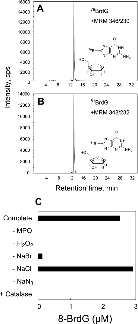
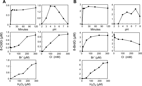
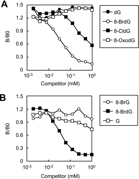
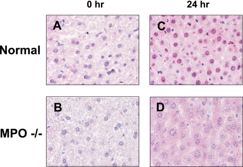
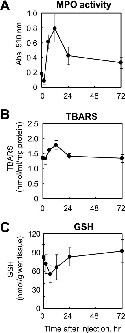
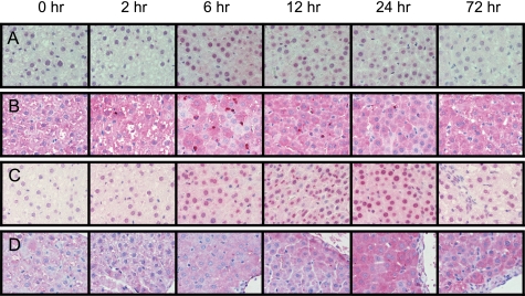
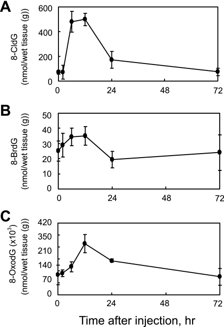
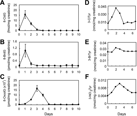
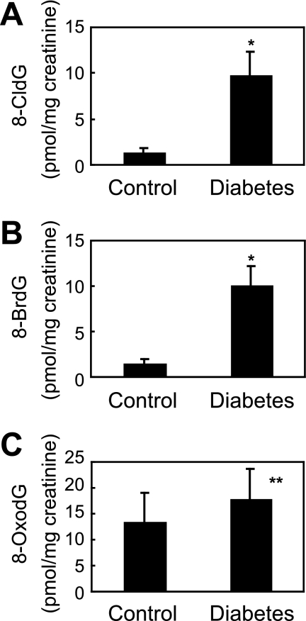
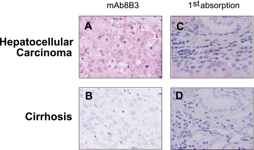
Myeloperoxidase (MPO) generates reactive halogenating species that can modify DNA. The aim of this study was to investigate the formation of 8-halogenated 2'-deoxyguanosines (8- halo-dGs) during inflammatory events. 8-Bromo-2'-dG (8-BrdG) and 8-chloro-2'-dG (8-CldG) were generated by treatment of MPO with hydrogen peroxide at physiological concentrations of Cl(-) and Br(-). The formation of 8-halo-dGs with other oxidative stress biomarkers in lipopolysaccharide-treated rats was assessed by liquid chromatography tandem mass spectrometry and immunohistochemistry using a novel monoclonal antibody (mAb8B3) to 8-BrdG-conjugated keyhole limpet hemocyanin. The antibody recognized both 8-BrdG and 8-CldG. In the liver of lipopolysaccharide-treated rats, immunostaining for 8-halo-dGs, halogenated tyrosines, and MPO were increased at 8 h, whereas those of 8-oxo-2'-dG (8-OxodG) and 3-nitrotyrosine were increased at 24 h. Urinary excretion of both 8-CldG and 8-BrdG was also observed earlier than those of 8-OxodG and modified tyrosines (3-nitrotyrosine, 3-chlorotyrosine, and 3- bromotyrosine). Moreover, the levels of the 8-halo-dGs in urine from human diabetic patients were 8-fold higher than in healthy subjects (n = 10, healthy and diabetic, p < 0.0001), whereas there was a moderate difference in 8-OxodG between the two groups (p < 0.001). Interestingly, positive mAb8B3 antibody staining was observed in liver tissue from hepatocellular carcinoma patients but not in liver tissue from human cirrhosis patients. These data suggest that 8-halo-dGs may be potential biomarkers of early inflammation.


Figures

FIGURE 1.
FIGURE 1.
8-BrdG generation by the MPO-H2O2-Cl-Br system. dG (1 mm) was incubated with 20 nm MPO, 100 μm H2O2, 100 mm NaCl, and 100 μm NaBr in 50 mm sodium phosphate buffer (pH 7.4) for 60 min at 37 °C. The reaction conditions were varied by adding or removing components as indicated. 8-BrdG was identified and quantified by LC-MS/MS. The scans of multiple reaction monitoring (MRM) for 79BrdG (346/230) and 81BrdG (348/232) generated from the MPO-H2O2-Cl-Br system were performed separately (A and B). Quantification of 8-BrdG was performed as described under “Experimental Procedures” (C). NaN3, 10 mm sodium azide; Catalase, 10 mg/ml catalase.
FIGURE 2.
FIGURE 2.
Characterization of halogenation of dG by the MPO-H2O2-Cl-Br system. dG (1 mm) was incubated with 20 nm MPO, 100 μm H2O2, 100 mm NaCl, and 100 μm NaBr in 50 mm sodium phosphate buffer (pH 7.4) for 60 min at 37 °C. The reaction conditions were varied by altering the concentration of hydrogen ions or the length of the reaction time. 8-CldG (A) and 8-BrdG (B) were quantified by LC-MS/MS.
FIGURE 3.
FIGURE 3.
Characterization of the specificity of the monoclonal antibody against 8-halo-dGs. The cross-reactivity of the antibody (mAb8B3) with the 8-modified dG (A) or 8-substituted G (B) was estimated by competitive indirect ELISA. 8-BrG, 8-bromoguanine; G, guanosine.
FIGURE 4.
FIGURE 4.
Immunohistochemical detection of 8-halo-dGs with mAb8B3 in the liver of wild type or MPO−/− mice treated with LPS. A, wild type normal mouse liver. B, control MPO−/− mouse liver. C, LPS 24 h wild type mouse liver. D, LPS 24 h MPO−/− mouse liver. All of the sections were counterstained with hematoxylin. Magnification was ×400.
FIGURE 5.
FIGURE 5.
Intraperitoneal LPS administration to rats affects activity of MPO, TBARS, and GSH in the liver. The activity levels of MPO (A), TBARS (B), and GSH (C) in the liver of rats after LPS treatment were measured as described under “Experimental Procedures.”
FIGURE 6.
FIGURE 6.
Immunohistochemical detection of time-dependent generation of oxidative stress markers. 8-Halo-dGs (A), MPO (B), 8-OxodG (C), and 3-NO2Tyr (D) in the liver of rats treated with LPS were immunostained. All of the sections were counterstained with hematoxylin. Magnification was ×400.
FIGURE 7.
FIGURE 7.
LPS-induced change of 8-modified dG in the rat liver. Quantification of 8-modified dG (A, 8-CldG; B, 8-BrdG; C, 8-OxodG) in the livers of LPS-treated rats was performed by LC-MS/MS using internal standards. Briefly, 8-CldG, 8-BrdG, and 8-OxodG were detected in the liver after DNA extraction, DNA digestion, and fractionated by HPLC supplemented with the internal standards 15N5-8-CldG, 15N5-8-BrdG, and 15N5-8-OxodG. The multiple reaction monitoring for each compound was: 15N5-8-CldG (m/z 307.0 → 190.9), 8-CldG (m/z 302.1 → 185.9), 15N5-8-BrdG (m/z 350.9 → 235.0), 8-BrdG (m/z 345.9 → 229.9), 15N5-8-OxodG (m/z 288.9 → 173.1), and 8-OxodG (m/z 284.0 → 168.1), respectively.
FIGURE 8.
FIGURE 8.
LPS-induced change of modified dGs and tyrosines in rat urines. A–C, urine of rats was diluted 5× by water. The fraction containing 8-modified dG for the samples was collected by HPLC. Quantification of 8-modified dG (A, 8-CldG; B, 8-BrdG; C, 8-OxodG) in fractions was performed by LC-MS/MS using internal standards as described in legend to Fig. 7. D–F, urine was supplemented with each internal standard before solid phase extraction. After the extraction, 3-chlorotyrosine (3-ClTyr), 3-bromotyrosine (3-BrTyr), and 3-NO2Tyr were derivatized with butanol and HCl. Quantification of 3-chlorotyrosine (D), 3-bromotyrosine (E), and 3-NO2Tyr (F) in the urine of rats treated with LPS was performed by LC-MS/MS using internal standards.
FIGURE 9.
FIGURE 9.
Quantification of 8-CldG, 8-BrdG, and 8-OxodG in control and diabetic human urine using LC-MS/MS. Human urine with protein removed was fractionated by HPLC and then dried. After resolving in 50 μl of diluted solution (water:acetnitrile = 1:1), 10 μl of samples was analyzed by LC-MS/MS. *, p < 0.0001; **, p < 0.001.
FIGURE 10.
FIGURE 10.
Immunohistochemical detection of 8-halo-dGs in human livers with cirrhosis or hepatocellular carcinoma. The sections of hepatocellular carcinoma (A) and cirrhosis (B) livers were immunostained with mAb8B3. Competitive experiments were performed by preincubation with respective antigens (C and D). All of the sections were counterstained with hematoxylin. Magnification was ×400.

Similar articles

Cited by

References

    1. Hazen S. L., Heinecke J. W. (1997) J. Clin. Invest. 99, 2075–2081 - PMC - PubMed
    1. Ohshima H., Bartsch H. (1994) Mutat. Res. 305, 253–264 - PubMed
    1. Takeshita J., Byun J., Nhan T. Q., Pritchard D. K., Pennathur S., Schwartz S. M., Chait A., Heinecke J. W. (2006) J. Biol. Chem. 281, 3096–3104 - PubMed
    1. Weitzman S. A., Gordon L. I. (1990) Blood 76, 655–663 - PubMed
    1. Henderson J. P., Byun J., Heinecke J. W. (1999) J. Biol. Chem. 274, 33440–33448 - PubMed

Publication types

MeSH terms