Review
. 2010 Mar;74(3):390-404.
doi: 10.1253/circj.cj-09-0923. Epub 2010 Jan 18.

Cardiac progenitor cells and bone marrow-derived very small embryonic-like stem cells for cardiac repair after myocardial infarction

Affiliations
Review

Cardiac progenitor cells and bone marrow-derived very small embryonic-like stem cells for cardiac repair after myocardial infarction

Xian-Liang Tang et al. Circ J. 2010 Mar.

Abstract

Heart failure after myocardial infarction (MI) continues to be the most prevalent cause of morbidity and mortality worldwide. Although pharmaceutical agents and interventional strategies have contributed greatly to therapy, new and superior treatment modalities are urgently needed given the overall disease burden. Stem cell-based therapy is potentially a promising strategy to lead to cardiac repair after MI. An array of cell types has been explored in this respect, including skeletal myoblasts, bone marrow (BM)-derived stem cells, embryonic stem cells, and more recently, cardiac progenitor cells (CPCs). Recently studies have obtained evidence that transplantation of CPCs or BM-derived very small embryonic-like stem cells can improve cardiac function and alleviate cardiac remodeling, supporting the potential therapeutic utility of these cells for cardiac repair. This report summarizes the current data from those studies and discusses the potential implication of these cells in developing clinically-relevant stem cell-based therapeutic strategies for cardiac regeneration.


Figures

Figure 1
Figure 1
Administration of cardiac progenitor cells (CPCs) promotes myocardial regeneration in rats after myocardial infarction. (A) Large transverse section halfway between the base and the apex of the left ventricle, showing an infarct in a CPC-treated rat. (Inset) Regenerated infarcted myocardium (EGFP and α-sarcomeric actin, yellow-green). (Bars, 1 mm and 100 µm.) (B) Another example of regenerated infarcted myocardium (arrows) is shown first by α-sarcomeric actin staining (red), then by EGFP labeling (green) and then by the combination of EGFP and α-sarcomeric actin (yellow-green). (C) EGFPpos (green), α-sarcomeric actinpos (red), small newly farmed myocytes with in the infarcted region express in their nuclei GATA-4 (white) and MEF2C (magenta). (D) EGFPpos (green), cardiac myosin heavy chainpos (red) small newly formed myocytes within the infarcted region express in their plasma membrane connexin 43 (magenta, arrowheads). (E) Newly formed myocytes in the surviving non-infarcted left ventricular myocardium (arrows) are shown first by EGFP labeling (green) and then by the combination of EGFP and α-sarcomeric actin (yellow-green). Bar (BE)=10 µm. (Reprinted from Dawn et al.35)
Figure 2
Figure 2
Administration of cardiac stem cells improves left ventricular (LV) systolic function in rats after myocardial infarction: echocardiographic measurements in untreated and treated animals. *P<0.05 between untreated and treated rats. Data are mean±SEM. BSL, baseline. (Modified from Dawn et al.35)
Figure 3
Figure 3
Administration of cardiac progenitor cells improves postinfarction ventricular remodeling in rats after myocardial infarction: multiple anatomical parameters obtained in hearts arrested in diastole. *P<0.05 between untreated and treated rats. Data are mean±SEM. LV, left ventricular. (Reprinted from Dawn et al.35)
Figure 4
Figure 4
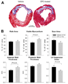
Morphometric analysis in rats after myocardial infarction. (A) Representative Masson's trichrome-stained myocardial sections from a vehicle-treated and a CPC-treated rat. Scar tissue and viable myocardium are blue and red, respectively. (B) Quantitative analysis of LV morphometric parameters. Data are means±SEM. CPC, cardiac progenitor cell; LV, left ventricular. (Reprinted from Tang et al.44)
Figure 5
Figure 5
Cardiac function assessed by echocardiography and hemodynamics in rats after myocardial infarction (MI). (A) Quantitative echocardiographic parameters including systolic thickness of the anterior (infarcted) wall, thickening fraction in the anterior (infarcted) wall, left ventricular (LV) fractional shortening, and LV ejection fraction in vehicle-treated (n=14) and cardiac progenitor cell (CPC)-treated group (n=17) at baseline (BSL), at 30 days (d) after MI (before vehicle or CPC treatment) (MI), and at 35 d after treatment. (B) Quantitative analysis of hemodynamic variables including LV end-diastolic pressure, dP/dt, ejection fraction, end-systolic elastance (Ees, preload-adjusted maximal power, and preload-recruitable stroke work from normal (n = 4), vehicle-treated (n=15), and CPC-treated rats (n=15) at 35 d after treatment. Data are means±SEM. (Reprinted from Tang et al.44)
Figure 6
Figure 6
Correlation between the amount of viable myocardium in the risk region and hemodynamic variables, including left ventricular (LV) pressure, dP/dt, ejection fraction, end-systolic elastance, preload-adjusted maximal power, and preload recruitable stroke work in rats after myocardial infarction. Correlation analyses were performed by pooling all rats together, regardless of treatment, although the distribution is illustrated separately for vehicle-treated (solid circles) and cardiac progenitor cell (CPC)-treated rats (open circles). (Reprinted from Tang et al.44)
Figure 7
Figure 7
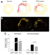
Myocardial collagen content in rats after myocardial infarction. Representative microscopic images of a normal, vehicle-treated, and cardiac progenitor cell (CPC)-treated heart obtained using (A) regular and (B) polarized light. (C) Collagen content expressed as percent of total area in the scarred and noninfarcted region, left ventricular sections were stained with picrosirius red and collagen content was quantitated under polarized light. Data are means±SEM. (Reprinted from Tang et al.44)
Figure 8
Figure 8
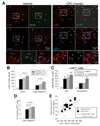
Effect of cardiac progenitor cell (CPC) transplantation on c-kitpos cells in rats after myocardial infarction. (A) Representative confocal microscopic images from a vehicle- treated and a CPC-treated rat showing c-kitpos cells (red) and BrdUpos cells (green) in the risk region. Yellow arrows indicate c-kitpos cells that are BrdU positive; while arrows show c-kitneg cells that are BrdU positive. (B) Quantitative analysis of c-kitpos cells and double positive (c-kitpos/BrdUpos) cells in normal, vehicle-treated, and CPC-treated hearts (the last group is subdivided into 2 subgroups: with EGFPpos cells [EGFPpos] and without EGFPpos cells [EGFPneg]). (C) Quantitative analysis of c-kitpos cells in the risk and noninfarcted regions in normal, vehicle-treated, and CPC-treated hearts [as in (B), the last group is subdivided into EGFPpos and EGFPneg subgroups]. (D) Quantitative analysis of double positive (c-kitpos/BrdUpos) cells expressed as a percent of all c-kitpos cells. (E) Correlation between the number of c-kitpos cells and the percent of viable myocardium in the risk region (r=0.59, P<0.01). Data are means±SEM. (Reprinted from Tang et al.44)
Figure 9
Figure 9
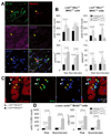
Cardiac commitment of endogenous cardiac progenitor cells (CPCs) after trasplantation of exogenous CPCs in rats after myocardial infarction. The commitment of endogenous CPCs (c-kitpos/EGFPneg cells) to the cardiac lineage was assessed by quantitating endogenous CPCs that expressed Nkx2.5 and MHC in vehicle- and CPC-treated hearts. New myocytes formed in the last 2 weeks of life were identified by measuring α-sarcomeric actinpos and BrdUpos cells. (A) Representative confocal microscopic image of a c-kitpos/EGFPneg/Nkx2.5pos/MHCpos cell obtained in serial sections of a CPC-treated heart. (B) Quantitative analysis of c-kitpos/EGFPneg/Nkx2.5pos/MHCpos cells in the risk and noninfarcted regions of vehicle- and CPC-treated hearts (the CPC-treated group is subdivided into 2 subgroups: with EGFPpos cells [EGFPpos] and without EGFPpos cells [EGFPneg]). The number of c-kitpos/EGFPneg/Nkx2.5pos/MHCpos cells is normalized to both number of cells (104 nuclei, Upper panels) and area (mm2, Lower panels). Because cell density in the CPC-treated, EGFPpos hearts was higher (because of clusters of EGFPpos cells), the magnitude of the increase in endogenous CPCs in CPC-treated EGFPpos hearts was greater when the number of cells was normalized to area (mm2) than to the number of nuclei. (C) Representative confocal microscopic image of α-sarcomeric actinpos/BrdUpos cells in a CPC-treated heart. (D) Quantitative analysis of the number of α-sarcomeric actinpos/BrdUpos cells in the risk and noninfarcted regions of vehicle- and CPC-treated hearts (subdivided into EGFPneg and EGFPpos subgroups). Data are means±SEM. (Reprinted from Tang et al.44)
Figure 10
Figure 10
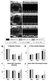
Echocardiographic assessment of left ventricular (LV) function in mice after myocardial infarction. Representative two-dimensional (A,C,E) and M-mode (B,D,F) images from vehicle-treated (A,B), CD45+ cell-treated (C,D), and very small embryonic-like stem cell (VSEL)-treated (E,F) mice 35 days (d) after coronary occlusion/reperfusion. The infarct wall is delineated by arrowheads (A,C,E). Compared with the vehicle-treated and CD45+ cell-treated hearts, the VSEL-treated heart had a smaller LV cavity, thicker infarct wall, and improved motion of the infarct wall. (GJ) Transplantation of VSEL improved echocardiographic measurements of LV systolic function 35 d after myocardial infarction. Data are mean±SEM. n=11–14 mice per group. *P<0.05 vs group II at 35 d; #P<0.05 vs group I at 35 d; §P<0.05 vs values at 96 h in respective groups. BSL, baseline. (Reprinted from Dawn et al.50)
Figure 11
Figure 11
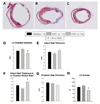
Morphometric assessment of left ventricular (LV) remodeling in mice after myocardial infarction. Representative Masson's trichrome-stained myocardial sections from Vehicle-treated (A), CD45+ hematopoietic stem cell-treated (B), and very small embryonic-like stem cell (VSEL)-treated (C) hearts. Scar tissue and viable myocardium are blue and red, respectively. Note that the LV cavity is smaller and the infarct wall thicker in the VSEL-treated heart. (DH) Morphometric measurements of LV structural parameters. Data are mean±SEM. n=11–14 mice per group. *P<0.05 vs group II. (Reprinted from Dawn et al.50)
Figure 12
Figure 12
Assessment of cardiomyocyte and left ventricular (LV) hypertrophy in mice after myocardial infarction. (AC) Representative images of cardiomyocytes in the viable myocardium from Masson's trichrome-stained vehicle-treated (A), CD45+ hematopoietic stem cell-treated (B), and very small embryonic-like stem cell (VSEL)-treated hearts (C). Scale bars=50 µm. In contrast to CD45+ hematopoietic stem cell-treated hearts, VSEL-treated hearts do not have an increased myocyte cross-sectional area compared with noninfarcted control hearts (D). Echocardiographically estimated LV mass was significantly less in VSEL-treated hearts (E). Data are mean±SEM. n=11–14 mice per group. (D) *P<0.05 vs group II; #P<0.05 vs control; (E) *P<0.05 vs group II and III (final); #P<0.05 vs respective baseline values. (Reprinted from Dawn et al.50)

Similar articles

Cited by

References

    1. Dimmeler S, Zeiher AM, Schneider MD. Unchain my heart: The scientific foundations of cardiac repair. J Clin Invest. 2005;115:572–583. - PMC - PubMed
    1. Prockop DJ. Marrow stromal cells as stem cells for nonhematopoietic tissues. Science. 1997;276:71–74. - PubMed
    1. Orlic D, Kajstura J, Chimenti S, Jakoniuk I, Anderson SM, Li B, et al. Bone marrow cells regenerate infarcted myocardium. Nature. 2001;410:701–705. - PubMed
    1. Kajstura J, Rota M, Whang B, Cascapera S, Hosoda T, Bearzi C, et al. Bone marrow cells differentiate in cardiac cell lineages after infarction independently of cell fusion. Circ Res. 2005;96:127–137. - PubMed
    1. Rota M, Kajstura J, Hosoda T, Bearzi C, Vitale S, Esposito G, et al. Bone marrow cells adopt the cardiomyogenic fate in vivo. Proc Natl Acad Sci USA. 2007;104:17783–17788. - PMC - PubMed

Publication types