Review
. 2010 Feb 1;9(3):487-97.
doi: 10.4161/cc.9.3.10508.

Regulation of Akt signaling activation by ubiquitination

Affiliations
Review

Regulation of Akt signaling activation by ubiquitination

Wei-Lei Yang et al. Cell Cycle. .

Abstract

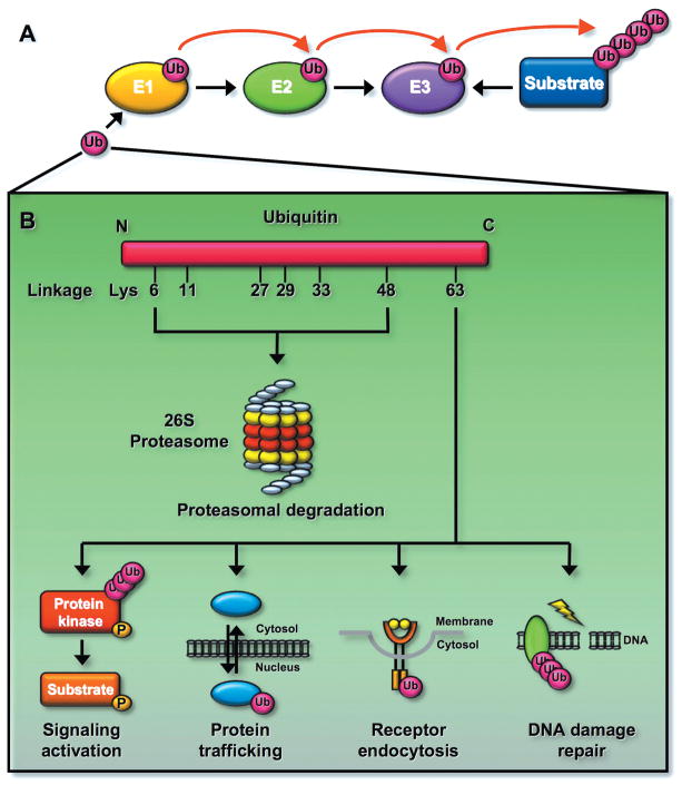
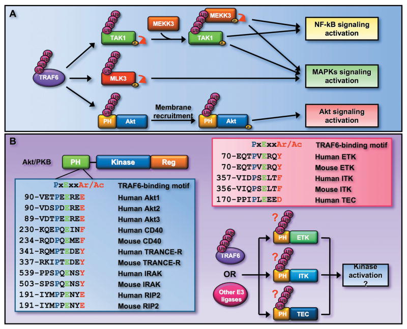
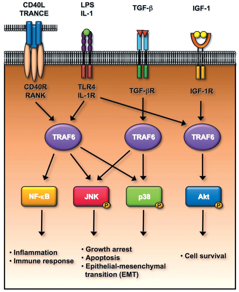
Akt (also known as PKB) signaling orchestrates many aspects of biological functions and, importantly, its deregulation is linked to cancer development. Akt activity is well-known regulated through its phosphorylation at T308 and S473 by PDK1 and mTOrC2, respectively. Although in the last decade the research has been primarily focused on Akt phosphorylation and its role in Akt activation and functions, other posttranslational modifications on Akt have never been reported. Until very recently, a novel posttranslational modification on Akt termed ubiquitination was identified and shown to play an important role in Akt activation. The cancer-associated Akt mutant recently identified in a subset of human cancers displays enhanced Akt ubiquitination, in turn contributing to Akt hyperactivation, suggesting a potential role of Akt ubiquitination in cancers. Thus, this novel posttranslational modification on Akt reveals an exciting avenue that has advanced our current understandings of how Akt signaling activation is regulated.


Figures

Figure 1
Figure 1
Ubiquitination can regulate protein degradation or activation. (A) Ubiquitination reaction involves three enzymes. Ubiquitin is activated by the E1 and is transferred to the E2. The E3 recognizes its protein substrates and brings them to the E2, resulting in protein ubiquitination. (B) Ubiquitination regulates the fate of proteins. Ubiquitin consists of seven lysine (K) residues. The K48-linked ubiquitination is recognized by the 26S proteasome and results in protein degradation. In contrast, the K63-linked ubiquitination does not induce protein degradation since it is not recognized by the proteasome, instead it regulates signaling activation involved in distinct biological functions including DNA damage response, receptor endocytosis, and protein trafficking. Although the function for other types of ubiquitnation is less clear, it is proposed that these types of ubiquitination may also regulate protein degradation.
Figure 2
Figure 2
Akt regulates numerous biological functions by phosphorylating distinct protein substrates. For instance, Akt protects cells from apoptosis by phosphorylating and inactivating proapoptotic proteins, such as Bad and Foxo3a. Akt regulates cell growth and protein translation by phosphorylating and inactivating TSC2, resulting in activation of the mTOR pathway. Akt regulates cell cycle progression and tumorigenesis by phosphorylating and activating oncogenic proteins, such as Skp2, Mdm2, and IKKα. Akt can orchestrate glucose metabolism by regulating the activity of GSK3β. Akt may also regulate cell migration by inducing the phosphorylation and activation of Skp2, Girdin/APE, ACAP1, and PAK1.
Figure 3
Figure 3
The activity of Akt is regulated by phosphorylation and dephosphorylation of Akt. β-arrestin-2 recruits Src and Akt to the activated insulin receptor, and phosphoryaltion of Akt at Y315 and Y326 by Src is a prerequisite for Akt T308 and S473 phosphorylaiton induced by PDK1 and mTOC2, respectively, leading to the full activation of Akt. Akt T308 dephosphorylation is triggered by PP2A phosphatase. PML or β-arrestin-2 recruits PP2A and facilitates Akt T308 dephosphorylation. Dephosphorylation of Akt at S473 is induced by PHLPP phosphates. The adaptor protein FKBP51 recruits PHLPP to elicit Akt dephosphorylation at S473.
Figure 4
Figure 4
Akt membrane localization and activation is regulated by ubiquitination of Akt. TRAF6 E3 ligase is activated by engagement of IGF-1, IL-1, and LPS to their cognate receptors. The activated TRAF6 then interacts with Akt and triggers K63-linked ubiquitination of Akt, which facilitates Akt membrane recruitment and subsequent phosphorylation by PDK1 and mTORC2 through an unknown mechanism. It is possible that the ubiquitinated Akt may recruit the essential adaptors to facilitate Akt membrane localization.
Figure 5
Figure 5
K63-linked ubiquitination may serve as a general mechanism for kinase activation. (A) TRAF6 induces the activation of TAK1 and MLK3 by triggering their K63-linked ubiquitination. TRAF6 also induces K63 ubiquitination of Akt within its PH domain and regulates Akt activation. (B) TRAF6 consensus binding motif is conserved among all three Akt isoforms and is found in several other TRAF6 interacting proteins. The PH domain is also found in several other kinases, such as ETK, ITK, and TEC, which are important downstream effectors for growth factor receptor and TCR signaling. Interestingly, these kinases also contain the TRAF6 consensus binding motif, suggesting that TRAF6 or other E3 ligases may induce the K63-linked ubiquitinaiton of these kinases and in turn regulates their activation.
Figure 6
Figure 6
TRAF6 E3 ligase regulates multiple signal transduction pathways involved in the plethora of biological functions including the innate immune response, cell survival, apoptosis, and epithelial-mesenchymal transition (EMT). TRAF6 is a critical mediator for NF-κB activation upon the engagement of CD40L, TRANCE, LPS and IL-1 to their receptors, in turn regulating inflammation and innate immune response. TRAF6 is activated by TGF-β/TGF-β receptor and is required for TGF-β-mediated p38 and JNK activation. The activated p38 and JNK positively regulate cell apoptosis and EMT. Interestingly, TRAF6 is also activated upon the binding of IGF-1 to IGF-1 receptor and regulates cell survival by inducing Akt activation.

References

    1. Bellacosa A, Testa JR, Staal SP, Tsichlis PN. A retroviral oncogene, akt, encoding a serine-threonine kinase containing an SH2-like region. Science. 1991;254:274–7. - PubMed
    1. Coffer PJ, Woodgett JR. Molecular cloning and characterisation of a novel putative protein-serine kinase related to the cAMP-dependent and protein kinase C families. Eur J Biochem. 1991;201:475–81. - PubMed
    1. Jones PF, Jakubowicz T, Pitossi FJ, Maurer F, Hemmings BA. Molecular cloning and identification of a serine/threonine protein kinase of the second-messenger subfamily. Proc Natl Acad Sci U S A. 1991;88:4171–5. - PMC - PubMed
    1. Hennessy BT, Smith DL, Ram PT, Lu Y, Mills GB. Exploiting the PI3K/AKT pathway for cancer drug discovery. Nat Rev Drug Discov. 2005;4:988–1004. - PubMed
    1. Liu P, Cheng H, Roberts TM, Zhao JJ. Targeting the phosphoinositide 3-kinase pathway in cancer. Nat Rev Drug Discov. 2009;8:627–44. - PMC - PubMed

Publication types

Substances

LinkOut - more resources