

In vivo endotoxin synchronizes and suppresses clock gene expression in human peripheral blood leukocytes
- PMID: 20081528
- PMCID: PMC2929957
- DOI: 10.1097/CCM.0b013e3181cd131c
In vivo endotoxin synchronizes and suppresses clock gene expression in human peripheral blood leukocytes
Abstract
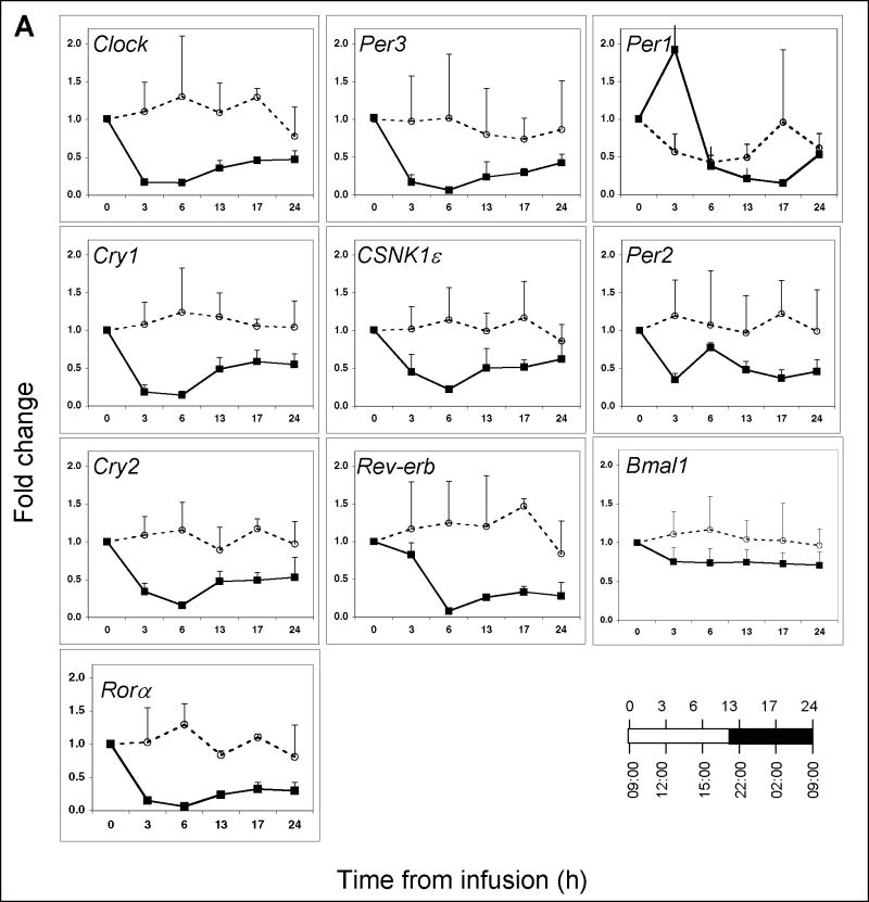
Objectives: The intravenous administration of a bolus dose of endotoxin to healthy human subjects triggers acute systemic inflammatory responses that include cytokine production and dynamic changes in gene expression in peripheral blood leukocytes. This study sought to determine the state of clock gene expression in human peripheral blood leukocytes, and leukocyte subpopulations, challenged with in vivo endotoxin at two circadian/diurnal phases of the clock.
Design: Clinical and laboratory investigation.
Setting: University-based research laboratory and clinical research center.
Subjects: Human volunteers.
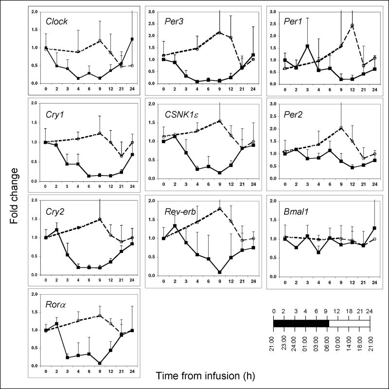
Interventions: Human subjects were administered a standard dose of endotoxin (2 ng/kg) or saline at either 0900 or 2100 hrs. Blood samples were collected at selected time points pre- and postinfusion.
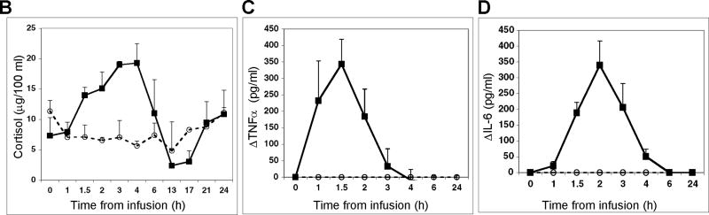
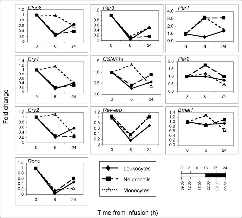
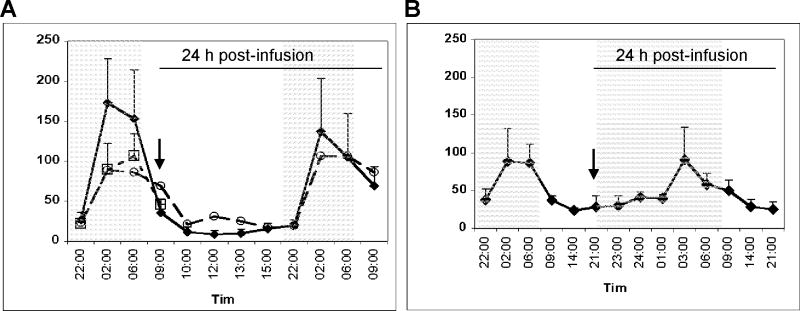
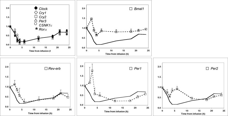
Measurements and main results: Clock gene expression was determined in human peripheral blood leukocytes, neutrophils, and monocytes by quantitative real-time polymerase chain reaction. The fold change for each gene was determined by use of the 2 method. We show that endotoxin causes profound suppression of circadian clock gene expression, clearly manifested in human peripheral blood leukocytes, neutrophils, and monocytes. Clock, Cry1-2, Per3, CSNK1epsilon, Rora, and Rev-erb gene expression were all reduced by 80% to 90% with the nadir between 3 and 6 hrs postinfusion. Per1 and Per2 reached an expression nadir between 13 and 17 hrs postinfusion. The levels of plasma interleukin-6 and tumor necrosis factor peaked and then returned to baseline within 6 hrs. In contrast, clock gene expression remained suppressed for up to 17 hrs irrespective of the phase of the clock at the time of the endotoxin challenge. Endotoxin did not perturb the melatonin secretory rhythm.
Conclusions: Circadian clock gene expression in peripheral blood leukocytes is dramatically altered and possibly uncoupled from the activity of the central clock during periods of acute systemic inflammation. The realignment of the central and peripheral clocks may constitute a previously unappreciated key factor affecting recovery from disease in humans.
Conflict of interest statement
The authors have no potential conflicts of interest to disclose.
Figures
Comment in
-
Endotoxin desynchronizes biological clocks.Crit Care Med. 2010 Mar;38(3):977-8. doi: 10.1097/CCM.0b013e3181cfb33d. Crit Care Med. 2010. PMID: 20168150 No abstract available.
References
-
- Turek FW. Are the suprachiasmatic nuclei the location of the biological clock in mammals? Nature. 1981;292(5821):289–290. - PubMed
-
- Albrecht U, Eichele G. The mammalian circadian clock. Curr Opin Genet Dev. 2003;13(3):271–277. - PubMed
-
- Reppert SM, Weaver DR. Coordination of circadian timing in mammals. Nature. 2002;418(6901):935–941. - PubMed
Publication types
MeSH terms
Substances
Grants and funding
LinkOut - more resources
Full Text Sources
Other Literature Sources
