. 2010 Feb;6(2):117-24.
doi: 10.1038/nchembio.293.

The p110 delta structure: mechanisms for selectivity and potency of new PI(3)K inhibitors

Affiliations

The p110 delta structure: mechanisms for selectivity and potency of new PI(3)K inhibitors

Alex Berndt et al. Nat Chem Biol. 2010 Feb.

Abstract

Deregulation of the phosphoinositide-3-OH kinase (PI(3)K) pathway has been implicated in numerous pathologies including cancer, diabetes, thrombosis, rheumatoid arthritis and asthma. Recently, small-molecule and ATP-competitive PI(3)K inhibitors with a wide range of selectivities have entered clinical development. In order to understand the mechanisms underlying the isoform selectivity of these inhibitors, we developed a new expression strategy that enabled us to determine to our knowledge the first crystal structure of the catalytic subunit of the class IA PI(3)K p110 delta. Structures of this enzyme in complex with a broad panel of isoform- and pan-selective class I PI(3)K inhibitors reveal that selectivity toward p110 delta can be achieved by exploiting its conformational flexibility and the sequence diversity of active site residues that do not contact ATP. We have used these observations to rationalize and synthesize highly selective inhibitors for p110 delta with greatly improved potencies.


Figures

Figure 1
Figure 1
Domain organization, construct design and overall crystal structure of p110δ. A) A TEV protease cleavage site was introduced between residues 105 and 106 of the p110δ ABD-RBD linker. The numbers below the boxes correspond to the indicated domain boundaries. After purification of the p110δ/iSH2 complex, the catalytic core is released by cleavage with TEV protease. B) Cartoon representation of the overall co-crystal structure of the ΔABDp110δ/PIK-39 complex. Linker regions are colored in white, the RBD in salmon, the C2 domain in cyan, the helical domain in green, the kinase domain N-lobe in red and the kinase domain C-lobe in yellow. PIK-39 is shown in light blue as a ball and stick representation. Selected secondary structure elements of the kinase domain are labeled.
Figure 2
Figure 2
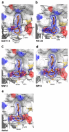
The propeller-shaped p110δ-selective inhibitors induce the formation of the “specificity” pocket. Shown are the active sites of p110δ in complex with the inhibitors IC87114 (a), PIK-39 (b), SW13 (c), SW14 (d) and SW30 (e). Key residues that outline the active site and interact with the compounds and the 2mFo-DFc electron densities (contouring level 1σ) are presented. Selected water molecules in the active sites are shown as gray spheres. Note, that IC87114 and PIK-39 do not fill the “affinity” pocket, whereas SW13, SW14 and SW30 do. Dashed black lines represent hydrogen bonds.
Figure 3
Figure 3
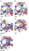
The flat inhibitors DL06, DL07, ZSTK474, AS5 and GDC-0941 are multi- to pan-selective class I PI3K inhibitors that do not induce the opening of the “specificity” pocket. Shown are the binding modes of DL06 (a), DL07 (b), ZSTK474 (c), AS5 (d) and GDC-0941 (e) in the active site of p110δ. Met752 is in its “in” position for all these compounds. For panel (d), the structure of the p110γ/ATP complex (PDB entry 1e8x) was superimposed on the Cα-backbone of p110δ to show the proximity of the sulfonyl group of AS5 to the alpha phosphate group of ATP (purple). This sulfonyl group is a hydrogen bond acceptor to Ser754 located in the P-loop of p110δ. (e) GDC-0941 is a pan-class IA PI3K inhibitor that (like AS15) interacts with residues outside the active site. GDC-0941 occupies the “adenine” pocket and the “affinity” pocket within the active site of p110δ and engages there in hydrogen bonds with Val828, Tyr813 and Asp787. Additionally, the substituted piperazine group of GDC-0941 extends out of the ATP-binding site where its methylsulfonyl moiety acts as a hydrogen bond acceptor for Asp753 of the P-loop and Lys708 at the beginning of kα2. The contouring level of the 2mFo-DFc electron densities is 1σ for each compound.
Figure 4
Figure 4
Binding mode of the p110δ-selective PI3K inhibitor AS15 and comparison of AS15 with the propeller-shaped inhibitor PIK-39 (2mFo-DFc contouring level 1σ). (a) The highly p110δ-selective compound AS15 does not open the “specificity” pocket and makes extensive use of a hydrophobic patch between Trp760, Thr750 and Met752 adjacent to the adenine-binding pocket. (b) Chemical structures of the highly p110δ-selective inhibitors AS15 and PIK-39. (d) Superposition of the AS15 and PIK-39 to demonstrate their different mode of binding within the active site of p110δ.

Comment in

Similar articles

Cited by

References

    1. Vanhaesebroeck B, et al. Synthesis and function of 3-phosphorylated inositol lipids. Annu Rev Biochem. 2001;70:535–602. - PubMed
    1. Zhao L, Vogt PK. Class I PI3K in oncogenic cellular transformation. Oncogene. 2008;27:5486–5496. - PMC - PubMed
    1. Manning BD, Cantley LC. AKT/PKB signaling: navigating downstream. Cell. 2007;129:1261–1274. - PMC - PubMed
    1. Sundstrom TJ, Anderson AC, Wright DL. Inhibitors of phosphoinositide-3-kinase: a structure-based approach to understanding potency and selectivity. Org Biomol Chem. 2009;7:840–850. - PubMed
    1. Miled N, et al. Mechanism of two classes of cancer mutations in the phosphoinositide 3-kinase catalytic subunit. Science. 2007;317:239–242. - PubMed

Publication types

Substances