. 2010 Feb;12(2):153-63.
doi: 10.1038/ncb2015. Epub 2010 Jan 17.

Muscle injury activates resident fibro/adipogenic progenitors that facilitate myogenesis

Affiliations

Muscle injury activates resident fibro/adipogenic progenitors that facilitate myogenesis

Aaron W B Joe et al. Nat Cell Biol. 2010 Feb.

Abstract

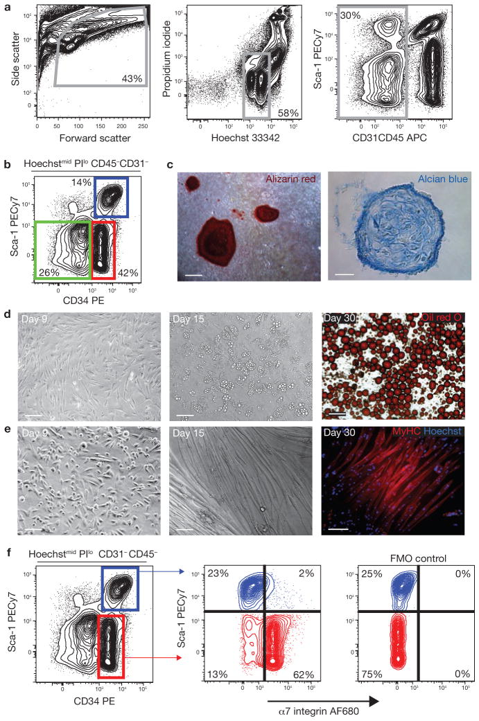
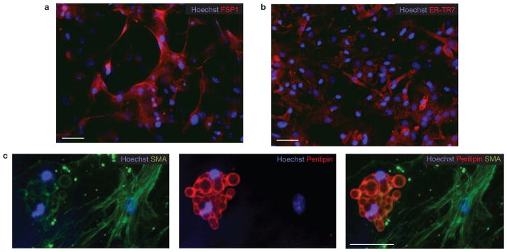
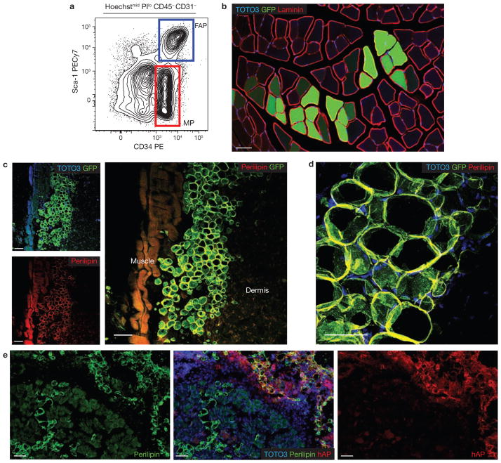
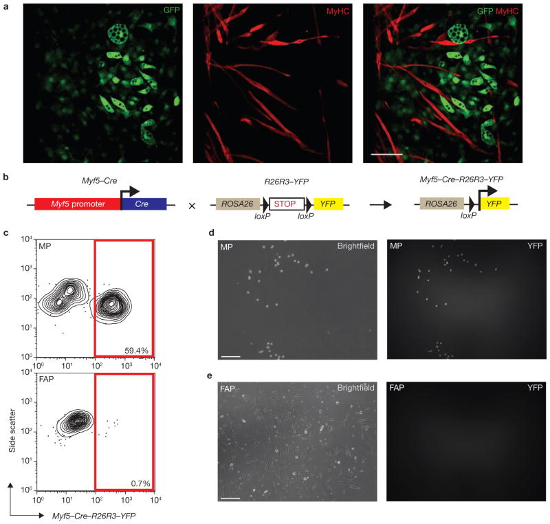
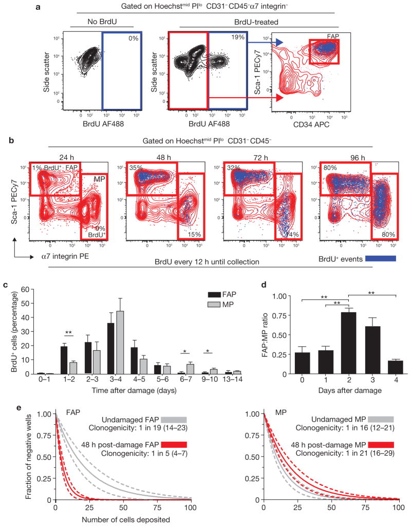
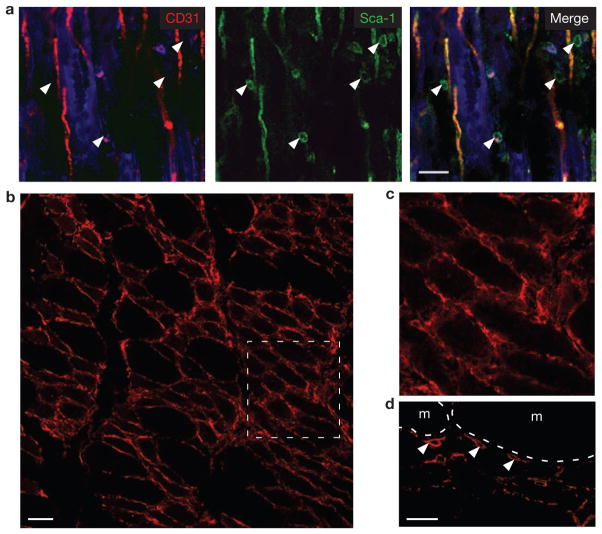
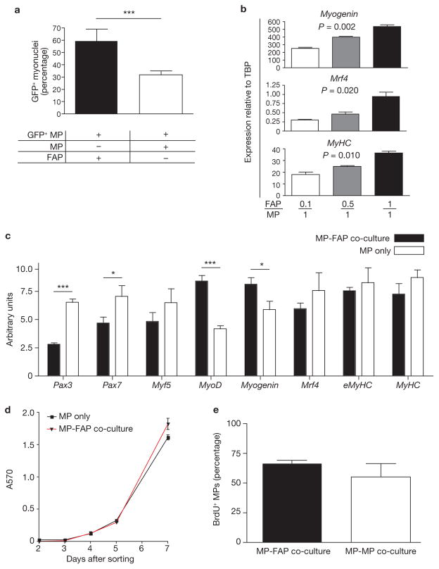
Efficient tissue regeneration is dependent on the coordinated responses of multiple cell types. Here, we describe a new subpopulation of fibro/adipogenic progenitors (FAPs) resident in muscle tissue but arising from a distinct developmental lineage. Transplantation of purified FAPs results in the generation of ectopic white fat when delivered subcutaneously or intramuscularly in a model of fatty infiltration, but not in healthy muscle, suggesting that the environment controls their engraftment. These cells are quiescent in intact muscle but proliferate efficiently in response to damage. FAPs do not generate myofibres, but enhance the rate of differentiation of primary myogenic progenitors in co-cultivation experiments. In summary, FAPs expand upon damage to provide a transient source of pro-differentiation signals for proliferating myogenic progenitors.


Conflict of interest statement

COMPETING FINANCIAL INTERESTS

The authors declare no competing financial interests.

Figures

Figure 1
Figure 1
Prospective isolation of progenitor populations from skeletal muscle. (a) Viable cells were identified based on forward/side scatter, Hoechst staining to exclude anuclear debris and low propidium iodide (PI) staining to exclude dead cells. Haematopoietic (CD45) and endothelial (CD31) cells were also excluded from analysis. (b) Expression of CD34 and Sca-1 in Hoechstmid PIlo CD45CD31(lin) cells. Sca-1 CD34, Sca-1CD34+ (MP) and Sca-1+CD34+ cells were sorted and characterized. PE, phycoerythrin. (c) LinSca-1CD34 cells contain osteogenic and chondrogenic activity. Mineralized, multilayered nodules in cultures grown in osteogenic conditions for 10 weeks, and stained with alizarin red (left, scale bar, 100 μm). Alcian blue-positive cartilage in cryosections of cell pellets grown in chondrogenic conditions (right; scale bar, 25 μm). (d) LinSca-1+CD34+ cells contain adipogenic progenitors. Sorted cells spontaneously gave rise to multilocular adipocytes (centre). Triglycerides were detected by oil red O staining in unilocular mature adipocytes after 30 days (right). Scale bars, 50 μm (left and centre) and 100 μm (right). (e) MP cells spontaneously differentiate in culture. MyHC-expressing myotubes were observed after 15 days in culture (centre and right). Scale bars, 50 μm (left) and 100 μm (centre and right). (f) Sca-1CD34+ cells (MP; red), but not Sca-1+CD34+ adipogenic cells (blue), express α7 integrin. The specificity of the α7 staining was confirmed by the ‘fluorescence minus one’ (FMO) control.
Figure 2
Figure 2
LinSca-1+CD34+ cells generate both fibroblasts and adipocytes. (ac) Linα7Sca-1+ cultures were grown for three weeks in growth media and then immunostained using antibodies against fibroblast markers FSP-1 (a) and ER-TR7 (b). Scale bars, 50 μm (a) and 100 μm (b). (c) Single linSca-1+α7 cells were deposited into individual wells of a 96-well plate directly from the sorter. After three weeks culture in growth medium, cells were immunostained for smooth muscle actin (SMA) and perilipin (a mature adipocyte marker). Scale bar, 50 μm.
Figure 3
Figure 3
Developmental potential of sorted progenitor populations in vivo. (a) LinSca-1CD34+ (MP; red) and LinSca-1+CD34+ (FAP; blue) cells were isolated from transgenic mice ubiquitously expressing GFP. (b) MP cells engraft in skeletal muscle. Freshly isolated MP cells (5 × 10⁴) from GFP-expressing mice were injected into the tibialis anterior muscle of syngeneic hosts. Three weeks later, we observed GFP-expressing myofibres along the needle tract (n = 6). Laminin staining identified the basement membranes of myofibres. (c) Subcutaneous transplantation of FAP cells. Freshly isolated FAP cells (4 × 10⁴) from GFP-expressing mice were injected subcutaneously into syngeneic GFP recipients (n = 6). Three weeks later, confocal microscopy revealed GFP+, perilipin-expressing adipocytes located between the skeletal muscle (muscle) and dermis. GFP expression was detected by immunostaining. (d) High magnification image of transplanted GFP+ FAPs shows colocalization of GFP with the mature adipocyte marker perilipin. A maximum intensity projection image from a confocal optical stack is shown. (e) FAPs from a donor ubiquitously expressing membrane-bound human alkaline phosphatase (hAP) were transplanted in skeletal muscle that was previously injected with glycerol to induce adipocytic infiltration (n = 4). In these conditions FAPs efficiently engrafted and gave rise to differentiated adipocytes. All scale bars, 50 μm.
Figure 4
Figure 4
Skeletal muscle-derived FAPs and MPs have distinct developmental potentials and do not arise from a common progenitor. (a) FAP and MP co-cultivation confirms that their developmental potentials are non-overlapping. FAPs (a) or MPs (Supplementary Information, Fig. S6) sorted from transgenic GFP+ animals were co-cultivated for 14 days with equal numbers of GFP MPs. Confocal microscopy revealed no contribution of GFP+ FAPs to MyHC+ cells, and no contribution of GFP MPs to lipid-laden adipocytes (n = 6; a). Scale bar, 100 μm. (b) Schematic of the lineage tracing strategy. Myf5–Cre–R26R3–YFP mice were generated by crossing Myf5-Cre mice with a reporter strain carrying YFP integrated into the ROSA26 locus downstream of a floxed transcriptional stop sequence. CRE expression results in the heritable and irreversible expression of YFP under control of the ROSA26 locus. (c) MPs, but not FAPs, arise from a Myf-5-expressing precursor cell. Analysis of MPs and FAPs from Myf5–Cre–R26R3–YFP mice revealed YFP expression in a large proportion of MPs (top), whereas over 99% of FAPs were YFP-negative (bottom). (d, e) YFP expression in sorted MPs and FAPs from Myf5–Cre–R26R3–YFP mice after five days in culture. Over 85% of cultured MPs expressed YFP (d); in contrast, no YFP was detected in FAP cultures (e). Scale bars, 50 μm
Figure 5
Figure 5
FAPs proliferate in response to muscle damage. (a) Detection of BrdU incorporation in endogenous FAPs. Myofibre damage was induced by intramuscular injection of notexin into the tibialis anterior muscle. Starting 12 h before damage, BrdU was administered by intraperitoneal (IP) injection (100 mg kg−1 every 12 h) and in drinking water (0.8 mg ml−1 in 2% sucrose). After two days, BrdU incorporation in FAPs was detected by flow cytometry. Graphs show overlaying of BrdU+ events (blue dots) onto Sca-1/CD34 plots (right). The specificity of BrdU staining was confirmed by staining cells from non-BrdU-treated animals (left). (b) Comparison of FAP and MP proliferation kinetics after damage. BrdU was administered as in a. Tibialis anteriors were collected every 24 h after damage induction, up to 96 h. The data are presented as BrdU+ events (blue) overlaid onto Sca-1/α7 integrin plots. Percentages indicate the frequency of BrdU+ cells within the gate. PE, phycoerythrin. (c) Daily analysis of damage-induced progenitor cell proliferation. Damage was induced at day 0 in all animals. BrdU was administered by IP injection (100 mg kg−1, 24 h pulse) at 24 and 12 h before collection. Time after damage indicates the period over which BrdU was administered. Data were analysed by flow cytometry as in b. (d) Dynamic changes in the ratio of FAP to MP cells after muscle damage. Ratios were determined by comparing the number of events falling into FAP or MP gates using flow cytometric analysis. Day 0 represents undamaged animals. Error bars in c and d represent the mean ± s.e.m., n = 45 (c) or 18 (d). (e) Regression analysis of limiting dilution data from FAPs and MPs sorted from undamaged and damaged tibialis anteriors. The indicated numbers of cells were deposited in replicate wells of 96-well plates directly from the FACS sorter. After 21 days, wells were scored for the presence of colonies. Data from damaged tibialis anteriors are highlighted in red. Data were analysed based on the single-hit Poisson model for limiting dilution analysis. 95% confidence intervals are represented by dashed lines and are located in brackets in the legend. *P < 0.05, **P < 0.01 (Student’s t-test).
Figure 6
Figure 6
FAPs are in close proximity to myofibres after damage. (a) CD31Sca-1+ cells are found adjacent to both regenerating myofibres and blood vessels after NTX damage. Tissues were collected three days after damage induction, fixed, cryosectioned and then immunostained for CD31 and Sca-1. A single optical slice from a confocal z-stack is shown. Background autofluorescence from myofibres is shown in the blue channel. White arrowheads indicate Sca-1+CD31 cells. Scale bar, 25 μm. (b) PDGFRα-positive cells surround the myofibres three days after damage. Scale bar, 50 μm. (c) Detail of the boxed area in b. (d) Single confocal optical section clearly confirms that PDGFRα (white arrowheads) is expressed on the surface of myofibre (m)-associated cells. Scale bar, 25 μm.
Figure 7
Figure 7
FAPs enhance myogenic differentiation. (a) Immunohistological analysis of co-cultures revealed increased MP differentiation in the presence of FAPs. MPs (5,000) were isolated from GFP+ mice and co-cultivated with 5,000 GFP FAPs or GFP MPs. After ten days, cultures were fixed and stained for MyHC. Data are expressed as the ratio of nuclei in GFP+MyHC+ cells (myonuclei) to total nuclei in GFP+ cells. (b) FAPs induced MP differentiation in a dose-dependent manner. GFP+ MPs were co-cultivated with increasing numbers of GFP FAPs and re-isolated by FACS after ten days. The expressions of markers of myogenic differentiation were analysed by qRT-PCR using Taqman probe and primer sets spanning exon–exon boundaries. TBP, TATA-binding protein. P values were determined using ANOVA. (c) FAPs increase MP commitment to terminal differentiation. GFP+ MPs were co-cultivated with equal numbers of GFP FAPs or GFP MPs and re-isolated by FACS after seven days. qRT-PCR analysis was performed as described in b. Gene expression is presented in arbitrary units. (d) MTT (3-(4,5-dimethylthiazol-2-yl)-2,5-diphenyltetrazolium bromide) proliferation assay revealed little difference in proliferation between MP-only or MP-FAP co-cultures. Assays were performed on freshly sorted cell cultures containing either 5,000 MPs, or 2,500 MPs co-cultured with 2,500 FAPs for seven days. Data are expressed as absorbance at 570 nm (A570). (e) No difference in BrdU incorporation in MPs exposed to equal numbers of GFP+ MPs or GFP+ FAPs. GFP MPs (5,000) were co-cultivated with either 5,000 GFP+ MPs or 5,000 GFP+ FAPs for a total of seven days. BrdU (10 μM) was applied during the last 24 h of co-culture. GFP+ cells were removed using FACS and immunostaining for BrdU was performed on the remaining GFP MPs. Error bars in ae represent the mean ± s.e.m., n = 3 (b, c and e), 4 (d) or 6 (a). *P < 0.05, ***P < 0.001 (Student’s t-test).

Comment in

References

    1. Charge SB, Rudnicki MA. Cellular and molecular regulation of muscle regeneration. Physiol Rev. 2004;84:209–238. - PubMed
    1. Dhawan J, Rando TA. Stem cells in postnatal myogenesis: molecular mechanisms of satellite cell quiescence, activation and replenishment. Trends Cell Biol. 2005;15:666–673. - PubMed
    1. Collins CA, et al. Stem cell function, self-renewal, and behavioral heterogeneity of cells from the adult muscle satellite cell niche. Cell. 2005;122:289–301. - PubMed
    1. Kuang S, Kuroda K, Le Grand F, Rudnicki MA. Asymmetric self-renewal and commitment of satellite stem cells in muscle. Cell. 2007;129:999–1010. - PMC - PubMed
    1. Sacco A, Doyonnas R, Kraft P, Vitorovic S, Blau HM. Self-renewal and expansion of single transplanted muscle stem cells. Nature. 2008;456:502–506. - PMC - PubMed

Publication types

MeSH terms