. 2010 Feb;42(2):175-80.
doi: 10.1038/ng.519. Epub 2010 Jan 17.

AHI1 is required for photoreceptor outer segment development and is a modifier for retinal degeneration in nephronophthisis

Affiliations

AHI1 is required for photoreceptor outer segment development and is a modifier for retinal degeneration in nephronophthisis

Carrie M Louie et al. Nat Genet. 2010 Feb.

Abstract

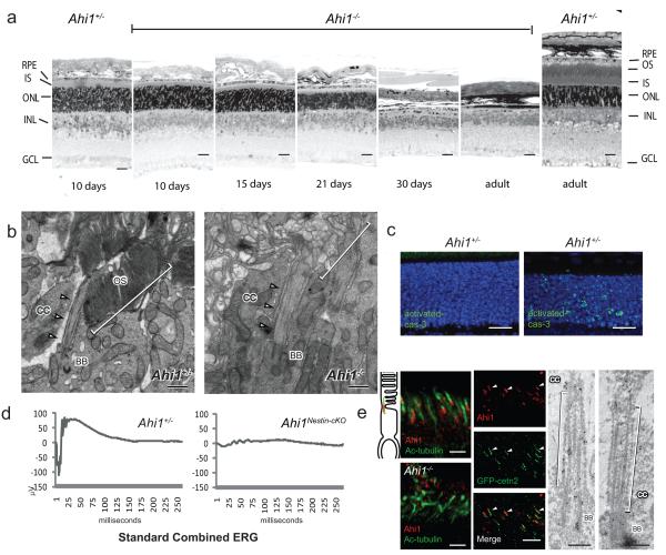
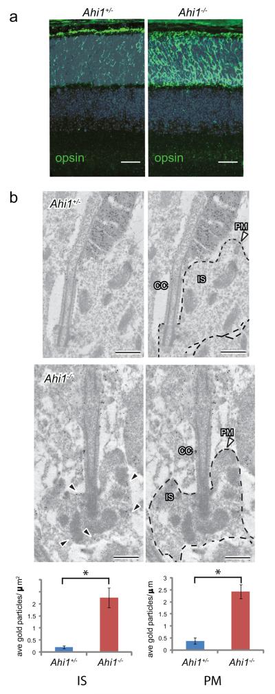
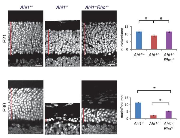
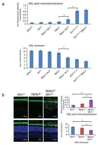
Degeneration of photoreceptors is a common feature of ciliopathies, owing to the importance of the specialized ciliary structure of these cells. Mutations in AHI1, which encodes a cilium-localized protein, have been shown to cause a form of Joubert syndrome that is highly penetrant for retinal degeneration. We show that Ahi1-null mice fail to form retinal outer segments and have abnormal distribution of opsin throughout their photoreceptors. Apoptotic cell death of photoreceptors occurs rapidly between 2 and 4 weeks of age in these mice and is significantly (P = 0.00175 and 0.00613) delayed by a reduced dosage of opsin. This phenotype also shows dosage-sensitive genetic interactions with Nphp1, another ciliopathy-related gene. Although it is not a primary cause of retinal blindness in humans, we show that an allele of AHI1 is associated with a more than sevenfold increase in relative risk of retinal degeneration within a cohort of individuals with the hereditary kidney disease nephronophthisis. Our data support context-specific roles for AHI1 as a contributor to retinopathy and show that AHI1 may explain a proportion of the variability in retinal phenotypes observed in nephronophthisis.


Figures

Figure 1
Figure 1
Degeneration of photoreceptor cells following failed outer segment development in Ahi1−/− mouse retina. (a) Semi-thin sections of retina stained with toluidine blue from P10 to adult (10 wks) showing acute loss of the outer nuclear layer (ONL) between P21 and P30. Scale=20μm (b) Transmission electron microscopy from P10. Outer segments (OS, brackets) are present in Ahi1+/−, but not in Ahi1−/−. Connecting cilia (CC, arrowheads) present in both. Scale=0.5 μm (c) Photoreceptor cell death is evident before three weeks of age, indicated by activated caspase-3 immunofluorescence (green) from 20μm cryosections. Nuclei are stained with Hoescht 33342 (blue). Shown are representative images from P19 mice, scale=20μm. (d) Full field dark-adapted electroretinograms (ERGs) from P19 Ahi1flox/−; Nes-Cre+ (Ahi1Nestin cKO) and Ahi1 heterozygous control mice. Shown are representative waveforms from n=3-4 mice/genotype. (e) Endogenous Ahi1 localization (red) to base of photoreceptor connecting cilium (acetylated-tubulin: green) in Ahi1+/− section is absent in Ahi1−/− cilium. Ahi1 distribution overlaps with centrin-2 (green) in GFP-Cetn-2 transgenic mouse retina. Arrowheads indicating example GFP-centrin2 labeled connecting cilia, 10μm cryosections, scale=5μm. Ahi1 immunoelectron microscopy from P10 retina, showing particles at the basal body (BB) and along the cilium (CC), scale=0.25μm
Figure 2
Figure 2
Opsin accumulation in Ahi1−/− photoreceptors (a) Opsin immunofluorescence (green) at P10 in Ahi1+/− and Ahi1−/− retina from cryosections. Nuclei are stained with Hoescht 33342. Scale=20μm (b) Opsin immunoEM and quantification of immunogold labeling from ultrathin sections of P10 Ahi1+/− and Ahi1−/− retina, n=20-23 photoreceptor connecting cilia/genotype. Data are expressed as number of gold particles per μm2 area within the inner segment (IS, defined as within inner segment and at least 30nm away from PM, P=8.0E-06), and as number of particles per μm length of inner segment membranes (PM, dashed lines, P=2.2E-07). Black arrowheads indicate examples of abnormally localized opsin, CC=connecting cilium, scale=0.5μm, error bars represent s.e.m.
Figure 3
Figure 3
Opsin contributes to cell death in Ahi1−/− mice. 10μm cryosections from P21 and P30 retina showing delay of cell loss associated with reduced opsin dosage (Ahi1−/−Rho+/−), measured as average number of cells/column (nuclei were stained with Hoescht 33342 and counts expressed as average of three counts across each section). Asterisk in top panel denotes significant difference from both control and rescue, n=3-7, P=0.00175 (P21) and 0.00613 (P30), scale=10μm, error bars represent s.e.m.
Figure 4
Figure 4
Genetic interaction of Ahi1 with Nphp1. (a) Increased cell loss and redistribution of opsin with increased load of deleterious Ahi1 and Nphp1 mutations in mouse retina. Quantification of opsin immunofluorescence(green) from ONL (outer nuclear layer, normalized as ratio of ONL:apical region encompassing IS and OS), and quantification of outer nuclear layer (ONL) thickness, expressed as averaged number of nuclei/column (indicated by Hoescht 33342 staining) from 10μm cryosections from indicated genotypes at P21. (b) From part (a), Ahi1 null allele modifies the Nphp1−/− phenotype, with Nphp1−/−Ahi1+/− showing increased opsin accumulation and decreased thickness of the ONL versus Nphp1−/− and Ahi1+/− controls. Asterisk denotes significant difference from both Ahi1+/− and Nphp1−/−, n=6-7, P(ONL thickness)=0.00315 and 0.000106, respectively; and P(fluorescence)=0.0454 and 0.0141, respectively, scale=20μm, error bars represent s.e.m.

Similar articles

Cited by

References

    1. Valente EM, et al. AHI1 gene mutations cause specific forms of Joubert syndrome-related disorders. Ann Neurol. 2006;59:527–34. - PubMed
    1. Parisi MA, et al. AHI1 mutations cause both retinal dystrophy and renal cystic disease in Joubert syndrome. J Med Genet. 2006;43:334–9. - PMC - PubMed
    1. Quinlan RJ, Tobin JL, Beales PL. Modeling ciliopathies: Primary cilia in development and disease. Curr Top Dev Biol. 2008;84:249–310. - PubMed
    1. Dixon-Salazar T, et al. Mutations in the AHI1 gene, encoding jouberin, cause Joubert syndrome with cortical polymicrogyria. Am J Hum Genet. 2004;75:979–987. - PMC - PubMed
    1. Ferland RJ, et al. Abnormal cerebellar development and axonal decussation due to mutations in AHI1 in Joubert syndrome. Nat Genet. 2004;36:1008–1013. - PubMed

Publication types

MeSH terms