. 2010 Feb;16(2):219-23.
doi: 10.1038/nm.2084. Epub 2010 Jan 17.

Neutrophil elastase-mediated degradation of IRS-1 accelerates lung tumor growth

Affiliations

Neutrophil elastase-mediated degradation of IRS-1 accelerates lung tumor growth

A McGarry Houghton et al. Nat Med. 2010 Feb.

Abstract

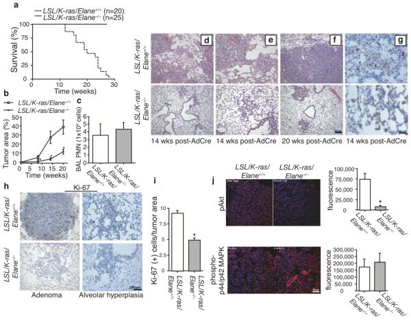
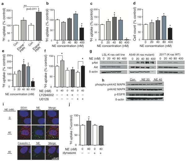
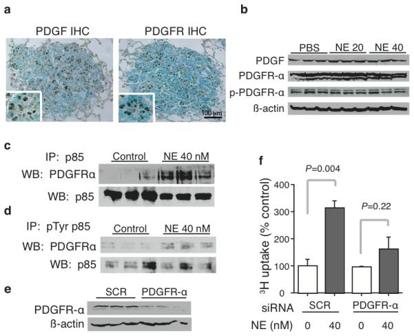
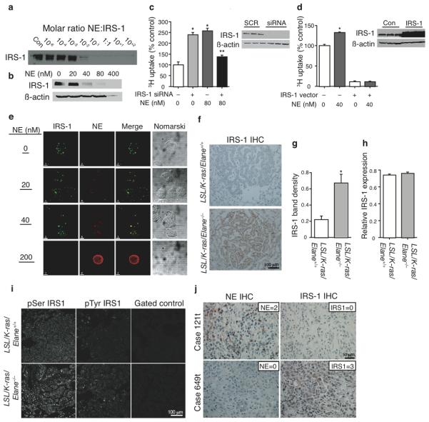
Lung cancer is the leading cause of cancer death worldwide. Recent data suggest that tumor-associated inflammatory cells may modify lung tumor growth and invasiveness. To determine the role of neutrophil elastase (encoded by Elane) on tumor progression, we used the loxP-Stop-loxP K-ras(G12D) (LSL-K-ras) model of mouse lung adenocarcinoma to generate LSL-K-ras-Elane(-/-) mice. Tumor burden was markedly reduced in LSL-K-ras-Elane(-/-) mice at all time points after induction of mutant K-ras expression. Kaplan-Meier survival analysis showed that whereas all LSL-K-ras-Elane(+/+) mice died, none of the mice lacking neutrophil elastase died. Neutrophil elastase directly induced tumor cell proliferation in both human and mouse lung adenocarcinomas by gaining access to an endosomal compartment within tumor cells, where it degraded insulin receptor substrate-1 (IRS-1). Immunoprecipitation studies showed that, as neutrophil elastase degraded IRS-1, there was increased interaction between phosphatidylinositol 3-kinase (PI3K) and the potent mitogen platelet-derived growth factor receptor (PDGFR), thereby skewing the PI3K axis toward tumor cell proliferation. The inverse relationship identified between neutrophil elastase and IRS-1 in LSL-K-ras mice was also identified in human lung adenocarcinomas, thus translating these findings to human disease. This study identifies IRS-1 as a key regulator of PI3K within malignant cells. Additionally, to our knowledge, this is the first description of a secreted proteinase gaining access to the inside of a cell and altering intracellular signaling.


Figures

Figure 1
Figure 1. NE promotes lung tumor growth in vivo
(a) Kaplan-Meier Survival curve for AdenoCre recipient LSL/K-ras/Elane+/+ and LSL/K-ras/Elane−/− mice; P=0.006, log-rank test. (b) Tumor area (%) for both groups at 8, 14, and 20 weeks post-AdenoCre. N=5 mice per group. Bars ± SEM. *P<0.01. (c) BALF neutrophil counts for AdenoCre recipient LSL/K-ras/Elane+/+ and LSL/K-ras/Elane−/− mice at the 14-week time point. N=5 mice per group. Bars ± SEM. P=NS. Representative H&E images at 14 weeks (d, e) and 20 weeks (f) post-AdenoCre. (g) Representative images for anti-p40phox (anti-neutrophil) IHC at 14 weeks post-AdenoCre. (h) Representative Ki-67 IHC 14 weeks post-AdenoCre. (i) Ki-67 (+) cells per tumor area for N=5 mice per group at the 14-week time point. Bars ± SEM. *P<0.01. (j) Representative IF images and quantification for pAkt and phospho-MEK/ERK from both groups of mice 14 weeks post-AdenoCre. Bars ± SEM. P<0.01 for pAkt.
Figure 2
Figure 2. Neutrophil Elastase induces tumor cell proliferation
(a) 3H uptake for LSL-K-ras tumor-derived cell lines co-incubated with WT and Elane−/− PMN for two hours. Results from a representative experiment in triplicate. Bars ± SEM. *P<0.05 from control. **P=0.011, ANOVA. (b) 3H uptake for LSL-K-ras cells stimulated with NE or vehicle for 60 min. Data from a representative experiment in triplicate. Bars ± SEM. *P<0.01 from NE=0. (c) 3H uptake and (d) cell counts for A549 cells and 3H uptake for (e) 201T cells (K-ras WT). Results from representative experiments in triplicate. Bars ± SEM, P<0.05 from NE=0. (f) 3H uptake for A549 cells stimulated with NE in presence or absence of 1.0 μM LY294002 or 10 μM U0126 for 60 min. Results from a representative experiment in triplicate. Bars +/− SEM. P<0.05. (g) Western blots of pAkt, Akt and B-actin for NE-exposed lysates of A549, 201T, and LSL-K-ras cells. (h) Western blot of phospho-p44/42-MAPK, p44-42-MAPK, pEGFR, and B-actin for NE-exposed A549 lysates. (i) Confocal images for EEA1, caveolin-1, and NE from A549 cells exposed to AlexaFluor488-conjugated NE or vehicle. Nuclei were counterstained using DAPI. (j) 3H uptake for A549 cells stimulated with NE ± 40 μM dynasore. Results from a representative experiment in triplicate. Bars +/− SEM. *P<0.05.
Figure 3
Figure 3. NE-induced proliferation is dependent upon PDGFR—PI3K signaling
(a) Representative images for PDGF and PDGFR IHC of LSL/K-ras tumors. Inset, high magnification showing PDGF and PDGFR staining within cells displaying tumor morphology. (b) Western blots for PDGF, PDGFRα, pPDGFRα and β-actin from NE-exposed A549 cell lysates. Results from a representative experiment in triplicate. Immunoprecipitation of (c) p85 and (d) phospho-tyrosine p85 from A549 cell lysates followed by Western blotting for PDGFRα. Membranes stripped and probed for p85. (e) Representative blot for PDGFRα following siRNA treatment with SCR siRNA vs. PDGFRα siRNA. (f) 3H uptake for PDGFRα-silenced A549 cells subsequently exposed to NE or vehicle. Results from a representative experiment in triplicate. Bars +/− SEM. *P<0.001.
Figure 4
Figure 4. NE co-localizes with and degrades IRS-1
(a) IRS-1 Western blot following incubation of recombinant IRS-1 protein with NE over a range of molar ratios. (b) IRS-1 and β-actin Western blots for NE-exposed A549 cell lysates. (c) 3H uptake for IRS-1 silenced (or SCR control) A549 cells subsequently exposed to NE or vehicle. Results from a representative experiment in triplicate. Bars ± SEM. *P<0.001 from NE=0 control. **P<0.05 from NE=80. Inset, IRS-1 blot of SCR and IRS-1 siRNA treated lysates. (d) 3H uptake for IRS-1 over-expressing A549 cells subsequently exposed to NE or vehicle. Results from a representative experiment in triplicate. Bars ± SEM. *P<0.01. Inset, IRS-1 blot of IRS-1 vector and control treated lysates. (e) Confocal images for IRS-1 and NE from A549 cells exposed to NE (or vehicle). (f) Representative IRS-1 IHC images from LSL/K-ras/Elane+/+ and LSL/K-ras/Elane−/− mice 14 weeks post-AdenoCre. (g) IRS-1 Western blot for 14-week post-AdenoCre tumor homogenates from both groups (N=5). Results presented as relative band density ± SEM. P<0.001. (h) IRS-1 real-time PCR for 14-week post-AdenoCre tumor homogenates from both groups (N=4). Results expressed as GAPDH CT/IRS-1 CT ± SEM. (i) Representative IF images for pSer and pTyr IRS-1 from LSL/K-ras/Elane+/+ and LSL/K-ras/Elane−/− tumors 14-wks post-AdenoCre. (j) Representative images of human lung adenocarcinoma specimens for NE=2 and IRS-1=0 (Case 121t) and NE=0 and IRS-1=3 (Case 649t). NE and IRS-1 were considered discordant when either one was present but the other absent/faint in the same view. The empirical probability of discordance was 0.88, which was significantly greater than chance (0.5), P<0.001.

Comment in

Similar articles

Cited by

References

    1. Jemal A, et al. Cancer statistics 2009. CA Cancer J. Clin. 2009;59:225–249. - PubMed
    1. Karin M. Inflammation and cancer: the long reach of Ras. Nat. Med. 2005;11:20–21. - PubMed
    1. Coussens LM, Werb Z. Inflammation and cancer. Nature. 2002;420:860–867. - PMC - PubMed
    1. Jackson EL, et al. Analysis of lung tumor initiation and progression using conditional expression of oncogenic K-ras. Genes Dev. 2001;15:3243–3248. - PMC - PubMed
    1. Sparmann A, Bar-Sagi D. Ras-induced interleukin-8 expression plays a critical role in tumor growth and angiogenesis. Cancer Cell. 2004;6:447–458. - PubMed

Reference for Methods

    1. Rice WR, et al. Maintenance of the mouse type II cell phenotype in vitro. Am. J. Physiol. 2002;283:L256–64. - PubMed
    1. Kliment CR, et al. Oxidative stress alters syndecan-1 distribution in lungs with pulmonary fibrosis. J Biol Chem. 2009;284:407–10. - PMC - PubMed
    1. Yasuoka H, et al. Insulin-like growth factor-binding protein-5 induces pulmonary fibrosis and triggers mononuclear cellular infiltration. Am. J. Pathol. 2006;169:1633–42. - PMC - PubMed
    1. Houghton AM, et al. Macrophage elastase (MMP-12) suppresses the growth of lung metastases. Cancer Res. 2006;66:6149–6155. - PubMed
    1. Tu Y, Liang L, Frank SJ, Wu C. Src homology 3 domain-dependent interaction of Nck-2 with insulin receptor substrate-1. Biochem J. 2001;354:315–322. - PMC - PubMed

Publication types

Substances