. 2010 Feb;16(2):224-7.
doi: 10.1038/nm.2078. Epub 2010 Jan 17.

Epidermal injury and infection during poxvirus immunization is crucial for the generation of highly protective T cell-mediated immunity

Affiliations

Epidermal injury and infection during poxvirus immunization is crucial for the generation of highly protective T cell-mediated immunity

Luzheng Liu et al. Nat Med. 2010 Feb.

Abstract

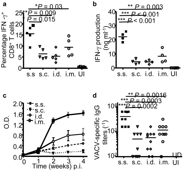
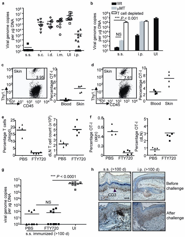
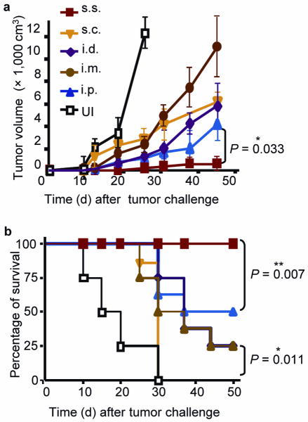
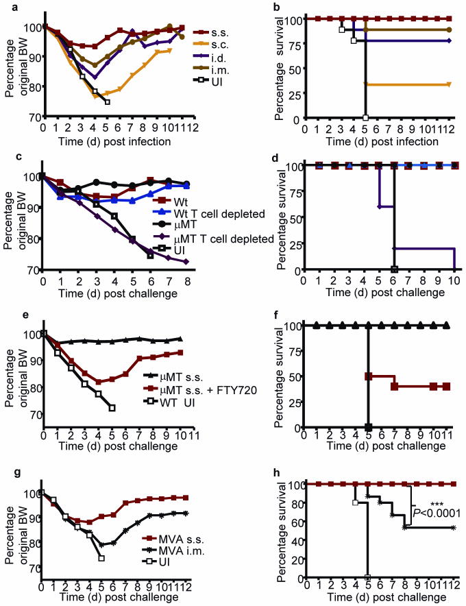
Variola major (smallpox) infection claimed hundreds of millions lives before it was eradicated by a simple vaccination strategy: epicutaneous application of the related orthopoxvirus vaccinia virus (VACV) to superficially injured skin (skin scarification, s.s.). However, the remarkable success of this strategy was attributed to the immunogenicity of VACV rather than to the unique mode of vaccine delivery. We now show that VACV immunization via s.s., but not conventional injection routes, is essential for the generation of superior T cell-mediated immune responses that provide complete protection against subsequent challenges, independent of neutralizing antibodies. Skin-resident effector memory T cells (T(EM) cells) provide complete protection against cutaneous challenge, whereas protection against lethal respiratory challenge requires both respiratory mucosal T(EM) cells and central memory T cells (T(CM) cells). Vaccination with recombinant VACV (rVACV) expressing a tumor antigen was protective against tumor challenge only if delivered via the s.s. route; it was ineffective if delivered by hypodermic injection. The clinically safer nonreplicative modified vaccinia Ankara virus (MVA) also generated far superior protective immunity when delivered via the s.s. route compared to intramuscular (i.m.) injection as used in MVA clinical trials. Thus, delivery of rVACV-based vaccines, including MVA vaccines, through physically disrupted epidermis has clear-cut advantages over conventional vaccination via hypodermic injection.


Figures

Figure 1
Figure 1. Inoculation with rVACV via barrier-disrupted epidermis generates significantly stronger cellular and humoral responses compared to s.c., i.d. and i.m. injection
(a) Quantification of the frequencies of IFN-γ+ CD8+ T cells in spleens on d 7 post immunization (p.i.) by intracellular IFN-γ staining. (b) Quantification of IFN-γ production from memory splenocytes at 5 weeks p.i. by in vitro restimulation assay. (c) Serum VACV-specific IgG determined by ELISA at the indicated time points p.i.. Each data point represents the average OD450nm value ± s.e.m. n = 5 in each group. (d) Serum VACV-specific IgG titer determined 11 weeks p.i.. n = 8–10 in each group. Data are representative results from 2–3 independent experiments. UI: unimmunized control.
Figure 2
Figure 2. rVACV scarification generates superior protective immunity against cutaneous viral challenge that is mediated by skin-targeted TEM
(a, b) Skin viral load measured by real-time PCR on d 6 after cutaneous challenge of Wt or μMT immune mice (immunized with VACV 6 weeks prior to challenge). In some Wt memory mice, T cells were depleted before and during the challenge (b). Data is pooled results from 3 independent experiments. (c, d) The frequencies of OT-I T cells in CD45+ leukocyte population in skin and blood either before (c) or after (d) challenge. FACS plots were gated on CD45+ leukocyte populations in skin. Numbers on the plots represent the frequencies of OT-I T cells in CD45+ populations. (e) T cell frequency in blood and absolute number in dLN measured immediately before secondary challenge to assess the efficiency of FTY720 blockage of T cell egress from lymphoid tissues. (f) Frequencies of OT-I cells in total viable cell populations in skin tissue and dLN at d 4 after challenge. (g) Cutaneous viral load at d 6 after secondary challenge determined by real-time PCR. Data are pooled results from 2 independent experiments. (h) Immunohistochemistry (IHC) staining showing the presence of CD3+ T cells in skin tissue of s.s. or i.p. immunized mice before or after secondary cutaneous rVACV challenge. Scale bar represents 5 μm. Photographs shown are the representative of 9 slides from 3 mice per group.
Figure 3
Figure 3. Immunization with rVACV via s.s. route provided superior protection against cutaneous melanoma challenge
(a) Tumor volumes measured at the indicated time points after MO5 melanoma challenge of mice immunized with rVACV via various routes. Each data point represents the mean tumor volume of different groups. (b) Survival rate of mice after MO5 implantation. Data is the pooled results of two independent experiments. A total of 8 mice were used for each group in the two experiments.
Figure 4
Figure 4. Immunization with rVACV as well as non-replicative MVA via s.s. route is highly effective to protect mice against lethal respiratory WR-VACV challenge
Change of body weight (BW) (a) and survival rate (b) of rVACV-immune Wt mice following lethal intranasal WR-VACV challenge. Change of BW (c) and survival rate (d) of rVACV s.s. immunized and WR-VACV challenged Wt or μMT mice, with or without T cell depletion at the time of challenge. Change of BW (e) and survival rate (f) of rVACV s.s. immune μMT mice following WR-VACV challenge, with or without FTY720 treatment. Change of BW (g) and survival rate (h) of MVA immune Wt mice following intranasal WR-VACV challenge. Immune mice were challenged with WR-VACV intranasally at 6 weeks (a–d, g,h) or 16 weeks (e,f) after immunization. Unimmunized (UI) controls were included in all the experiments. Data are representative results from 2–3 independent experiments (n = 10 per group).

Similar articles

Cited by

References

    1. Stewart AJ, Devlin PM. The history of the smallpox vaccine. J Infect. 2006;52:329–334. - PubMed
    1. Hammarlund E, et al. Duration of antiviral immunity after smallpox vaccination. Nat Med. 2003;9:1131–1137. - PubMed
    1. Demkowicz WE, Jr, Littaua RA, Wang J, Ennis FA. Human cytotoxic T-cell memory: long-lived responses to vaccinia virus. J Virol. 1996;70:2627–2631. - PMC - PubMed
    1. Liu L, Fuhlbrigge RC, Karibian K, Tian T, Kupper TS. Dynamic programming of CD8+ T cell trafficking after live viral immunization. Immunity. 2006;25:511–520. - PubMed
    1. Tian T, et al. Overexpression of IL-1alpha in skin differentially modulates the immune response to scarification with vaccinia virus. J Invest Dermatol. 2009;129:70–78. - PMC - PubMed

Publication types