. 2010 Feb;28(2):161-6.
doi: 10.1038/nbt.1605. Epub 2010 Jan 17.

Expansion and maintenance of human embryonic stem cell-derived endothelial cells by TGFbeta inhibition is Id1 dependent

Affiliations

Expansion and maintenance of human embryonic stem cell-derived endothelial cells by TGFbeta inhibition is Id1 dependent

Daylon James et al. Nat Biotechnol. 2010 Feb.

Abstract

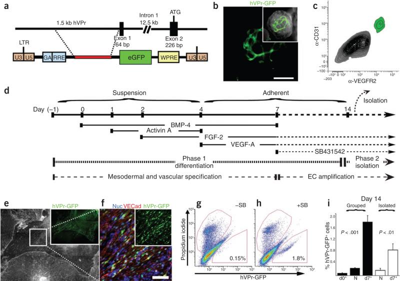
Previous efforts to differentiate human embryonic stem cells (hESCs) into endothelial cells have not achieved sustained expansion and stability of vascular cells. To define vasculogenic developmental pathways and enhance differentiation, we used an endothelial cell-specific VE-cadherin promoter driving green fluorescent protein (GFP) (hVPr-GFP) to screen for factors that promote vascular commitment. In phase 1 of our method, inhibition of transforming growth factor (TGF)beta at day 7 of differentiation increases hVPr-GFP(+) cells by tenfold. In phase 2, TGFbeta inhibition maintains the proliferation and vascular identity of purified endothelial cells, resulting in a net 36-fold expansion of endothelial cells in homogenous monolayers, which exhibited a transcriptional profile of Id1(high)VEGFR2(high)VE-cadherin(+) ephrinB2(+). Using an Id1-YFP hESC reporter line, we showed that TGFbeta inhibition sustains Id1 expression in hESC-derived endothelial cells and that Id1 is required for increased proliferation and preservation of endothelial cell commitment. Our approach provides a serum-free method for differentiation and long-term maintenance of hESC-derived endothelial cells at a scale relevant to clinical application.


Figures

Figure 1
Figure 1
Sequential TGFβ activation followed by inhibition during phase 1 differentiation promotes a tenfold expansion of hVPr-GFP+ heSC-derived cells. (a) A 1.5-kb fragment of the putative human Ve-cadherin promoter (hVPr) region was isolated from a BAC clone and placed upstream of GFP in a lentiviral expression vector (hVPr-GFP). (b) Spontaneously differentiating embryoid bodies exhibited expression of hVPr-GFP in tubular structures. Inset, merge of GFP and brightfield views. (c) Flow cytometric analysis showed hVPr-GFP+ cells were positive for the vascular markers CD31 and VeGFR2. (d) Schematic diagram showing the sequence in which BMP4, activinA, FGF-2, VeGF-A and SB431542 were added and removed from differentiation cultures. eC, endothelial cell. (e) Adherent hVPr-GFP cultures stimulated with SB431542 (10 μM) resulted in differentiation of heSCs into monolayers of hVPr-GFP+ adherent cells. Inset, hVPr-GFP+ cells alone. (f) Human VPr-GFP+ cells were immuno-positive for Ve-cadherin. Blue, nuclear counterstain. Inset, hVPr-GFP+ cells alone. (g,h) The proportion of hVPr-GFP+ cells was measured by flow cytometry at day 14 after culture in the absence (–SB; g) and presence (+SB; h) of SB431542. (i) Measurement of hVPr-GFP+ cells at day 14 when embryoid bodies were cultured either in groups or as isolated embryoid bodies and SB431542 was added at day 0, day 7 or not at all (N). error bars represent s.d. of experimental values performed in triplicate. Scale bars, 100 μm.
Figure 2
Figure 2
TGFβ inhibition after endothelial cell isolation during phase 2 increases yield and preserves vascular identity of purified endothelial cells. (a–c) Human VPr-GFP heSCs were sequentially stimulated with cytokines (–SB) and SB431542 (+SB) (Fig. 1d and Online Methods) and cultures were assessed for the prevalence of pluripotency (Oct3/4) and mesodermal transcripts (brachyury) (a), CD31 and α-SMA transcripts (b) and endothelial cell markers hVPr-GFP and CD31 (c) at multiple time points during differentiation. The secondary axis in b shows values for cells shown in solid bars. (d) Isolated endothelial cells that were cultured in the absence of SB431542 were stained for both Ve-cadherin and α-SMA and showed rare cells that were positive for both markers (arrowhead in the inset). Inset, α-SMA alone. (e–i) Human VPr-GFP+ cells were isolated from differentiation cultures at day 14 by FACS and further cultured in the absence (e) or presence (f) of SB431542. (g) Flow cytometric assessment of CD31 was performed after 5 d of isolated culture (total cells are shown in white and CD31+ cells are shown in black in the bar graph). (h) After isolation and 5 d of culture in the presence or absence of SB431542, the incidence of α-SMA+ cells was measured. (i) After 5 d of culture following isolation, unstimulated cultures showed reduced incidence of cells positive for phospho-histoneH3 (PHH3+), relative to SB431542-stimulated cultures. The mean incidences of α-SMA and phospho-histoneH3 positive cells were obtained by counting positively stained cells in multiple parallel wells. (j) The yield of endothelial cells (eCs) from heSCs is schematized relative to a 50,000 heSC input at day 0. The relative difference in endothelial cell (eCs) number is indicated at day 14 (upon isolation from differentiation cultures), and day 20 (after expansion in isolated conditions). The ratio of input heSCs to committed heSC-derived endothelial cells after 20 d is also shown. Relative transcript abundance was measured by QPCR and normalized to the housekeeping gene β-actin (ACTB). error bars in (a–c and g–i) represent s.d. of experimental values performed in triplicate. Scale bars, 100 μm.
Figure 3
Figure 3
Molecular profiling of heSC-derived endothelial cells reveals a signature defined by high Id1 expression. Human VPr-GFP embryoid bodies and highly purified hVPr-GFP+ cells were compared to mature vascular cells by microarray analysis. (a) RNA was extracted for microarray analysis from human VPr-GFP embryoid bodies cultured in the presence of recombinant cytokines alone until day 14; isolated endothelial cells (99.8% pure) from hVPr-GFP embryoid bodies cultured in the presence of recombinant cytokines and the TGFβ inhibitor SB431542 until day 14; isolated endothelial cells (>95% pure) from hVPr-GFP embryoid bodies cultured in the presence of recombinant cytokines and the TGFβ inhibitor SB431542 until day 14, followed by 10 d additional culture in the presence of cytokines and SB431542; HUVeCs; human umbilical vein smooth muscle cells; and CD34+ umbilical cord blood cells. expressed factors are displayed in an ordered array, as labeled, with highly expressed factors shown in red, minimally expressed factors shown in blue and factors for which transcripts are below a significant expression level shown in gray. (b–f) Following the endothelial cell differentiation protocol (Fig. 1d), Id1-YFP heSC-derived cells were sorted by FACS, separating the CD31+ population into Id1-YFPhigh-expressing cells (b (green) and c) and Id1-YFPlow-expressing cells (b (red) and d). Insets, brightfield views on the day after isolation of Id1-YFPhigh (c) and Id1-YFPlow (d) cells. (e) After 3 d culture in the presence of SB431542, both populations were transferred to conditions with and without SB431542 for an additional 4 d (+SB and –SB, respectively). (f) Total cells and mean fluorescence intensity (MFI) measurements of Id1:YFP (black) and CD31+ (white) were measured for: CD31+Id1low (I) and CD31+Id1high (II) populations upon isolation; and for four populations following culture conditions (as shown in e, III–VI). Scale bars, 100 μM.
Figure 4
Figure 4
TGFβ inhibition upregulates Id1 expression and is necessary for the increased yield of functional endothelial cells capable of in vivo neo-angiogenesis. (a,b) Human VPr-GFP heSCs that were stably transduced with control (a) or Id1-specific (b) shRNAs were differentiated according to the protocol shown in Figure 1d and assessed at day 14 for the prevalence of VeGFR2+ (blue) and hVPr-GFP+ (green) cells. The insets show plots of side scatter on the y axis and hVPr-GFP on the x axis. (c) Control and Id1-specific shRNAs were added to HUVeC or freshly isolated (at day 14) hVPr-GFP+ cells, and the relative Id1 transcript levels were measured after 3 d. *, P < 0.05. error bars, s.d. of experimental values performed in triplicate. (d) Control and Id1-specific shRNAs were added to freshly isolated hVPr-GFP+ cells, which were cultured in the absence or presence of SB431542. After 5 d, the total cell number and proportion of CD31+ cells was measured by flow cytometry. error bars, s.d. of experimental values performed in triplicate. Scr, scrambled control shRNA. (e–g) Human VPr-GFP+ cells were isolated by FACS at day 14 and expanded in monolayer culture (e) for 8 d while retaining expression of both the endogenous Ve-cadherin (f) and the hVPr-GFP transgene (g). Panel e shows a mosaic view of one well of a 24-well dish. A magnified view of the boxes in e and f are shown in f and g, respectively. (h,i) expanded cells were injected in Matrigel plugs into immunodeficient mice and excised after 10 d following intravital labeling of functional vasculature with lectin (GIB4, blue). h, View of hVPr-GFP+ cells alone; i, view of hVPr-GFP+ cells merged with GIB4+ cells. Scale bars, 100 μM.

References

    1. Thomson JA, et al. Embryonic stem cell lines derived from human blastocysts. Science. 1998;282:1145–1147. - PubMed
    1. Yamahara K, et al. Augmentation of neovascularization in hindlimb ischemia by combined transplantation of human embryonic stem cells-derived endothelial and mural cells. PLoS ONE. 2008;3:e1666. - PMC - PubMed
    1. Sone M, et al. Pathway for differentiation of human embryonic stem cells to vascular cell components and their potential for vascular regeneration. Arterioscler. Thromb. Vasc. Biol. 2007;27:2127–2134. - PubMed
    1. Lu SJ, et al. Generation of functional hemangioblasts from human embryonic stem cells. Nat. Methods. 2007;4:501–509. - PMC - PubMed
    1. Goldman O, et al. A boost of BMP4 accelerates the commitment of human embryonic stem cells to the endothelial lineage. Stem Cells. 2009;27:1750–1759. - PubMed

Publication types

MeSH terms

Substances

Associated data