. 2010 Feb;17(2):165-72.
doi: 10.1038/nsmb.1765. Epub 2010 Jan 17.

Structural basis for dsRNA recognition and interferon antagonism by Ebola VP35

Affiliations

Structural basis for dsRNA recognition and interferon antagonism by Ebola VP35

Daisy W Leung et al. Nat Struct Mol Biol. 2010 Feb.

Abstract

Ebola viral protein 35 (VP35), encoded by the highly pathogenic Ebola virus, facilitates host immune evasion by antagonizing antiviral signaling pathways, including those initiated by RIG-I-like receptors. Here we report the crystal structure of the Ebola VP35 interferon inhibitory domain (IID) bound to short double-stranded RNA (dsRNA), which together with in vivo results reveals how VP35-dsRNA interactions contribute to immune evasion. Conserved basic residues in VP35 IID recognize the dsRNA backbone, whereas the dsRNA blunt ends are 'end-capped' by a pocket of hydrophobic residues that mimic RIG-I-like receptor recognition of blunt-end dsRNA. Residues critical for RNA binding are also important for interferon inhibition in vivo but not for viral polymerase cofactor function of VP35. These results suggest that simultaneous recognition of dsRNA backbone and blunt ends provides a mechanism by which Ebola VP35 antagonizes host dsRNA sensors and immune responses.


Figures
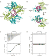
Figure 1
Figure 1
Overall structure of the VP35 IID-dsRNA complex. (a) Crystallographic asymmetric unit (P212121) contains four VP35 IID molecules and one 8 base pair dsRNA. Two-fold non-crystallographic symmetry relates molecules A and C (green) and molecules B and D (cyan). The phosphate backbone of the dsRNA is represented by a magenta ribbon and the bases are shown in stick representation. (b) VP35 IID displays two different sets of interactions in the complex, where molecule A is mostly engaged in protein-protein interactions, while molecule B makes direct contacts with dsRNA. ITC binding isotherms and corresponding raw data for wildtype VP35 IID binding to (c) 8 base pair dsRNA and (d) 8 base single stranded RNA.
Figure 2
Figure 2
Conserved basic residues are important for protein-protein and protein-dsRNA interactions. (a-b), VP35 IID molecule A interacts with molecule B in a head to tail orientation, where residues Arg312 and Arg322 are involved in hydrogen bonding with the Asp271 and Glu262, respectively. (c-d) VP35 molecule B interacts with the dsRNA in a sequence independent manner with the sidechains of Arg312 and Arg322 forming hydrogen bonds with the phosphodiester backbone.
Figure 3
Figure 3
The VP35 IID central basic patch residues are critical for dsRNA recognition. Electrostatic surface potential (scale of -10 to +10 kT e-1) of VP35 IID structures for (a) wildtype, (b) R312A, and (c) K339A. Locations of mutated residues are identified by green dashed circles. (d) Sites that are required for RNA binding and sites that enhance RNA binding are identified by red solid and dashed circles, respectively. The α-helical and β-sheet subdomains of VP35 IID are colored orange and yellow, respectively.
Figure 4
Figure 4
Intersubdomain interface of VP35 IID forms an “end-cap” that recognizes blunt ends of duplex RNA. (a) Surface representation of VP35 IID (molecule B, cyan) and 8 base pair dsRNA (magenta) to highlight the surface complementarity between the 5′ ends of dsRNA and VP35 IID β-sheet subdomain residues. Asterisks denote the complementary strand in the RNA duplex. (b) Residues Phe239 and Ile340 form van der Waals contacts with the 5′ end of the C1 base (double arrow).
Figure 5
Figure 5
Residues from the central basic patch and the “end-cap” of VP35 IID play key roles in the IFN-antagonist function. Sendai virus (SeV) infection-induced IFN-β promoter activity of VP35 IID wildtype (WT) and mutants are reported relative to IFN-β induction of an empty vector (EV) infected with SeV. (a) Residues from the first basic patch. (b) Residues from the “end-cap” (Phe235 is near Phe239, but is not part of the residues that form the “end-cap”.). (c-d) Residues from the central basic patch. Error bars represent s.d. Plasmid concentration was 500 ng and the corresponding western blots are shown in Supplementary Fig. 9.
Figure 6
Figure 6
VP35 competes with RIG-I for dsRNA binding and inhibits RIG-I dependent IFN-β promoter activation by dsRNA. 1H/15N HSQC of 15N-labeled VP35 IID (black) and in the presence of unlabeled (a) IVT-8 bp dsRNA (red), (b) IVT-8 bp dsRNA + RIG-I C-terminal domain (green), and (c) RIG-I C-terminal domain (blue). NMR signal for residue Arg312 is shown in the inset. (d) Comparison of VP35 IID residue number versus relative peak intensities of chemical shifts in a-c. (e) IVT-hairpin dsRNA activates IFN-β promoter in a RIG-I dependent manner. (f) VP35 can inhibit IFN-β promoter activated by RIG-I upon transfection of IVT-8 bp dsRNA and IVT-hairpin dsRNA. Error bars represent s.d.

References

    1. Bosio CM, et al. Ebola and Marburg viruses replicate in monocyte-derived dendritic cells without inducing the production of cytokines and full maturation. J Infect Dis. 2003;188:1630–8. - PubMed
    1. Mahanty S, et al. Protection from lethal infection is determined by innate immune responses in a mouse model of Ebola virus infection. Virology. 2003;312:415–24. - PubMed
    1. Bray M, Geisbert TW. Ebola virus: the role of macrophages and dendritic cells in the pathogenesis of Ebola hemorrhagic fever. Int J Biochem Cell Biol. 2005;37:1560–6. - PubMed
    1. Baize S, et al. Defective humoral responses and extensive intravascular apoptosis are associated with fatal outcome in Ebola virus-infected patients. Nat Med. 1999;5:423–6. - PubMed
    1. Feldmann H, Wahl-Jensen V, Jones SM, Stroher U. Ebola virus ecology: a continuing mystery. Trends Microbiol. 2004;12:433–7. - PubMed

Publication types

MeSH terms