. 2010 Apr;49(4):398-409.
doi: 10.1002/mc.20612.

Opposing actions of insulin and arsenite converge on PKCdelta to alter keratinocyte proliferative potential and differentiation

Affiliations

Opposing actions of insulin and arsenite converge on PKCdelta to alter keratinocyte proliferative potential and differentiation

Tatiana V Reznikova et al. Mol Carcinog. 2010 Apr.

Abstract

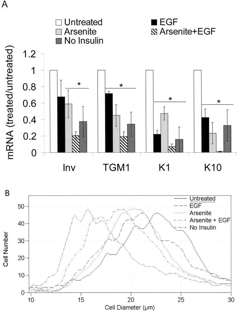
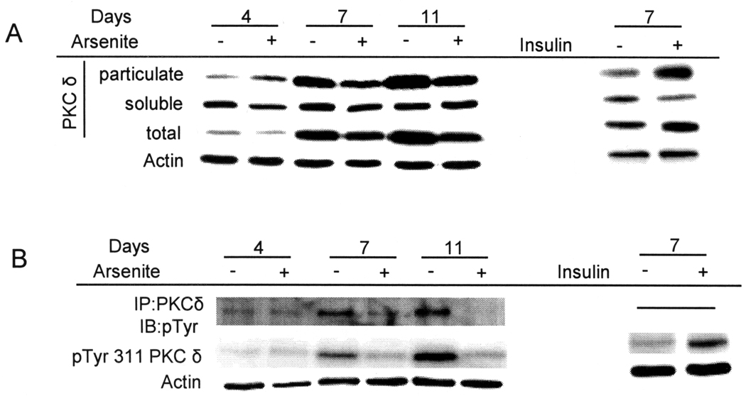
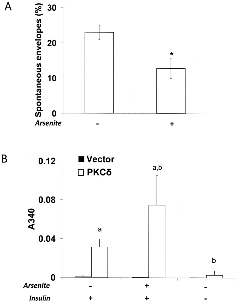
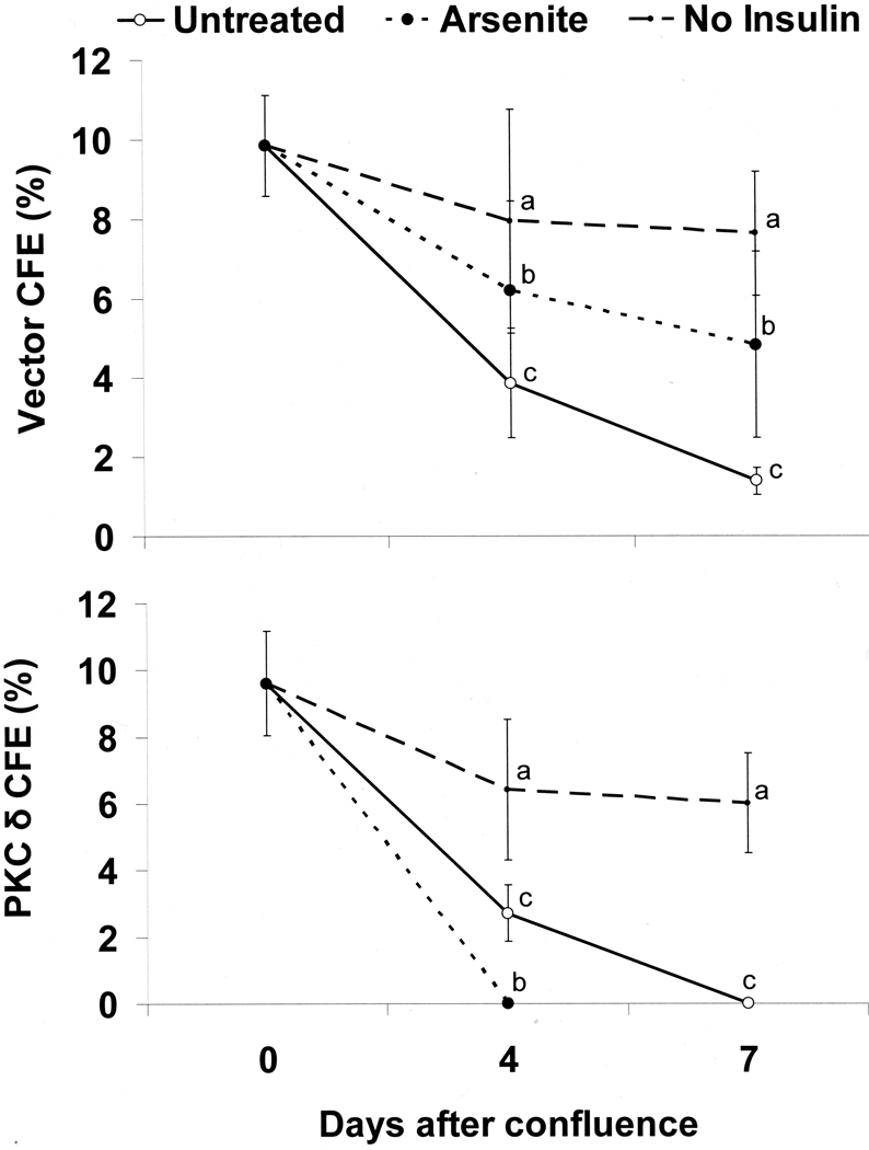
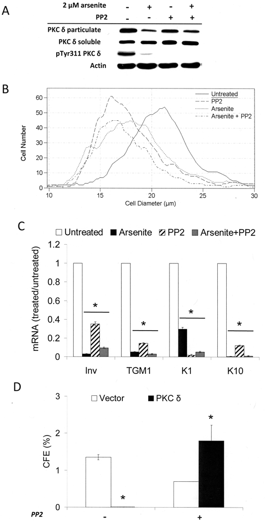
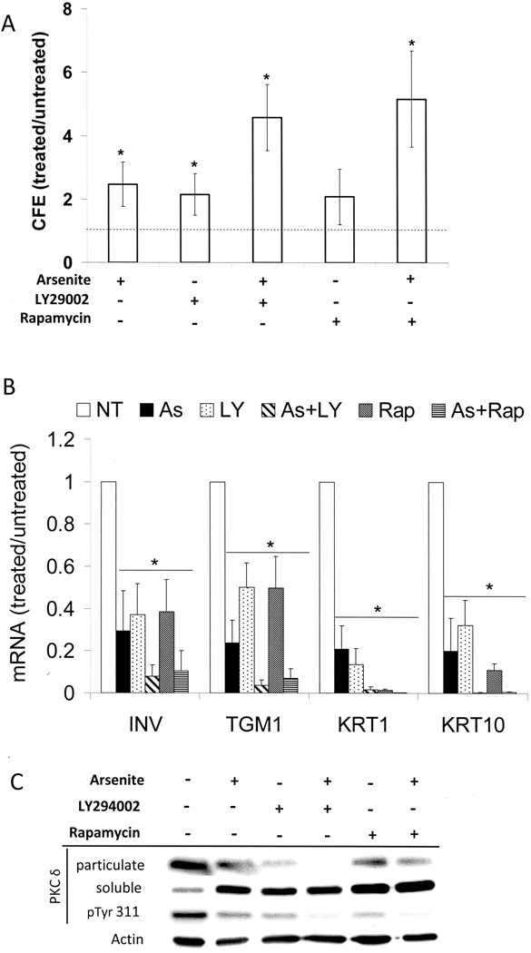
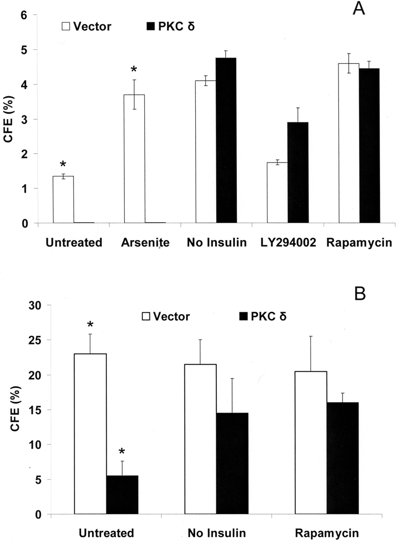
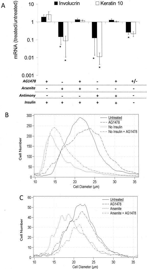
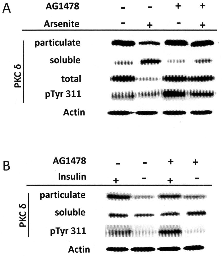
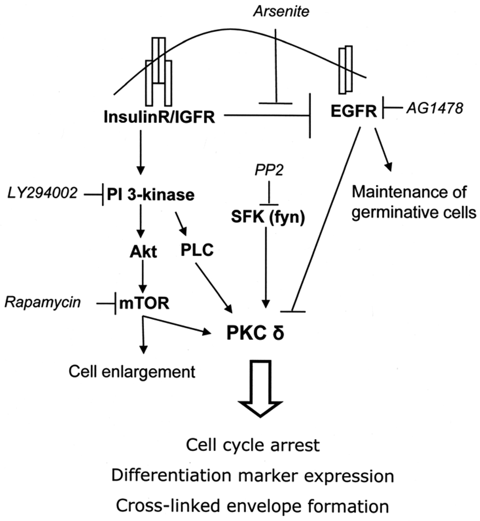
When cultured human keratinocytes reach confluence, they undergo a program of changes replicating features of differentiation in vivo, including exit from the proliferative pool, increased cell size, and expression of specialized differentiation marker proteins. Previously, we showed that insulin is required for some of these steps and that arsenite, a human carcinogen in skin and other epithelia, opposes the differentiation process. In present work, we show that insulin signaling, probably through the IGF-I receptor, is required for the increase in cell size accompanying differentiation and that this is opposed by arsenite. We further examine the impact of insulin and arsenite on PKCdelta, a known key regulator of keratinocyte differentiation, and show that insulin increases the amount, tyrosine phosphorylation, and membrane localization of PKCdelta. All these effects are prevented by exposure of cells to arsenite or to inhibitors of downstream effectors of insulin (phosphotidylinositol 3-kinase and mammalian target of rapamycin). Retrovirally mediated expression of activated PKCdelta resulted in increased loss of proliferative potential after confluence and greatly increased formation of cross-linked envelopes, a marker of keratinocyte terminal differentiation. These effects were prevented by removal of insulin, but not by arsenite addition. We further demonstrate a role for src family kinases in regulation of PKCdelta. Finally, inhibiting epidermal growth factor receptor kinase activity diminished the ability of arsenite to prevent cell enlargement and to suppress insulin-dependent PKCdelta amount and tyrosine 311 phosphorylation. Thus suppression of PKCdelta signaling is a critical feature of arsenite action in preventing keratinocyte differentiation and maintaining proliferative capability.


Figures

Figure 1
Figure 1
Keratinocyte size and differentiation after treatment with arsenite and EGF. Starting at confluence, cultures were treated in medium supplemented with the indicated combinations of 2 µM arsenite and 10 ng/ml EGF. Unless otherwise indicated, the medium contained 5 µg/ml insulin. After 7 days of treatment, cells were trypsinized and mRNA levels of involucrin, keratin 1, keratin 10, and TGM1 (A) or cell sizes (B) were measured. In (A), values are given relative to untreated cultures in the presence of insulin, normalized to 1. Data were compiled from three independent experiments. Significant differences from untreated (p < 0.01) are indicated by an asterisk (p = 0.01 for involucrin, p < 0.01 for TGM1, p < 0.001 for KRT1 and p = 0.001 for KRT10).
Figure 2
Figure 2
Regulation of PKCδ level and phosphorylation. SIK cultures were treated with 2 µM arsenite for the indicated number of days or switched to medium without insulin for 7 days. (A) Levels of PKCδ were measured in soluble or particulate fractions, or in unfractionated cell lysates (total). (B) PKCδ was immunoprecipitated and examined for total tyrosine phosphorylation in precipitates. In parallel, PKCδ Tyr 311 phosphorylation (pTyr 311) was measured in total cell lysates. Data are representative of four (A) or two (B) independent experiments. β-Actin was used as a loading control.
Figure 3
Figure 3
Spontaneous envelope formation after PKCδ overexpression. (A) SIK cultures near confluence were pre-treated with 2 µM arsenite for one day before suspension and with continued treatment for 4 days in suspension culture before cells were recovered and envelopes were counted. (B) Surface cultures of SIK, retrovirally infected with vector only control (pBabe) or with activated PKCδ construct in pBABE, were treated near confluence with 2 µM arsenite or switched to medium without insulin and, after 8 days, envelopes were quantitated by light scattering (A340). Data were compiled from three independent experiments. In panel A, values of arsenite-treated cultures were significantly different from untreated (*p < 0.05 by 2 tailed Student’s t-test). In panel B, the values for cultures expressing constitutively active PKCδ and treated with arsenite and insulin were significantly greater than that from the PKCδ expressing cultures treated with insulin alone (a, p = 0.02) and not treated with either insulin or arsenite (b, *p < 0.001). Envelope formation in cultures infected with the control pBABE vector (A340 ≈ 0.002) was negligible (0.4% of the cells in two independent experiments).
Figure 4
Figure 4
Suppression of CFE by PKCδ overexpression. Nearly confluent SIK cultures were treated with 2 µM arsenite or switched to medium without insulin for the indicated number of days. CFE values were determined in cultures retrovirally infected with either the vector only (pBabe) (A) or a constitutively active PKCδ (B) expression construct. Data (mean ± SD) are shown for three independent experiments. In each case, the values for untreated (c), arsenite-treated (b) and no insulin (a) cultures were significantly different (p< 0.05) by 3 way ANOVA.
Figure 5
Figure 5
Alteration of proliferative potential, cell size and differentiation by inhibition of Src family kinases. SIK cultures were treated near confluence with 2 µM arsenite and/or 3 µM PP2, an inhibitor of Src family kinases. (A) After 4 days of treatment, protein levels of PKCδ in particulate and soluble fractions and phosphorylated Tyr 311 PKCδ (pTyr 311 PKCδ) levels in total cell lysates were assessed by immunoblotting. The data are representative of two independent experiments for PKCδ in particulate and soluble fractions and a single experiment for pTyr311 PKCδ. (B) SIK cultures were treated with the indicated agents for 8 days, then trypsinized for determination of cell size distribution. The graph is representative of three independent experiments. (C) Expression of keratin 1 and 10, involucrin, and TGM1 mRNAs measured by real-time PCR after 8 days of treatment. Values are given relative to untreated cultures, set at 1. For each gene, the values in treated cultures were significantly different from untreated (*p < 0.001). (D) CFE of cultures infected with activated PKCδ in pBABE or vector control with and without PP2 treatment for 7 days. The difference in values for PKCδ-expressing cultures in the presence and absence of PP2 was significantly different (*p < 0.03) by Student’s two tailed t-test.
Figure 6
Figure 6
Increased proliferative potential and decreased differentiation in the presence of PI 3-kinase or mTOR inhibitors. Starting near confluence, SIK cultures were treated with 2 µM arsenite, 10 µM LY294002, 5 nM rapamycin or co-treated with arsenite and LY294002 or rapamycin. (A) Fold increases in CFE are given after 7 days relative to those of untreated cultures (absolute values 1.9 + 0.9%), indicated by the dotted line. The results (mean ± SD) are representative of five independent experiments. Except for cultures treated with rapamycin alone, values are significantly different from untreated (*p < 0.02). (B) mRNA expression of involucrin, keratin 1, keratin 10, and TGM1 was measured using real-time PCR after 3 days of treatment and is presented relative to untreated, set as 1. Averages are given of 3–7 independent experiments, where significant differences from untreated for each differentiation marker are indicated by the asterisk (p < 0.001 for each marker). (C) PKCδ levels in particulate and soluble fractions were determined after 7 days of treatment. In parallel, Tyr 311 phosphorylation (pTyr 311) in total cell lysates was measured. β-Actin is used as a loading control. Data are representative of three independent experiments.
Figure 7
Figure 7
Effects of overexpressed PKCδ on colony forming ability: influence of inhibiting insulin action. (A) Starting at confluence, vector only (pBabe) or PKCδ overexpressing SIK cultures were treated or changed to medium without insulin for 7 days before measuring CFE. The differences between cultures expressing PKCδ or not were significant only for the untreated and arsenite-treated conditions (p < 0.01 in each case by Student’s t-test). (B) PKCδ was overexpressed in SCC9, and CFE was measured after 7 days in cultures with or without insulin, or with insulin and 5 nM rapamycin. Treatments began just before confluence. Without arsenite treatment, the values for cultures expressing and not expressing PKCδ were significantly different by Student’s t-test (*p < 0.02).
Figure 8
Figure 8
Requirement for EGFR function in regulation of differentiation. SIK cultures were pre-treated starting near confluence with AG1478 or solvent for 1 hr prior to treatment with 2 µM arsenite, 5 µM antimonite or switched to medium without insulin for 7 days. (A) Expression of involucrin and keratin 10 mRNAs were measured using real-time PCR; values are given relative to untreated cultures in the presence of insulin, set as 1. Averaged from 4 independent experiments, values significantly different from untreated are indicated (*p < 0.02). (B) Starting at confluence, cultures were grown in the presence or absence of insulin and AG1478 as indicated and analyzed for size after 7 days. (C) Starting near confluence, cultures in the presence of insulin were treated with AG1478 or solvent for 1 hr prior to treatment with 2 µM arsenite as indicated and sizes analyzed after 7 days. Graphs are representative of three (B) and four (C) independent experiments.
Figure 9
Figure 9
Effect of AG1478 on PKCδ processing. Cultures were harvested for total protein lysates or fractionated into soluble and particulate fractions and subjected to immunoblot analysis. PKCδ levels in particulate, soluble fractions and total cell lysates as well as phosphorylated Tyr 311 (pTyr 311) PKCδ in total cell lysates were measured. β-Actin was used as a loading control for total lysates. Data are representative of three (A) and two (B) independent experiments.
Figure 10
Figure 10
Model illustrating interrelations of insulin, PKCδ and arsenite. Activation of the insulin/IGF receptor pathway appears to suppress EGFR signaling, an action prevented by arsenite. Insulin/IGFR signaling also stimulates the PI3-kinase pathway, including mTOR signaling. These pathways converge on PKCδ, a critical determinant of cell state. Arsenite and PI3-kinase or mTOR inhibitor cotreatment thus has an additive response in maintaining proliferative potential and yields PKCδ levels equivalent to insulin deprivation. Arrows indicate activation, and bars indicate suppression. Chemical inhibitors are in italics.

Similar articles

Cited by

References

    1. Kitchin KT. Recent advances in arsenic carcinogenesis: Modes of action, animal model systems and methylated arsenic metabolites. Toxicol Sci. 2001;172:249–261. - PubMed
    1. Council NR. Arsenic in Drinking Water. Washington: National Academy Press; 2001.
    1. Smith AH, Marshall G, Yuan Y, et al. Increased mortality from lung cancer and bronchiectasis in young adults after exposure to arsenic in utero and in early childhood. Environ Hlth Perspect. 2006;114:1293–1296. - PMC - PubMed
    1. Tseng C-H. Cardiovascular disease in arsenic-exposed subjects living in the arseniasis-hyperendemic areas in Taiwan. Atherosclerosis. 2008;199:12–18. - PubMed
    1. Coronado-González JA, Del Razo LM, García-Vargas G, Sanmiguel-Salazar F, Escobedo-de la Peña J. Inorganic arsenic exposure and type 2 diabetes mellitus in Mexico. Environ Res. 2007;104:383–389. - PubMed

Publication types

MeSH terms