

Review
doi: 10.3748/wjg.v16.i3.395.
Actinomycosis of the appendix mimicking appendiceal tumor: a case report
Affiliations
- PMID: 20082489
- PMCID: PMC2807964
- DOI: 10.3748/wjg.v16.i3.395
Item in Clipboard
Review
Actinomycosis of the appendix mimicking appendiceal tumor: a case report
World J Gastroenterol.
.
Abstract
Actinomycosis is an uncommon chronic infectious disease. Common sites of involvement include the cervicofacial, thoracic and abdominopelvic regions. In abdominopelvic actinomycosis, the ileocecal region, including the appendix, is the most commonly involved site. In some reports, limited appendiceal actinomycosis has revealed a thickened appendiceal wall with peri-appendiceal inflammation as acute appendicitis or perforated appendicitis. We experienced pathologically confirmed intraluminal limited appendiceal actinomycosis without peri-appendiceal infiltration. Here, we report the computed tomography and ultrasound findings.
Figures
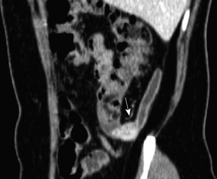
Contrast-enhanced CT revealed a well-defined solid mass with strong enhancement in the base of the appendix (arrow). Peri-appendiceal infiltration was not seen.
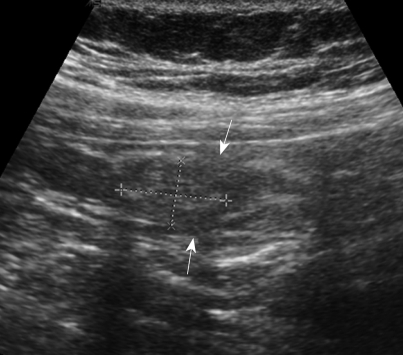
US showed a heterogeneous, hyperechoic, intraluminal mass at the base of the appendix, without peri-appendiceal infiltration. We also noted focal defects at the echogenic inner mucosal layer (arrows).
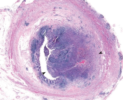
Microscopy of appendiceal actinomycosis. An abscess composed of chronic and acute inflammatory cells was observed in a mass-like lesion (arrow), from the mucosal surface to the superficial submucosa (arrowhead) (HE, × 10).
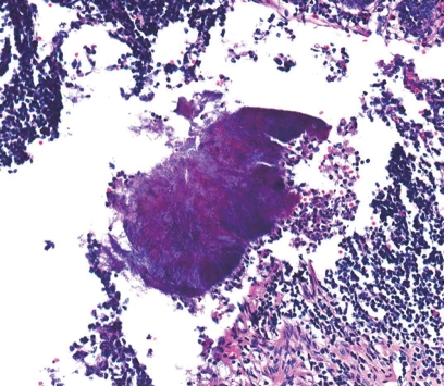
Higher magnification showed a typical sulfur granule surrounded by neutrophils in the clefted abscess center (HE, × 200).
Similar articles
-
Appendiceal Tumor or Something More?Gastroenterology. 2018 Jun;154(8):e14-e15. doi: 10.1053/j.gastro.2017.09.041. Epub 2017 Oct 6. Gastroenterology. 2018. PMID: 28989065 No abstract available.
-
[Pathogenesis of abdominal actinomycosis].Vestn Khir Im I I Grek. 1977 Oct;119(10):63-7. Vestn Khir Im I I Grek. 1977. PMID: 929881 Russian.
-
Actinomyces appendicitis: diagnostic dilemma--malignancy or infection?Am Surg. 2014 Jan;80(1):E33-5. Am Surg. 2014. PMID: 24401512 Review. No abstract available.
-
[Actinomycosis of the appendix. Case report].Rev Med Chir Soc Med Nat Iasi. 2004 Jul-Sep;108(3):640-3. Rev Med Chir Soc Med Nat Iasi. 2004. PMID: 15832990 Romanian.
-
Appendicitis: When there is more than meets the eye.Clin Res Hepatol Gastroenterol. 2011 Nov;35(11):765-7. doi: 10.1016/j.clinre.2011.05.014. Epub 2011 Jul 16. Clin Res Hepatol Gastroenterol. 2011. PMID: 21763232 Review.
Cited by
-
Pelvic Actinomycosis.Can J Infect Dis Med Microbiol. 2017;2017:9428650. doi: 10.1155/2017/9428650. Epub 2017 Jun 8. Can J Infect Dis Med Microbiol. 2017. PMID: 28684963 Free PMC article. Review.
-
Appendiceal actinomycosis mimicking malignant tumor: a rare case report.Ann Med Surg (Lond). 2024 Jan 3;86(2):1076-1079. doi: 10.1097/MS9.0000000000001564. eCollection 2024 Feb. Ann Med Surg (Lond). 2024. PMID: 38333266 Free PMC article.
-
Histopathological correlations of appendectomies: a clinical audit of a single center.Ann Transl Med. 2015 Jun;3(9):119. doi: 10.3978/j.issn.2305-5839.2015.05.02. Ann Transl Med. 2015. PMID: 26207247 Free PMC article.
-
Appendicular actinomycosis: The first reported case of an uncommon finding of a common ailment from Nepal.Clin Case Rep. 2023 Sep 30;11(10):e7996. doi: 10.1002/ccr3.7996. eCollection 2023 Oct. Clin Case Rep. 2023. PMID: 37786458 Free PMC article.
-
Large bowel obstruction in a young woman simulating a malignant neoplasm: a case report of actinomyces infection.Case Rep Obstet Gynecol. 2013;2013:756768. doi: 10.1155/2013/756768. Epub 2013 Jun 27. Case Rep Obstet Gynecol. 2013. PMID: 23936699 Free PMC article.
References
-
- Lee IJ, Ha HK, Park CM, Kim JK, Kim JH, Kim TK, Kim JC, Cho KS, Auh YH. Abdominopelvic actinomycosis involving the gastrointestinal tract: CT features. Radiology. 2001;220:76–80. - PubMed
-
- Shah HR, Williamson MR, Boyd CM, Balachandran S, Angtuaco TL, McConnell JR. CT findings in abdominal actinomycosis. J Comput Assist Tomogr. 1987;11:466–469. - PubMed
-
- Ha HK, Lee HJ, Kim H, Ro HJ, Park YH, Cha SJ, Shinn KS. Abdominal actinomycosis: CT findings in 10 patients. AJR Am J Roentgenol. 1993;161:791–794. - PubMed
-
- Yiğiter M, Kiyici H, Arda IS, Hiçsönmez A. Actinomycosis: a differential diagnosis for appendicitis. A case report and review of the literature. J Pediatr Surg. 2007;42:E23–E26. - PubMed
Publication types
MeSH terms
LinkOut - more resources
Full Text Sources
Medical
