

An automatic occlusion device for remote control of tumor tissue ischemia
- PMID: 20082532
- PMCID: PMC4332819
- DOI: 10.1177/153303461000900108
An automatic occlusion device for remote control of tumor tissue ischemia
Abstract
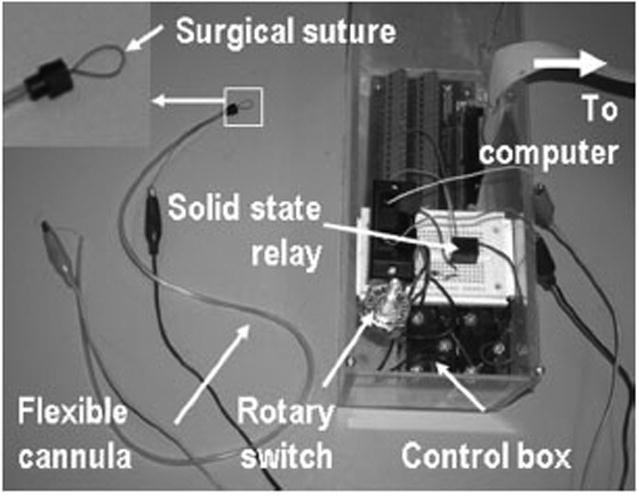
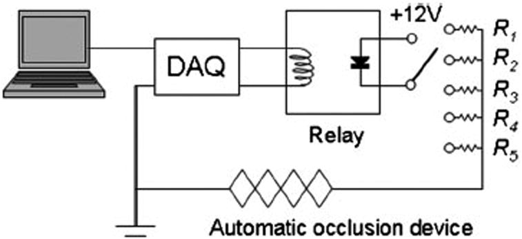
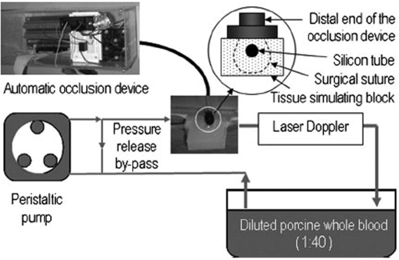
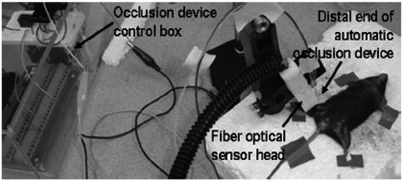
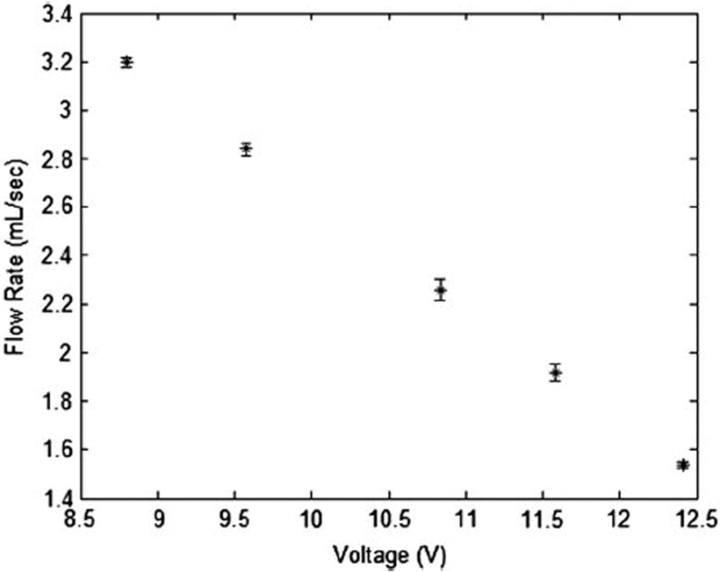
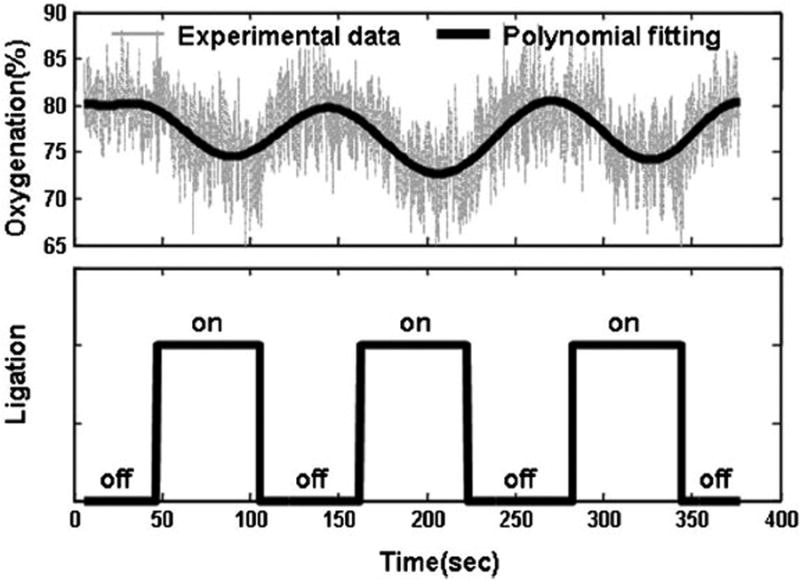
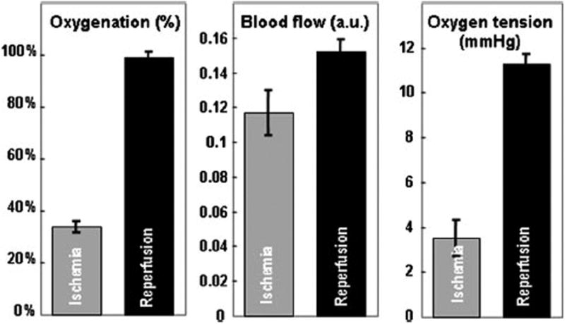
We developed an automatic occlusion device for remote control of tumor tissue ischemia. The device consists of a flexible cannula encasing a shape memory alloy wire with its distal end connected to surgical suture. Regional tissue occlusion was tested on both the benchtop and the animal models. In the benchtop test, the occlusion device introduced quantitative and reproducible changes of blood flow in a tissue simulating phantom embedding a vessel simulator. In the animal test, the device generated a cyclic pattern of reversible ischemia in the right hinder leg tissue of a black male C57BL/6 mouse. We also developed a multimodal detector that integrates near infrared spectroscopy and electron paramagnetic resonance spectroscopy for continuous monitoring of tumor tissue oxygenation, blood content, and oxygen tension changes. The multimodal detector was tested on a cancer xenograft nude mouse undergoing reversible tumor ischemia. The automatic occlusion device and the multimodal detector can be potentially integrated for closed-loop feedback control of tumor tissue ischemia. Such an integrated occlusion device may be used in multiple clinical applications such as regional hypoperfusion control in tumor resection surgeries and thermal ablation processes. In addition, the proposed occlusion device can also be used as a research tool to understand tumor oxygen transport and hemodynamic characteristics.
Figures

Similar articles
-
Cerebral ischemia caused by obstructed superior vena cava cannula is detected by near-infrared spectroscopy.J Cardiothorac Vasc Anesth. 2004 Jun;18(3):293-303. doi: 10.1053/j.jvca.2004.03.008. J Cardiothorac Vasc Anesth. 2004. PMID: 15232808
-
Online detection of myocardial ischemia by near infrared spectroscopy with a fiberoptic catheter.Thorac Cardiovasc Surg. 2001 Jun;49(3):162-6. doi: 10.1055/s-2001-14294. Thorac Cardiovasc Surg. 2001. PMID: 11432475
-
Tissue Monitoring with Three-Wavelength Light Emitting Diode-Based Near-Infrared Spectroscopy.J Reconstr Microsurg. 2016 Nov;32(9):712-718. doi: 10.1055/s-0036-1586256. Epub 2016 Aug 19. J Reconstr Microsurg. 2016. PMID: 27542109 Clinical Trial.
-
Near-Infrared Spectroscopy and Vascular Occlusion Test for Predicting Clinical Outcome in Pediatric Cardiac Patients: A Prospective Observational Study.Pediatr Crit Care Med. 2018 Jan;19(1):32-39. doi: 10.1097/PCC.0000000000001386. Pediatr Crit Care Med. 2018. PMID: 29140967
-
Applications of functional near-infrared spectroscopy (fNIRS) to Neurorehabilitation of cognitive disabilities.Clin Neuropsychol. 2007 Jan;21(1):38-57. doi: 10.1080/13854040600878785. Clin Neuropsychol. 2007. PMID: 17366277 Review.
References
-
- Kloner RA, Jennings RB. Consequences of brief ischemia: stunning, preconditioning, and their clinical implications: part 1. Circulation. 2001;104:2981–2989. - PubMed
-
- Weise ES, Winfield HN. Laparoscopic partial nephrectomy. J Endourol. 2005;19:634–642. - PubMed
-
- Novick AC. Renal hypothermia: in vivo and ex vivo. Urol Clin North Am. 1983;10:637–644. - PubMed
-
- Cadeddu JA, Corwin TS, Traxer O, Collick C, Saboorian HH, Pearle MS. Hemostatic laparoscopic partial nephrectomy: cable-tie compression. Urology. 2001;57:562–566. - PubMed
MeSH terms
Substances
Grants and funding
LinkOut - more resources
Full Text Sources
