

Human cardiac tissue in a microperfusion chamber simulating extracorporeal circulation--ischemia and apoptosis studies
- PMID: 20082695
- PMCID: PMC2821307
- DOI: 10.1186/1749-8090-5-3
Human cardiac tissue in a microperfusion chamber simulating extracorporeal circulation--ischemia and apoptosis studies
Abstract
Background: After coronary artery bypass grafting ischemia/reperfusion injury inducing cardiomyocyte apoptosis may occur. This surgery-related inflammatory reaction appears to be of extreme complexity with regard to its molecular, cellular and tissue mechanisms and many studies have been performed on animal models. However, finding retrieved from animal studies were only partially confirmed in humans. To investigate this phenomenon and to evaluate possible therapies in vitro, adequate human cardiomyocyte models are required. We established a tissue model of human cardiomyocytes preserving the complex tissue environment. To our knowledge human cardiac tissue has not been investigated in an experimental setup mimicking extracorporeal circulation just in accordance to clinical routine, yet.
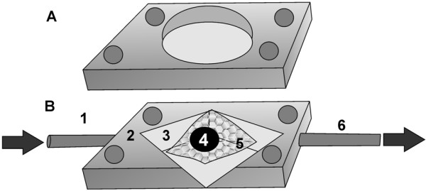
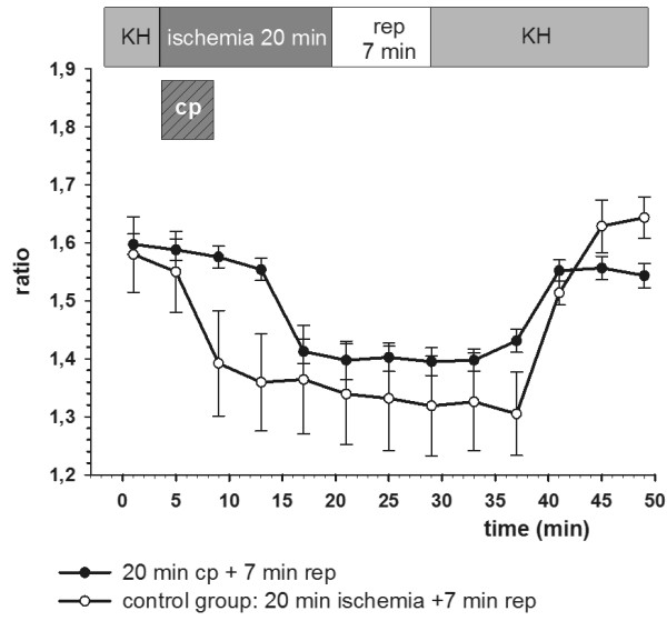
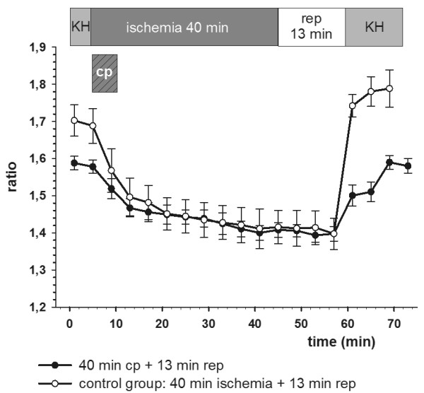
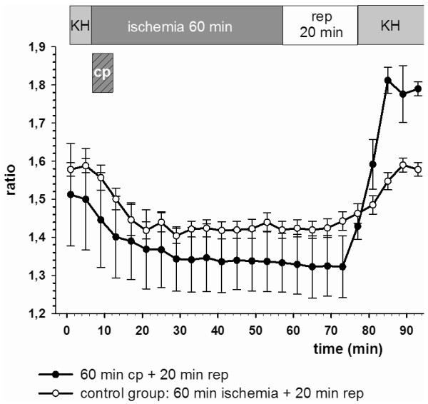
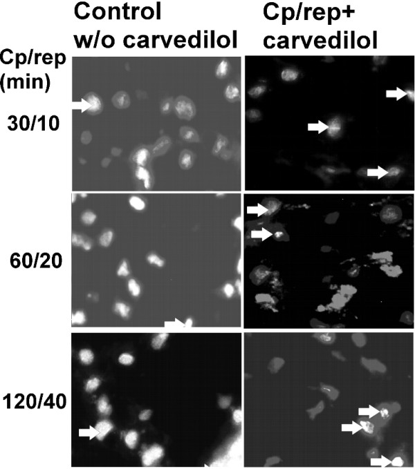
Methods: Cardiac biopsies were retrieved from the right auricle of patients undergoing elective coronary artery bypass grafting before cardiopulmonary bypass. The extracorporeal circulation was simulated by submitting the biopsies to varied conditions simulating cardioplegia (cp) and reperfusion (rep) in a microperfusion chamber. Cp/rep time sets were 20/7, 40/13 and 60/20 min. For analyses of the calcium homoeostasis the fluorescent calcium ion indicator FURA-2 and for apoptosis detection PARP-1 cleavage immunostaining were employed. Further the anti-apoptotic effect of carvedilol [10 microM] was investigated by adding into the perfusate.
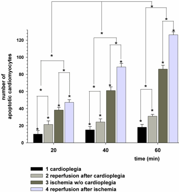
Results: Viable cardiomyocytes presented an intact calcium homoeostasis under physiologic conditions. Following cardioplegia and reperfusion a time-dependent elevation of cytosolic calcium as a sign of disarrangement of the calcium homoeostasis occurred. PARP-1 cleavage also showed a time-dependence whereas reperfusion had the highest impact on apoptosis. Cardioplegia and carvedilol could reduce apoptosis significantly, lowering it between 60-70% (p < 0.05).
Conclusions: Our human cardiac preparation served as a reliable cellular model tool to study apoptosis in vitro. Decisively cardiac tissue from the right auricle can be easily obtained at nearly every cardiac operation avoiding biopsying of the myocardium or even experiments on animals.The apoptotic damage induced by the ischemia/reperfusion stimulus could be significantly reduced by the cold crystalloid cardioplegia. The additional treatment of cardiomyocytes with a non-selective beta-blocker, carvedilol had even a significantly higher reduction of apoptotis.
Figures
Similar articles
-
The nonselective beta-blocker carvedilol suppresses apoptosis in human cardiac tissue: a pilot study.Heart Surg Forum. 2010 Aug;13(4):E218-22. doi: 10.1532/HSF98.20091179. Heart Surg Forum. 2010. PMID: 20719722
-
Resveratrol suppresses apoptosis in intact human cardiac tissue - in vitro model simulating extracorporeal circulation.J Cardiovasc Surg (Torino). 2011 Jun;52(3):399-409. J Cardiovasc Surg (Torino). 2011. PMID: 21577194
-
Carvedilol treatment after myocardial infarct decreases cardiomyocytic apoptosis in the peri-infarct zone during cardioplegia-induced cardiac arrest.Shock. 2013 Apr;39(4):343-52. doi: 10.1097/SHK.0b013e31828c588a. Shock. 2013. PMID: 23481492
-
Pharmacology of carvedilol: rationale for use in hypertension, coronary artery disease, and congestive heart failure.Cardiovasc Drugs Ther. 1997 May;11 Suppl 1:247-56. doi: 10.1023/a:1007735729121. Cardiovasc Drugs Ther. 1997. PMID: 9211017 Review.
-
Myocardial ischemia, stunning, inflammation, and apoptosis during cardiac surgery: a review of evidence.Eur J Cardiothorac Surg. 2004 Mar;25(3):304-11. doi: 10.1016/j.ejcts.2003.12.003. Eur J Cardiothorac Surg. 2004. PMID: 15019653 Review.
Cited by
-
The challenge to verify ceramide's role of apoptosis induction in human cardiomyocytes--a pilot study.J Cardiothorac Surg. 2011 Mar 28;6:38. doi: 10.1186/1749-8090-6-38. J Cardiothorac Surg. 2011. PMID: 21443760 Free PMC article.
References
Publication types
MeSH terms
Substances
LinkOut - more resources
Full Text Sources
Miscellaneous
