. 2010 Jan 18:11:10.
doi: 10.1186/1471-2474-11-10.

Characterization of differential properties of rabbit tendon stem cells and tenocytes

Affiliations

Characterization of differential properties of rabbit tendon stem cells and tenocytes

Jianying Zhang et al. BMC Musculoskelet Disord. .

Abstract

Background: Tendons are traditionally thought to consist of tenocytes only, the resident cells of tendons; however, a recent study has demonstrated that human and mouse tendons also contain stem cells, referred to as tendon stem/progenitor cells (TSCs). However, the differential properties of TSCs and tenocytes remain largely undefined. This study aims to characterize the properties of these tendon cells derived from rabbits.

Methods: TSCs and tenocytes were isolated from patellar and Achilles tendons of rabbits. The differentiation potential and cell marker expression of the two types of cells were examined using histochemical, immunohistochemical, and qRT-PCR analysis as well as in vivo implantation. In addition, morphology, colony formation, and proliferation of TSCs and tenocytes were also compared.

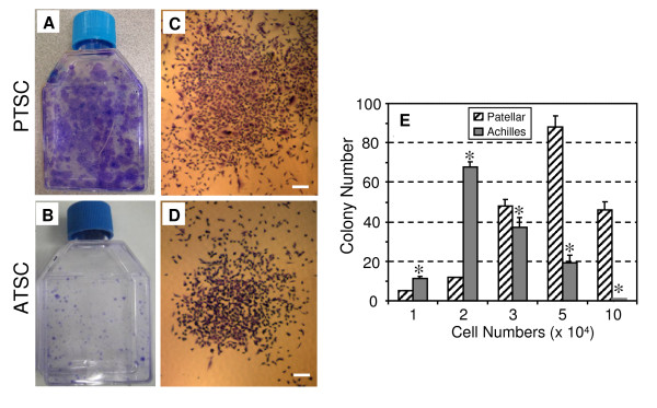
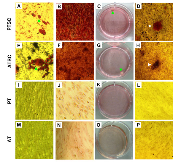
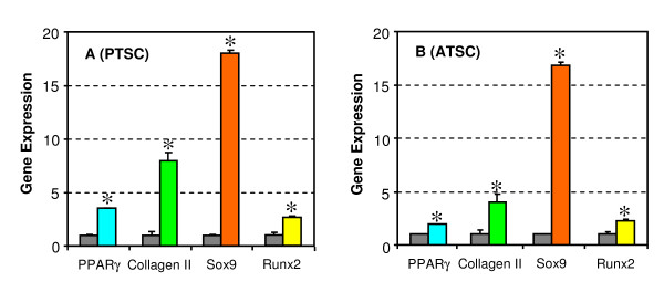
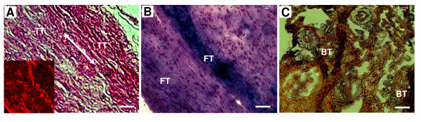
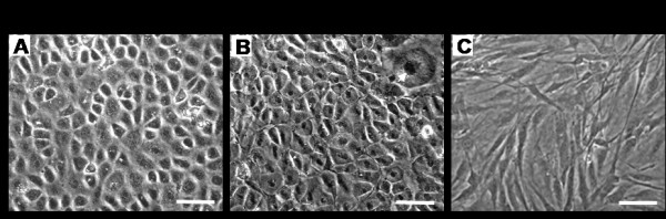
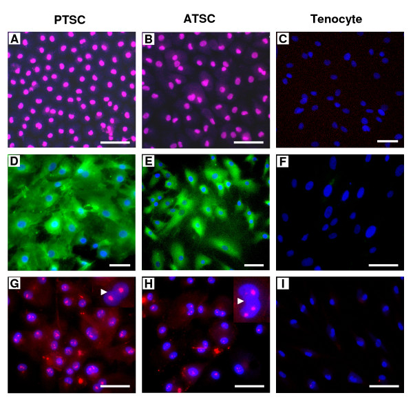
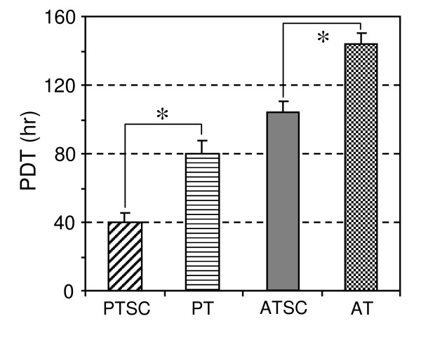
Results: It was found that TSCs were able to differentiate into adipocytes, chondrocytes, and osteocytes in vitro, and form tendon-like, cartilage-like, and bone-like tissues in vivo. In contrast, tenocytes had little such differentiation potential. Moreover, TSCs expressed the stem cell markers Oct-4, SSEA-4, and nucleostemin, whereas tenocytes expressed none of these markers. Morphologically, TSCs possessed smaller cell bodies and larger nuclei than ordinary tenocytes and had cobblestone-like morphology in confluent culture whereas tenocytes were highly elongated. TSCs also proliferated more quickly than tenocytes in culture. Additionally, TSCs from patellar tendons formed more numerous and larger colonies and proliferated more rapidly than TSCs from Achilles tendons.

Conclusions: TSCs exhibit distinct properties compared to tenocytes, including differences in cell marker expression, proliferative and differentiation potential, and cell morphology in culture. Future research should investigate the mechanobiology of TSCs and explore the possibility of using TSCs to more effectively repair or regenerate injured tendons.


Figures

Figure 1
Figure 1
The colony formation of rabbit tendon stem cells (TSCs). A, B. Total PTSC and ATSC colonies stained with Methyl violet at 10 days. C, D. Expanding colonies of PTSCs and ATSCs at 10 days, respectively. It is seen that more numerous and larger cell colonies were formed by PTSCs compared to ATSCs. E. Quantitative analysis of colonies formed by PTSCs and ATSCs. Colony number of PTSCs was significantly different from that of ATSCs (* p < 0.05). (Bars: 200 μm).
Figure 2
Figure 2
The testing of multi-differentiation potential of TSCs and tenocytes in vitro. A. Adipogenesis of PTSCs (arrows point to lipid droplets). B. Chondrogenesis of PTSCs. C. Cartilage-like pellet (arrow) formed from PTSCs. D. Osteogenesis of PTSCs (An arrow points to a clustered calcium droplet). Similar multi-differentiation potential is shown for ATSCs (E-H). Patellar tenocytes (PTs) and Achilles tenocytes (ATs) were not found to exhibit such a multi-differentiation potential (I-P, also see text for additional descriptions of experimental results). (Magnification of microscopy: 10×).
Figure 3
Figure 3
The qRT-PCR analysis of expression of marker genes. Rabbit PTSCs and ATSCs were differentiated into adipocytes (PPARγ), chondrocytes (collagen II and Sox9), and osteocytes (Runx2) using their respective differentiation induction media. Compared to non-differentiated, control cells (black columns) that were grown in regular growth medium, all these genes were significantly upregulated for both PTSCs (A) and ATSCs (B), albeit at different levels (* p < 0.05). Note that for real time RT-PCR analysis, the gene expression levels were normalized to GAPDH, obtained from at least three independent experiments and presented as 2(-ΔCT).
Figure 4
Figure 4
The testing of multi-differentiation of rabbit TSCs in vivo. A. Formation of tendon-like tissue (TT) revealed by H&E staining and immunohistochemical staining for collagen type I. The collagen fibers are parallel to each other (double arrow), indicative of formation of tendon-like tissue (inset: collagen type I staining). B. Formation of fibrocartilage-like tissue (FT) (Alcian blue staining); and C. formation of bone-like tissue (BT) (Alizarin Red S staining). (Bars: 50 μm).
Figure 5
Figure 5
The morphology of TSCs and tenocytes in culture. A, B. PTSCs and ATSCs at passage 10 were in culture for at least 63 days, respectively. These cells were cobblestone-like in a confluent culture. C. Morphology of tenocytes from the same rabbit patellar tendons; similar morphology was observed in tenocytes from the Achilles tendons (not shown). These tenocytes were highly elongated in a confluent culture. (Bar: 50 μm).
Figure 6
Figure 6
The testing of stem cell marker expression. A, B. PTSCs and ATSCs at passages 10 expressed Oct-4, respectively. C. No Oct-4 staining was detected on tenocytes. D, E. PTSCs and ATSCs expressed SSEA-4. F. Tenocytes were negative for SSEA-4 staining. G, H. PTSCs and ATSCs expressed nucleostemin. Insets show enlarged view of expressed nucleostemin in pink (arrows). I. Nucleostemin expression was not detected on tenocytes. (bar: 50 μm).
Figure 7
Figure 7
The population doubling time (PDT) of TSCs and tenocytes. Patellar TSCs (PTSCs), Achilles TSCs (ATSCs) at passage 2 proliferated faster than their counterparts: patellar tenocytes (PTs), and Achilles tenocytes (ATs).

Similar articles

Cited by

References

    1. Khan KM, Maffuli N. Tendinopathy: an Achilles' heel for athletes and clinicians. Clin J Sport Med. 1998;8:151–154. - PubMed
    1. Maffulli N, Khan KM, Puddu G. Overuse tendon conditions: time to change a confusing terminology. Arthroscopy. 1998;14(8):840–843. - PubMed
    1. Butler DL, Juncosa-Melvin N, Boivin GP, Galloway MT, Shearn JT, Gooch C, Awad H. Functional tissue engineering for tendon repair: A multidisciplinary strategy using mesenchymal stem cells, bioscaffolds, and mechanical stimulation. J Orthop Res. 2008;26(1):1–9. doi: 10.1002/jor.20456. - DOI - PubMed
    1. Dennis JE, Carbillet JP, Caplan AI, Charbord P. The STRO-1+ marrow cell population is multipotential. Cells Tissues Organs. 2002;170(2-3):73–82. doi: 10.1159/000046182. - DOI - PubMed
    1. Bajada S, Mazakova I, Richardson JB, Ashammakhi N. Updates on stem cells and their applications in regenerative medicine. J Tissue Eng Regen Med. 2008;2(4):169–183. doi: 10.1002/term.83. - DOI - PubMed

Publication types

MeSH terms