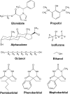
Numerous classes of general anesthetics inhibit etomidate binding to gamma-aminobutyric acid type A (GABAA) receptors
- PMID: 20083606
- PMCID: PMC2838283
- DOI: 10.1074/jbc.M109.074708
Numerous classes of general anesthetics inhibit etomidate binding to gamma-aminobutyric acid type A (GABAA) receptors
Abstract
Enhancement of gamma-aminobutyric acid type A receptor (GABA(A)R)-mediated inhibition is a property of most general anesthetics and a candidate for a molecular mechanism of anesthesia. Intravenous anesthetics, including etomidate, propofol, barbiturates, and neuroactive steroids, as well as volatile anesthetics and long-chain alcohols, all enhance GABA(A)R function at anesthetic concentrations. The implied existence of a receptor site for anesthetics on the GABA(A)R protein was supported by identification, using photoaffinity labeling, of a binding site for etomidate within the GABA(A)R transmembrane domain at the beta-alpha subunit interface; the etomidate analog [(3)H]azietomidate photolabeled in a pharmacologically specific manner two amino acids, alpha1Met-236 in the M1 helix and betaMet-286 in the M3 helix (Li, G. D., Chiara, D. C., Sawyer, G. W., Husain, S. S., Olsen, R. W., and Cohen, J. B. (2006) J. Neurosci. 26, 11599-11605). Here, we use [(3)H]azietomidate photolabeling of bovine brain GABA(A)Rs to determine whether other structural classes of anesthetics interact with the etomidate binding site. Photolabeling was inhibited by anesthetic concentrations of propofol, barbiturates, and the volatile agent isoflurane, at low millimolar concentrations, but not by octanol or ethanol. Inhibition by barbiturates, which was pharmacologically specific and stereospecific, and by propofol was only partial, consistent with allosteric interactions, whereas isoflurane inhibition was nearly complete, apparently competitive. Protein sequencing showed that propofol inhibited to the same extent the photolabeling of alpha1Met-236 and betaMet-286. These results indicate that several classes of general anesthetics modulate etomidate binding to the GABA(A)R: isoflurane binds directly to the site with millimolar affinity, whereas propofol and barbiturates inhibit binding but do not bind in a mutually exclusive manner with etomidate.
Figures


Similar articles
-
Neurosteroids allosterically modulate binding of the anesthetic etomidate to gamma-aminobutyric acid type A receptors.J Biol Chem. 2009 May 1;284(18):11771-5. doi: 10.1074/jbc.C900016200. Epub 2009 Mar 12. J Biol Chem. 2009. PMID: 19282280 Free PMC article.
-
Mapping general anesthetic binding site(s) in human α1β3 γ-aminobutyric acid type A receptors with [³H]TDBzl-etomidate, a photoreactive etomidate analogue.Biochemistry. 2012 Jan 31;51(4):836-47. doi: 10.1021/bi201772m. Epub 2012 Jan 23. Biochemistry. 2012. PMID: 22243422 Free PMC article.
-
Multiple propofol-binding sites in a γ-aminobutyric acid type A receptor (GABAAR) identified using a photoreactive propofol analog.J Biol Chem. 2014 Oct 3;289(40):27456-68. doi: 10.1074/jbc.M114.581728. Epub 2014 Aug 1. J Biol Chem. 2014. PMID: 25086038 Free PMC article.
-
GABA(A) receptors as molecular targets of general anesthetics: identification of binding sites provides clues to allosteric modulation.Can J Anaesth. 2011 Feb;58(2):206-15. doi: 10.1007/s12630-010-9429-7. Epub 2010 Dec 31. Can J Anaesth. 2011. PMID: 21194017 Free PMC article. Review.
-
Anesthetic sites and allosteric mechanisms of action on Cys-loop ligand-gated ion channels.Can J Anaesth. 2011 Feb;58(2):191-205. doi: 10.1007/s12630-010-9419-9. Epub 2011 Jan 7. Can J Anaesth. 2011. PMID: 21213095 Free PMC article. Review.
Cited by
-
The concept of allosteric interaction and its consequences for the chemistry of the brain.J Biol Chem. 2013 Sep 20;288(38):26969-26986. doi: 10.1074/jbc.X113.503375. Epub 2013 Jul 22. J Biol Chem. 2013. PMID: 23878193 Free PMC article. Review.
-
Specificity of intersubunit general anesthetic-binding sites in the transmembrane domain of the human α1β3γ2 γ-aminobutyric acid type A (GABAA) receptor.J Biol Chem. 2013 Jul 5;288(27):19343-57. doi: 10.1074/jbc.M113.479725. Epub 2013 May 15. J Biol Chem. 2013. PMID: 23677991 Free PMC article.
-
Structural models of ligand-gated ion channels: sites of action for anesthetics and ethanol.Alcohol Clin Exp Res. 2014 Mar;38(3):595-603. doi: 10.1111/acer.12283. Epub 2013 Oct 24. Alcohol Clin Exp Res. 2014. PMID: 24164436 Free PMC article. Review.
-
Awake nonhuman primate brain PET imaging with minimal head restraint: evaluation of GABAA-benzodiazepine binding with 11C-flumazenil in awake and anesthetized animals.J Nucl Med. 2013 Nov;54(11):1962-8. doi: 10.2967/jnumed.113.122077. Epub 2013 Oct 10. J Nucl Med. 2013. PMID: 24115528 Free PMC article.
-
Effects of inhaled anesthetic isoflurane on long-term potentiation of CA3 pyramidal cell afferents in vivo.Int J Gen Med. 2012;5:935-42. doi: 10.2147/IJGM.S30570. Epub 2012 Nov 9. Int J Gen Med. 2012. PMID: 23204857 Free PMC article.
References
-
- Harrison N. L., Krasowski M. D., Harris R. A. (2000) in GABA in the Nervous System: The View at Fifty Years (Martin D. L., Olsen R. W. eds.) pp. 167–189, Lippincott Williams & Wilkins, Philadelphia
-
- Ernst M., Bruckner S., Boresch S., Sieghart W. (2005) Mol. Pharmacol. 68, 1291–1300 - PubMed
-
- Franks N. P. (2008) Nat. Rev. Neurosci. 9, 370–386 - PubMed
Publication types
MeSH terms
Substances
Grants and funding
LinkOut - more resources
Full Text Sources
Other Literature Sources
Molecular Biology Databases

