. 2010 Feb 15;184(4):1710-20.
doi: 10.4049/jimmunol.0902796. Epub 2010 Jan 18.

IL-23 receptor regulates unconventional IL-17-producing T cells that control bacterial infections

Affiliations

IL-23 receptor regulates unconventional IL-17-producing T cells that control bacterial infections

Lorena Riol-Blanco et al. J Immunol. .

Abstract

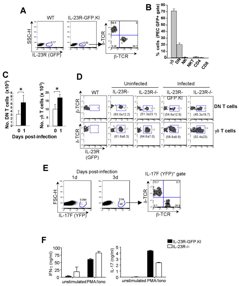
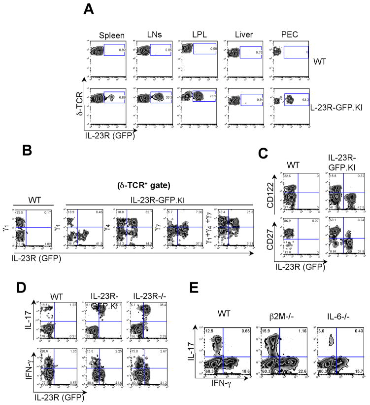
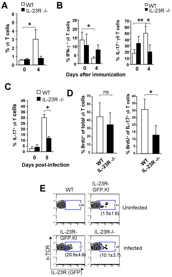
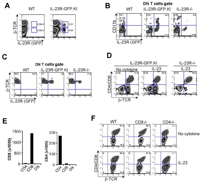
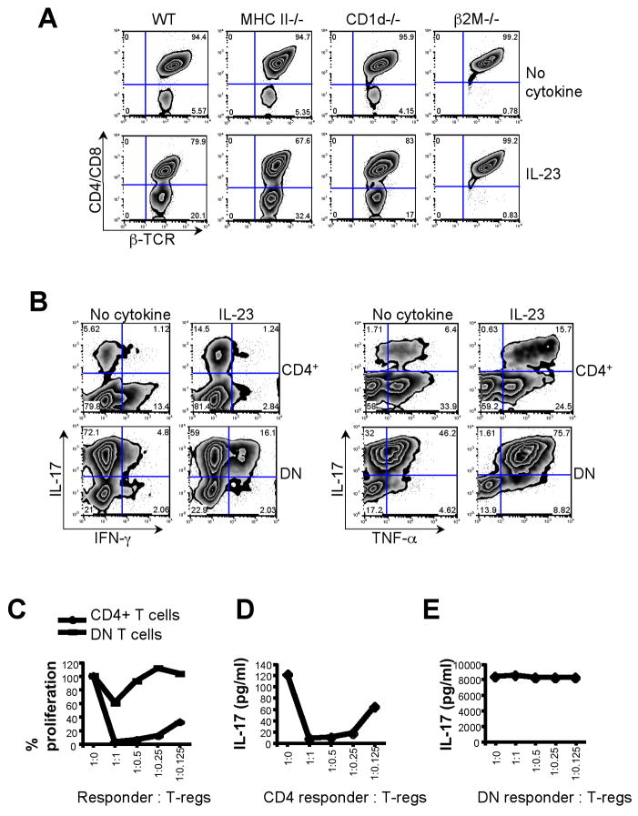
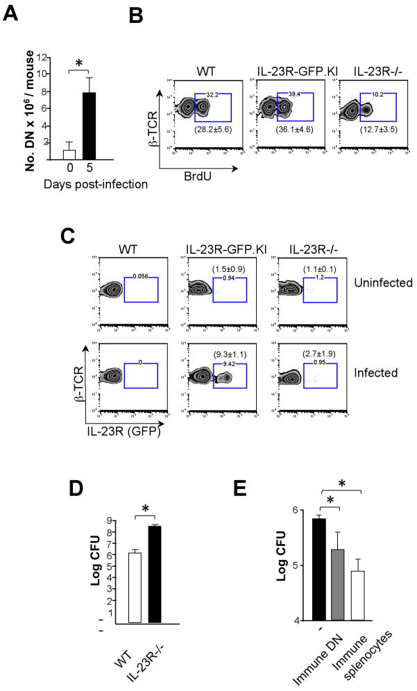
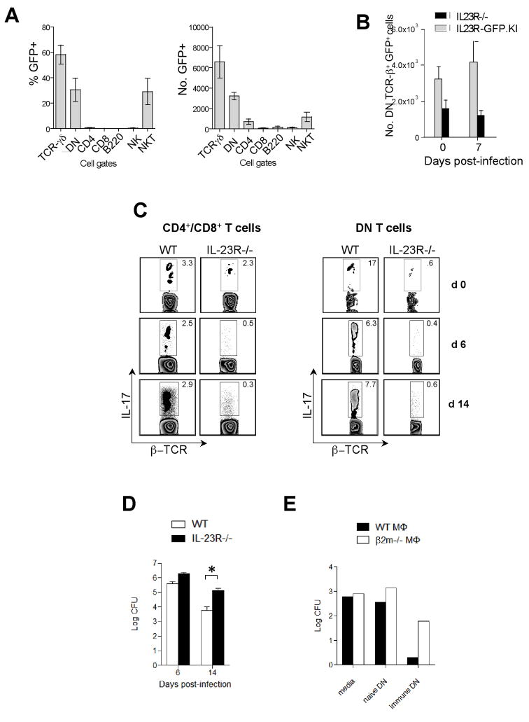
IL-23 plays an important role in autoimmune tissue inflammation and induces the generation of not fully characterized effector cells that mediate protection against pathogens. In this paper, we established the essential role of IL-23R in the host response against intracellular pathogens. IL-23 was critical for the expansion or maintenance of gammadelta and double negative (DN) alphabeta T cells. These cells were rapidly recruited to the site of infection and produced large amounts of IL-17, IFN-gamma, and TNF-alpha. Notably, DN T cells transferred into L. monocytogenes-infected RAG2(-/-) mice prevented bacterial growth, confirming their protective role against intracellular pathogens. Our results show that IL-23 regulates the function of IL-17-producing gammadelta and DN T cells, two essential components of the early protective immune response directed against intracellular pathogens.


Figures

Figure 1
Figure 1. Expression of IL-23R in the peritoneal cavity of L. monocytogenes-infected mice
Mice were infected via the intraperitoneal route with viable L. monocytogenes. (A, B, C, D and F) 24 h or 72 h (E) after infection. PEC were collected from WT, IL-23R-GFP.KI or IL-23R−/− (A, B, C, D and F) or IL-17F-CreEYFP (E) mice and β–TCR and δ–TCR surface stainings were performed. (A) IL-23R expression was analyzed in PEC of IL-23R-GFP.KI mice (B) Percentages of IL-23R (GFP)+ expressing cells were calculated within δ–TCR+, DN, CD4+, CD8+, NK1.1+, CD49b+ cell populations isolated from the peritoneal cavity of infected IL-23R-GFP.KI mice. The bars represent mean ± SD of 3 independent experiments (3 mice/group/experiment). (C) Histograms represent total numbers of γδ and DN collected from the peritoneum of IL-23R-GFP.KI mice 1d after infection. [*p<0.05 (two tail Student t-test)]. (D) The zebra plots represent the IL-23R (GFP) expression of peritoneal γδ or DN T cells from IL-23R-GFP.KI or IL-23R−/− mice 1 day after infection. In brackets, mean ± SD of 3 experiments with 3 mice/group is shown. (E) 1 d after infection, IL-23R-GFP.KI or IL-23R−/− PEC were isolated and stimulated or not with PMA/Ionomycin for an additional 18 h. Supernatants were collected and cytokine ELISA was performed for IL-17 and IFN-γ. Data shown are representative of 2 experiments with 3 mice/group. (F) 4× 10⁶ PEC where cultured in the presence of PMA/Ionomicin. After 24h the supernatants were collected and ELISA for IL-17 and INF-γ were performed.
Figure 2
Figure 2. Identification of IL-23R expressing γδ T cells
(A) IL-23R (GFP) expression was analyzed on γδ T cells from LNs, spleen, Lamina Propria (LP), liver and Peritoneal Exudate Cells (PEC) in IL-23R-GFP.KI naïve mice. (B, C) LN cells from IL-23R-GFP.KI naïve mice were collected and analyzed for δ-TCR and Vγ1 Vγ4 and Vγ7 (B) or CD27 or CD122 (C) expression on γδ T cells. (D) LN cells collected from IL-23R-GFP.KI or IL-23R−/− were stimulated with PMA/Ionomycin and intracellular cytokine staining for IFN-γ and IL-17 was performed. The quadrants represent intracellular cytokine staining and IL-23R (GFP) expression on γδ T cells. (E) Single cell suspensions were prepared from LNs from WT, β2M−/− or IL-6−/− mice. Quadrants represent intracellular cytokine staining for IL-17 and IFN-γ on γδ T cells after PMA/Ionomycin stimulation. The experiment was repeated 2 times, 3 mice/group.
Figure 3
Figure 3. IL-17-producing γδ T cells require Il-23R for expansion/survival during L. monocytogenes infection
(A, B) IL-23R-GFP.KI or WT mice were immunized with MOG35-55 emulsified in CFA. (A) Percentages of draining LN γδ T cells in naïve mice or 4 d after MOG35-55/CFA immunization of WT or IL23R−/− mice. The bars represent mean ± SD of 3 independent experiments [*p<0.05, **p<0.01 (two tail Student t-test)]. (B) Draining LN cells were stimulated with PMA/Ionomycin. Percentages of IFN-γ or IL-17+γδ T cells from naïve or MOG35-55/CFA-immunized mice after 4 d are represented. (C, D) Mice were i.p infected with viable L. monocytogenes. Mononuclear cells were isolated from the livers of IL- 23R−/− or WT mice and stimulated with PMA/Ionomycin. (C) Percentages of hepatic IL-17-producing γδ T cells from WT or IL-23R−/− during L. monocytogenes infection are represented. The bars represent mean ± SD of 3 independent experiments. (D) WT or IL23R−/− mice were infected with L. monocytogenes and received 2 mg of BrdU intraperitoneally on day 1 and 2 after infection. Intracellular staining of IL-17 and BrdU was performed in γδ T cells from livers of WT or IL-23R−/− mice 3 d after infection. The bars represent mean ± SD of 3 independent experiments [*p<0.05 (two tail Student t-test)]. (E). IL-23R (GFP) expression was assessed in intrahepatic γδ T cells from IL-23R-GFP.KI or IL-23R−/− mice during L. monocytogenes infection. In brackets, mean ± SD of 3 experiments with 3 mice/group is shown.
Figure 4
Figure 4. Expansion of DN T cells depends on IL-23R in vitro
LNs were collected from naïve WT and IL-23R-GFP.KI mice. (A) Single cell suspensions were prepared for surface staining of β–TCR, CD4 and CD8. (B) Quadrants show CD11b and IL-23R (GFP) expression in β–TCR+ CD4 CD8 DN T cells from LNs collected from WT, IL-23R-GFP.KI and IL-23R−/− mice. (C) Single cell suspensions from LNs of naïve WT, IL-23R-GFP.KI and IL-23R−/−mice were prepared and stained for β–TCR, CD4 and CD8. IL-23R (GFP) expression in the DN T cell gate is shown. (D) LNs from IL-23R-GFP.KI or IL-23R−/− mice were cultured with anti-CD3 alone or with rIL-23 (30 ng/ml). On day 6, cultured cells were stained for CD4, CD8 and β–TCR. The numbers in the quadrants represent percentages of β–TCR+ CD4 CD8 DN and β–TCR+ CD4+/CD8+ T cells. (E) Real time PCR was performed for CD4 and CD8 genes on FACS sorted DN T cells. (F) LNs were collected from CD8−/−, CD4 −/− and WT mice. Cells were activated with either anti-CD3 alone or in the presence of rIL-23. On day 6, cells were stained for β–TCR, CD4 and CD8. Cells were analyzed for generation of DN T cells.
Figure 5
Figure 5. Characterization of DN T cells
(A) LNs were collected from MHCII−/−, CD1d−/−, β2M−/− and WT littermate control mice. LN cells were activated with either anti-CD3 alone or in the presence of rIL-23. On day 6, cells were stained for β–TCR, CD4 and CD8 and analyzed for generation of DN T cells. (B) On day 6, CD4+ and DN T cells were FACS sorted and then re-stimulated with PMA and Ionomycin and intracellular cytokine staining was performed for IL-17, IFN-γ and TNF-α. (C, D, E) CD4+ and DN T cells generated as described were FACS sorted. CD4+ and DN T cells were used as effector cells with indicated ratio of Foxp3+ Tregs in an in vitro suppression assay. (E) Cell proliferation was measured by thymidine incorporation. (D, E) Culture supernatant was collected at 48 h and cytokine analysis was performed for IL-17.
Figure 6
Figure 6. DN T cells are involved in resistance to L. monocytogenes infection
(A) Absolute numbers of DN T cells from spleen and liver pooled cells before or 5 d after L. monocytogenes infection are shown. The bars represent mean ± SD of 3 independent experiments (5–10 mice pooled/group/experiment). (B, C) WT, IL-23R-GFP.KI or IL-23R−/− mice were infected with L. monocytogenes. After 3 d, BrdU incorporation (B) and IL-23R (GFP) expression (C) were analyzed in DN T cells. In brackets, mean ± SD of 2 experiments with 3 mice/group is shown. (D) After 3 d bacterial CFU counts were determined in livers of IL-23R−/− or WT mice infected with L. monocytogenes. (E) No cells, immune splenocytes or immune DNs isolated from WT mice 5 d after infection with L. monocytogenes were transferred intravenously into RAG2−/− mice. 24 h later mice were challenged i.p. with L. monocytogenes. After 3 d, the CFU in the liver was assessed to quantify bacterial burden. The bars represent mean ± SD of 3 independent experiments (5 mice pooled/group/experiment) [*p<0.05, **p<0.01 (two tail Student t-test)].
Figure 7
Figure 7. Role of IL-23R in F. tularensis LVS infection
(A) Percentages and absolute numbers of IL-23R (GFP) expressing cells within δ-TCR+, DN, CD4+, CD8+, B220+, NK1.1+, PBS57/CD1d tetramer+ cell populations isolated from the lungs of uninfected IL-23R-GFP.KI mice. The bars represent mean ± SEM of 3 independent experiments (3–5 mice pooled/group/experiment). (B) Absolute numbers of IL-23R (GFP) expressing DN T cells isolated from the lungs of uninfected and F. tularensis LVS-infected IL-23R−/− and IL-23R-GFP.KI mice. The bars represent mean ± SEM of 3 independent experiments (3–5 mice pooled/group/experiment). (C) The dot plots represent IL-17 production by β–TCR+ CD4+/CD8+ and β–TCR+ DN T cells isolated from uninfected and F. tularensis LVS infected lungs of IL-23R−/− and WT mice. The experiment was repeated twice with 3–4 mice/group pooled in each experiment. (D) Bacterial CFU was determined in the F. tularensis LVS infected lungs of IL-23R−/− and WT mice [*p<0.05, **p<0.01 (two tail Student t-test)]. (E) F. tularensis LVS infected β2M−/− and WT macrophages were incubated with DN T cells isolated from the spleens of naïve or immune mice and bacterial burden was determined 72 h after culture. The experiment was repeated 3 times.

Similar articles

Cited by

References

    1. Langrish CL, McKenzie BS, Wilson NJ, de Waal Malefyt R, Kastelein RA, Cua DJ. IL-12 and IL-23: master regulators of innate and adaptive immunity. Immunol Rev. 2004;202:96–105. - PubMed
    1. Langrish CL, Chen Y, Blumenschein WM, Mattson J, Basham B, Sedgwick JD, McClanahan T, Kastelein RA, Cua DJ. IL-23 drives a pathogenic T cell population that induces autoimmune inflammation. J Exp Med. 2005;201:233–240. - PMC - PubMed
    1. Park H, Li Z, Yang XO, Chang SH, Nurieva R, Wang YH, Wang Y, Hood L, Zhu Z, Tian Q, Dong C. A distinct lineage of CD4 T cells regulates tissue inflammation by producing interleukin 17. Nat Immunol. 2005;6:1133–1141. - PMC - PubMed
    1. McGeachy MJ, Chen Y, Tato CM, Laurence A, Joyce-Shaikh B, Blumenschein WM, McClanahan TK, O’Shea JJ, Cua DJ. The interleukin 23 receptor is essential for the terminal differentiation of interleukin 17-producing effector T helper cells in vivo. Nat Immunol. 2009;10:314–324. - PMC - PubMed
    1. Awasthi A, Riol-Blanco L, Jager A, Korn T, Pot C, Galileos G, Bettelli E, Kuchroo VK, Oukka M. Cutting edge: IL-23 receptor gfp reporter mice reveal distinct populations of IL-17-producing cells. J Immunol. 2009;182:5904–5908. - PMC - PubMed

Publication types

MeSH terms