. 2010 Feb 15;184(4):2107-15.
doi: 10.4049/jimmunol.0901208. Epub 2010 Jan 18.

A CD1d-dependent antagonist inhibits the activation of invariant NKT cells and prevents development of allergen-induced airway hyperreactivity

Affiliations

A CD1d-dependent antagonist inhibits the activation of invariant NKT cells and prevents development of allergen-induced airway hyperreactivity

Vincent Lombardi et al. J Immunol. .

Abstract

The prevalence of asthma continues to increase in westernized countries, and optimal treatment remains a significant therapeutic challenge. Recently, CD1d-restricted invariant NKT (iNKT) cells were found to play a critical role in the induction of airway hyperreactivity (AHR) in animal models and are associated with asthma in humans. To test whether iNKT cell-targeted therapy could be used to treat allergen-induced airway disease, mice were sensitized with OVA and treated with di-palmitoyl-phosphatidyl-ethanolamine polyethylene glycol (DPPE-PEG), a CD1d-binding lipid antagonist. A single dose of DPPE-PEG prevented the development of AHR and pulmonary infiltration of lymphocytes upon OVA challenge, but had no effect on the development of OVA-specific Th2 responses. In addition, DPPE-PEG completely prevented the development of AHR after administration of alpha-galactosylceramide (alpha-GalCer) intranasally. Furthermore, we demonstrate that DPPE-PEG acts as antagonist to alpha-GalCer and competes with alpha-GalCer for binding to CD1d. Finally, we show that DPPE-PEG completely inhibits the alpha-GalCer-induced phosphorylation of ERK tyrosine kinase in iNKT cells, suggesting that DPPE-PEG specifically blocks TCR signaling and thus activation of iNKT cells. Because iNKT cells play a critical role in the development of AHR, the inhibition of iNKT activation by DPPE-PEG suggests a novel approach to treat iNKT cell-mediated diseases such as asthma.


Figures

FIGURE 1
FIGURE 1
DPPE-PEG inhibits α-GalCer–dependent activation of iNKT cells in vitro. A, Splenocytes cells (5 × 10⁶ per ml) pooled from BALB/c mice (n = 4) were cultured in anti-CD3 mAb-precoated (2 µg/ml) 24-well plates in complete RPMI 1640 ± increasing concentrations of DPPE-PEG, or cultured in 24-well plates in complete RPMI ± α-GalCer (100 ng/ml) ± increasing concentrations of DPPE-PEG. After 48 h, supernatants were gathered and examined for IL-4 and IFN-γ by ELISA. ELISA data are representative of three separate experiments each and are shown as mean ± SD for triplicate samples. B, DN32 NKT hybridoma cells were incubated with 200 µg/ml of DPPE-PEG for 4 h and cultured with increasing concentrations of α-GalCer for 48 h. Culture supernatants were collected and IL-2 expression analyzed with IL-2 dependent cell line CTLL-2. Data are means ± SD of triplicate cultures.
FIGURE 2
FIGURE 2
AHR is inhibited by the administration of DPPE-PEG. A, A cohort of five BALB/c mice were immunized with OVA i.p. on day 0, followed by intranasal OVA challenges on days 9, 10, and 11. DPPE-PEG (250 µg) was injected i.v. on day 8, and AHR was measured on day 12. B, DPPE-PEG prevents AHR and eosinophilic airway inflammation. Methacholine-induced AHR was measured. Administration of DPPE-PEG completely inhibited AHR in OVA-immunized mice (measured by PenH). Data are the mean ± SEM PenH and are representative of three experiments. C, Invasive measurement of airway resistance was performed in BALB/c mice that received DPPE-PEG compared with the PBS-treated group. AHR was assessed by changes in airway resistance (RL, cm of H2O/ml/s) in response to methacholine in anesthetized, tracheostomized, intubated, and mechanically ventilated mice. Data represent the mean ± SEM of four mice per group. D, The increased cell number in the BAL fluid of OVA-immunized mice was almost completely abrogated by DPPE-PEG. BAL fluid from the mice was analyzed 3 h after airway measurements, shown as the number of cells per ml of BAL fluid. EOS, eosinophils; LYM, lymphocyte; MO, monocyte; NEU, neutrophils. E, DPPE-PEG inhibits airway inflammation. Left panel (H&E and PAS staining): lung tissue from an untreated control mouse showing normal airway and surrounding parenchyma. The airway mucosa is characterized by low cuboidal cells with minimal intracytoplasmic mucus and absence of peribronchiolar inflammatory infiltrates. Middle panel (H&E and PAS staining): numerous inflammatory cells surrounding the airways and streaks of mucus in the lumen characterize lung tissue from an OVA-treated mouse. The bronchiolar epithelium has hyperplastic columnar epithelial cells with abundant intracytoplasmic accumulations of mucus, as well as eosinophils and mononuclear cells in the peribronchial space. Right panel (H&E and PAS staining): lung parenchyma of an OVA-sensitized mouse that has received DPPE-PEG, showing minimal mucus production and negligible cellular infiltration. Bronchiolar mucosae consist of low cuboidal epithelium with an absence of peribronchiolar inflammatory infiltrates (original magnification ×400).
FIGURE 3
FIGURE 3
DPPE-PEG does not inhibit the development of OVA-specific Th2 responses. Bronchial lymph nodes were removed from DPPE-PEG treated or PBS treated mice and restimulated with 100 µg OVA in vitro. After 72 h, supernatants were collected and cytokine production was analyzed by ELISA. ELISA data are representative of three separate experiments (n = 4) and are shown as mean ± SD for triplicate samples.
FIGURE 4
FIGURE 4
DPPE-PEG inhibits the induction of α-GalCer induced AHR. A, A cohort of five BALB/c mice were intranasally challenged with 1 µg α-GalCer with or without 100 µg DPPE-PEG. After 24 h, increasing concentrations of methacholine were used to assess AHR. B, DPPE-PEG prevents AHR. Methacholine-induced AHR was measured by PenH on a cohort of BALB/c mice that received DPPE-PEG, compared with the PBS-treated group. Data are the mean ± SEM Penh and are representative of three experiments. C, Invasive measurement of airway resistance was performed in BALB/c mice that received DPPE-PEG, compared with the PBS-treated group. AHR was assessed by changes in airway resistance (RL, cm of H2O/ml/s) and Cdyn (ml/cm of H2O) in response to methacholine in anesthetized, tracheostomized, intubated, and mechanically ventilated mice. Data represent the mean ± SEM of five mice per group. D, DPPE-PEG inhibits airway inflammation. Left panel (H&E and PAS staining): lung parenchyma of a mouse treated with α-GalCer that has received DPPE-PEG, showing minimal mucus production and minor cellular infiltration. Bronchial mucosa consists of low cuboidal epithelium with an absence of peribronchiolar inflammatory infiltrates (original magnification ×400). Middle panel (H&E and PAS staining): lung tissue from vehicle control mouse showing normal airway and surrounding parenchyma. The airway mucosa is characterized by low intracytoplasmic mucus and absence of peribronchiolar inflammatory infiltrates. Right panel (H&E and PAS staining): lung tissue from an α-GalCer –treated mouse shows vast amounts of inflammatory cells surrounding the airways and strong mucus production in the lumen. The bronchiolar epithelium has hyperplastic columnar epithelial cells with abundant intracytoplasmic accumulations of mucus, as well as eosinophils and mononuclear cells in the peribronchial space.
FIGURE 5
FIGURE 5
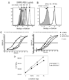
DPPE-PEG acts as CD1d antagonist. A, DN32 cells (1 × 10⁶) were incubated at 37°C with titrating concentration of DPPE-PEG (100, 50, 25, and 0 µg/ml) for 4 h. Subsequently, the cells were washed and stained with 10 ng/ml of BODIPY–α-GalCer as described in Materials and Methods. The histogram data are representative of three separate experiments. B, CD11c+ cells were positively selected by magnetic cell sorting from WT (thick line) or CD1d−/− (thin line) splenocytes and stained with BODIPY–α-GalCer as described in Materials and Methods. The results were compared with BODIPY isotype control (shaded histogram). The histogram data are representative of three separate experiments. C, Human CD1d:mIgG or mouse CD1d:mIgG dimers were incubated with plate-bound anti-mouse IgG1 Ab for 2 h. Next, biotinylated 18:1 PE lipid at 1 µg/ml was used as a competitor against serial-diluted DPPE-PEG or α-GalCer for the binding to hCD1d or mCD1d molecules. The amount of biotinylated 18:1 PE lipid bound was determined by adding streptavidin-HRP and TMB substrate. Inhibition curves were fitted with a sigmoidal dose-response formula. Results are representative of three independent assays and expressed as mean ± SD. Statistical analysis was done in each curve fitting, and p < 0.001 was considered significantly different. D, Histidine-tagged mCD1d was incubated with DPPE-PEG (5 µg/ml dissolved in ethyl alcohol) or vehicle (ethyl alcohol) overnight before the addition of a dose range of α-GalCer. The following day, the relative number of CD1d/α-GalCer complexes was detected by immobilizing the histidine-tagged CD1d/lipid-mixture onto a Ni-coated nitrilotriacetic acid sensor chip (Biacore). A saturating dose of L317 (10 µg/ml) was injected for 1 min at a flow rate of 25 µl/min. The extent of binding was analyzed 5 s after the end of the injection. Data are shown as concentration of α-GalCer versus L317 binding (Biacore Response Units) in triplicate injections ±SD. These data are representative of two independent experiments. For all of the concentrations of α-GalCer, p values were calculated using a one-way ANOVA Bonferroni multiple comparison test (p < 0.001).
FIGURE 6
FIGURE 6
DPPE-PEG blocks ERK phosphorylation pathway (FACS histogram for p-ERK1/2 expression in intracellularly stained iNKT lines activated as described in Materials and Methods). The FACS profile shows p-ERK1/2 activation of iNKT cells by APCs pulsed with α-GalCer alone (upper panel) or α-GalCer plus DPPE-PEG (lower panel) at basal rates (time point 0, shaded histograms) and at various time points after incubation (thick lines).

Similar articles

Cited by

References

    1. Centers for Disease Control and Prevention (CDC) Forecasted state-specific estimates of self-reported asthma prevalence—United States, 1998. MMWR Morb. Mortal. Wkly. Rep. 1998;47:1022–1025. - PubMed
    1. Wills-Karp M. Immunologic basis of antigen-induced airway hyperresponsiveness. Annu. Rev. Immunol. 1999;17:255–281. - PubMed
    1. Robinson DS, Hamid Q, Ying S, Tsicopoulos A, Barkans J, Bentley AM, Corrigan C, Durham SR, Kay AB. Predominant TH2-like bronchoalveolar T-lymphocyte population in atopic asthma. N. Engl. J. Med. 1992;326:298–304. - PubMed
    1. Grünig G, Warnock M, Wakil AE, Venkayya R, Brombacher F, Rennick DM, Sheppard D, Mohrs M, Donaldson DD, Locksley RM, Corry DB. Requirement for IL-13 independently of IL-4 in experimental asthma. Science. 1998;282:2261–2263. - PMC - PubMed
    1. Wills-Karp M, Luyimbazi J, Xu X, Schofield B, Neben TY, Karp CL, Donaldson DD. Interleukin-13: central mediator of allergic asthma. Science. 1998;282:2258–2261. - PubMed

Publication types

MeSH terms