. 2010 Feb 15;184(4):1909-17.
doi: 10.4049/jimmunol.0902778. Epub 2010 Jan 18.

Mechanisms of spatial and temporal development of autoimmune vitiligo in tyrosinase-specific TCR transgenic mice

Affiliations

Mechanisms of spatial and temporal development of autoimmune vitiligo in tyrosinase-specific TCR transgenic mice

Randal K Gregg et al. J Immunol. .

Abstract

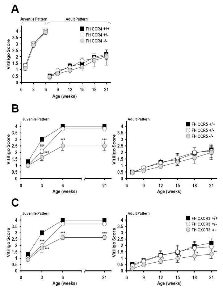
Generalized vitiligo is thought to have an autoimmune etiology and has been correlated with the presence of CD8 T cells specific for melanocyte differentiation Ag. However, limited animal models for the disease have hampered its understanding. Thus, we generated TCR transgenic mice that recognize an epitope of the melanocyte protein, tyrosinase. These animals develop vitiligo with strikingly similar characteristics to the human disease. Vitiligo develops temporally and spatially, with juvenile lesions forming bilaterally in head and facial areas, and only arising later in the body of adult animals. Vitiligo is entirely dependent on CD8 T cells, whereas CD4 T cells exert a negative regulatory effect. Importantly, CD8 T cells can be pervasively present in the skin in the steady state without inducing vitiligo in most areas. This points to developmental differences in melanocyte susceptibility and/or immunological effector mechanisms over time, or in different body locations. Disease is strongly dependent on both IFN-gamma and CXCR3, whereas dependence on CCR5 is more limited, and both CCR4 and perforin are dispensable. Genetic ablation of CXCR3 or IFN-gamma also resulted in scarce CD8 T cell infiltration into the skin. Our results identify unexpected complexity in vitiligo development and point toward possible therapeutic interventions.


Conflict of interest statement

Disclosures The authors have no financial conflict of interest.

Figures

Figure 1
Figure 1
Spatial and temporal development of vitiligo in FH mice. (A) Progression of depigmentation in FH mice as shown at the indicated ages; shown with an unaffected littermate nontransgenic control (top right panel). Note ears and eye ring depigmentation at 3 weeks, tail depigmentation at 5-7 weeks (juvenile vitiligo) and body hair depigmentation (adult vitiligo) at 15-21 weeks as outlined in Table 1. Mice shown at 7, 15 and 21 weeks of age had vitiligo scores of 1.5, 2 and 3, respectively. (B) Development of juvenile vitiligo in individual FH mice (circles, n=5) as compared to nontransgenic C57BL/6 controls (boxes, n=5). (C) Development of adult vitiligo in FH mice and nontransgenic controls (n=5 for both groups). Vitiligo scores were determined according to Table 1.
Figure 2
Figure 2
Role of CD4 and CD8 T cells in FH vitiligo development. (A) FH mice were intraperitoneally injected with anti-CD4 (open circles, n=8), anti-CD8 (shaded circles, n=6) or IgG isotype control (boxes, n=8) weekly from birth through 15 weeks of age and assessed for vitiligo development through 21 weeks. *, P<0.05; ***, P<0.0001 anti-CD4-treated mice compared with IgG-treated animals. (B) FH mice (n=8) were injected with anti-CD25 (shaded circles) or IgG isotype control Ab weekly from birth through 20 weeks (left panel, (n=8) or from 20 weeks through 27 weeks of age (right panel, (n=6) and scored for vitiligo. *, P<0.01; **, P<0.005; ***, P<0.0001 anti-CD25-treated mice compared with IgG-treated animals (left panel). *, P<0.05; **, P<0.005 anti-CD25-treated mice compared with IgG-treated animals (right panel). (C) FH mice (n=6) were intraperitoneally injected with anti-CD8 or IgG isotype control Ab weekly from birth through 15 weeks and assessed for juvenile (left panel) and adult (right panel) vitiligo development through 30 weeks. ***, P<0.0001 anti-CD8-treated mice compared with IgG-treated animals. Results are shown as mean vitiligo scores with error bars indicating SD.
Figure 3
Figure 3
Vitiligo is associated with increased infiltration of CD8, but not CD4 cells, into perilesional skin. (A) CD8 T cells were identified by immunohistochemical staining of nonlesional, perilesional (gray bordering fully depigmented), and lesional (fully depigmented) sections of back skin from 15 week old FH mice and age-matched controls as described in Methods. All images are 40X. (B) Quantitation of hair follicle-associated CD8 (left panel) and CD4 (right panel) T cells in skin sections. Ten histological sections from 2 animals were prepared for each category, [NonTg, FH (nonlesional), FH (perilesional) and FH (lesional)], and two hair follicle regions were analyzed from each section. Cell counts were determined from images magnified to 40X. *, P<0.02 FH (lesional) compared to FH (nonlesional); ***, P < 0.0001 FH (perilesional) compared to FH (nonlesional or lesional) mice.
Figure 4
Figure 4
Development of vitiligo depends upon CCR5 and CXCR3, but not CCR4. (A) CCR4+/+ (boxes, n=10), CCR4+/- (open circles, n=15) and CCR4-/- (shaded circles, n=17) FH mice were scored for vitiligo through 21 weeks. (B) CCR5+/+ (boxes, n=10), CCR5+/- (open circles, n=15) and CCR5-/- (shaded circles, n=15) FH mice were scored for juvenile (left panel) and adult (right panel) vitiligo. ***, P < 0.0005 compared with CCR5+/+ FH mice. (C) CXCR3+/+ (boxes, n=10), CXCR3+/- (open circles, n=15) and CXCR3-/- (shaded circles, n=17) FH mice were scored for juvenile (left panel) and adult (right panel) vitiligo. *, P<0.05; **, P<0.002; ***, P<0.0001 compared with CXCR3+/+ FH mice. Results are shown as mean vitiligo scores with error bars indicating SD.
Figure 5
Figure 5
Representative vitiligo appearance in FH mice lacking CCR4, CCR5 or CXCR3. Left panels depict juvenile pattern vitiligo at 21 weeks of age. Right panels depict adult pattern vitiligo at 18 weeks of age with the exception of the CCR4-/- FH mice, which are 12 weeks.
Figure 6
Figure 6
Development of vitiligo depends upon expression of IFNγ but not perforin. (A) Perforin+/+ (n=7), perforin+/- (n=7) and perforin-/- (n=8) FH mice were scored for juvenile and adult vitiligo. **, P<0.008 compared with Perforin+/+ FH mice. Results are shown as mean vitiligo scores with error bars indicating SD. (B) IFNγ+/+ (n=10) and IFNγ-/- (n=12) FH mice were scored for juvenile and adult vitiligo. *, P<0.01; **, P<0.001; ***, P<0.0001 compared with IFNγ+/+ FH mice. Results are shown as mean vitiligo scores with error bars indicating SD. (C) Sublethally irradiated (600 rads) NonTg mice were injected intravenously with 3×10⁶ purified IFNγ+/+ or IFNγ-/- FH CD8 T cells. Mice were also given intraperitoneal injections of 1,500 CU IL-2 at the time of T cell transfer and every other day through 10 days. A group of irradiated NonTg mice that did not receive T cells but were given exogenous IL-2 were included as a control. *, P<0.05; ***, P<0.0001 compared with recipient mice of IFNγ-/- FH CD8 T cells. Results are shown as mean vitiligo scores with error bars indicating SD.
Figure 7
Figure 7
Differential dependence of skin and ear cellular infiltrates upon CXCR3 and IFNγ. CD8 T cells (top and middle panels) and F4/80+ cells (bottom panels) from 2 day old juvenile (left panels) and 49 days adult (right panels) skin of the indicated mouse strains was quantitated by immunohistochemistry. Ten skin and 10 ear histological sections from 2 animals of each strain were prepared, and two independent regions per section were examined. Cell counts were determined from images magnified to 40X for CD8 T cells and 20X for F4/80+ cells. **, P<0.001 CXCR3-/- FH compared to FH; ***, P < 0.0001 CXCR3-/- FH or IFNγ-/- FH compared to FH.

References

    1. Handa S, Kaur I. Vitiligo: clinical findings in 1436 patients. J Dermatol. 1999;26:653–657. - PubMed
    1. Huggins RH, Schwartz RA, Janniger CK. Vitiligo. Acta Dermatovenerol Alp Panonica Adriat. 2005;14:137–142. - PubMed
    1. Taieb A, Picardo M. Clinical practice. Vitiligo. N Engl J Med. 2009;360:160–169. - PubMed
    1. Ogg GS, Rod DP, Romero P, Chen JL, Cerundolo V. High frequency of skin-homing melanocyte-specific cytotoxic T lymphocytes in autoimmune vitiligo. J Exp Med. 1998;188:1203–1208. - PMC - PubMed
    1. Palermo B, Campanelli R, Garbelli S, Mantovani S, Lantelme E, Brazzelli V, Ardigo M, Borroni G, Martinetti M, Badulli C, Necker A, Giachino C. Specific cytotoxic T lymphocyte responses against Melan-A/MART1, tyrosinase and gp100 in vitiligo by the use of major histocompatibility complex/peptide tetramers: the role of cellular immunity in the etiopathogenesis of vitiligo. J Invest Dermatol. 2001;117:326–332. - PubMed

Publication types

MeSH terms