Oxidative stress in the progression of Alzheimer disease in the frontal cortex
- PMID: 20084018
- PMCID: PMC2826839
- DOI: 10.1097/NEN.0b013e3181cb5af4
Oxidative stress in the progression of Alzheimer disease in the frontal cortex
Abstract
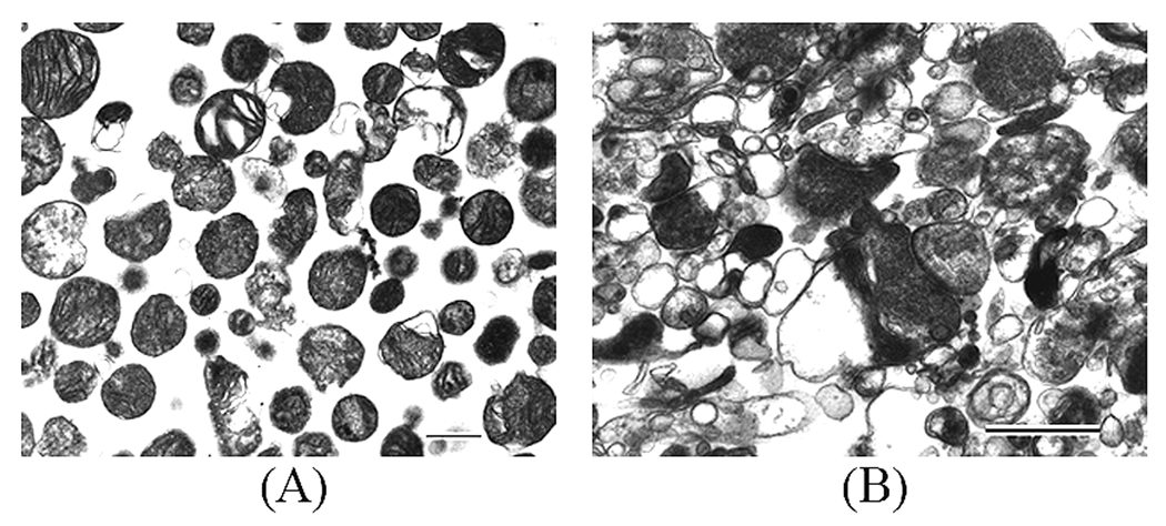
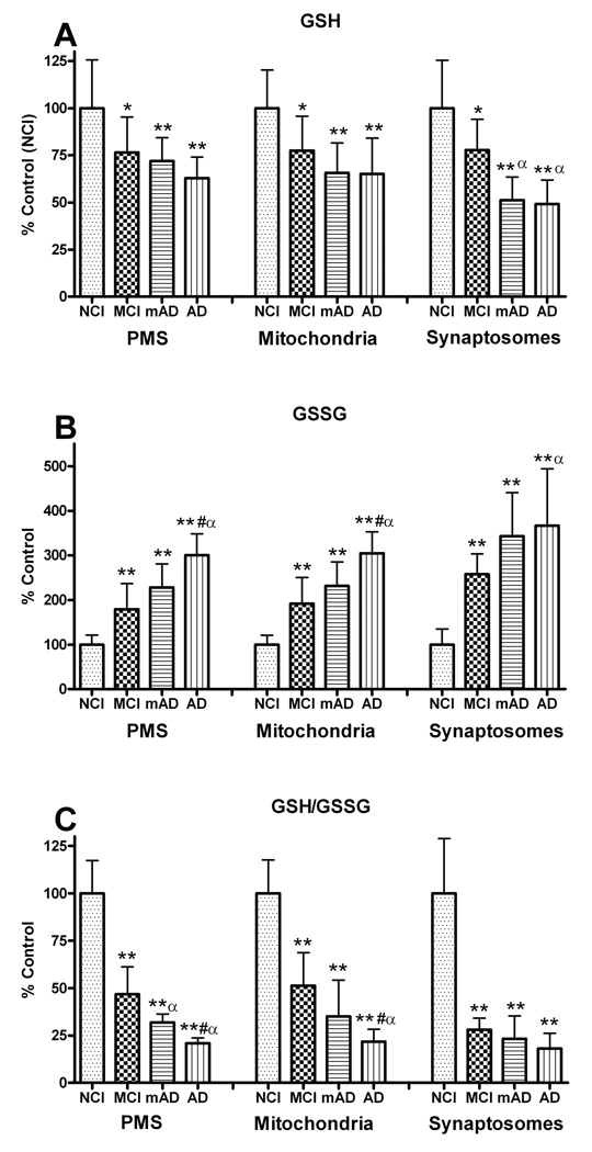
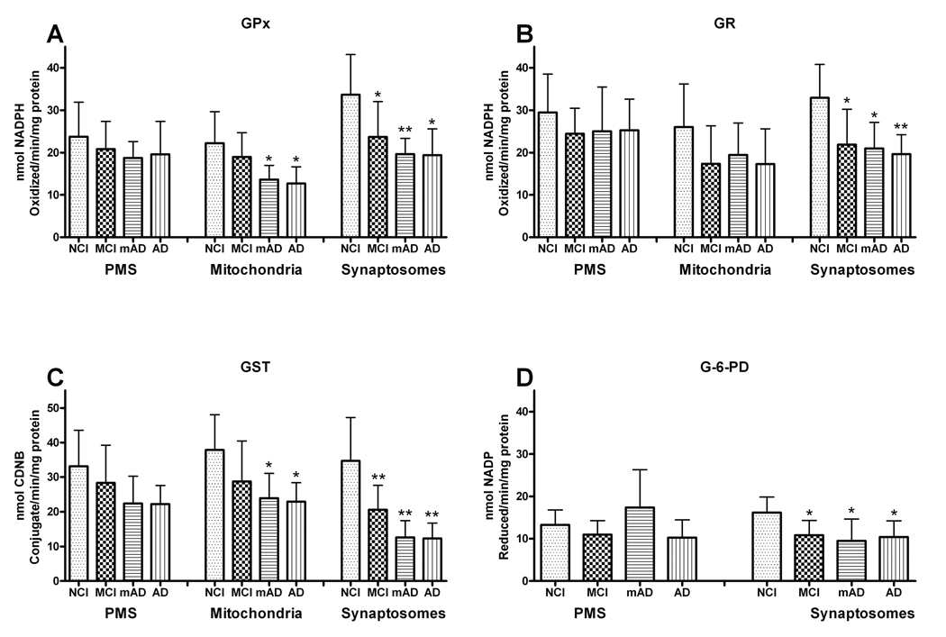
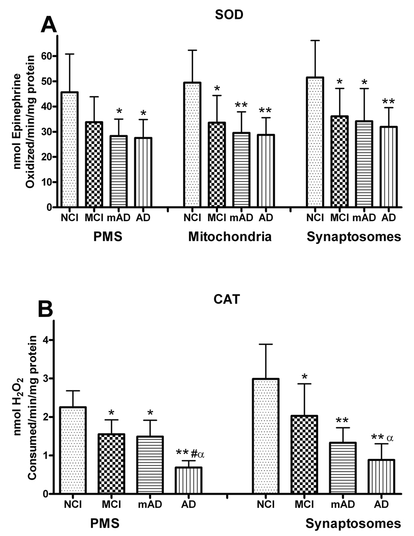
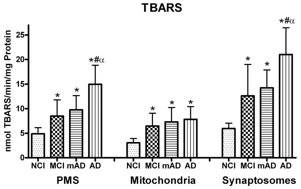
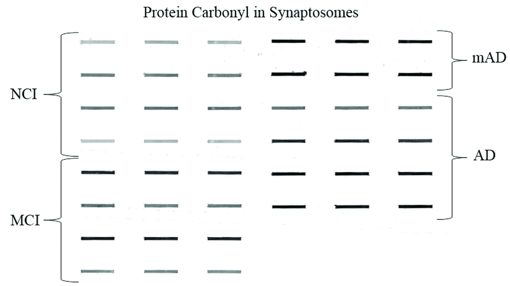
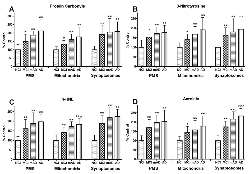
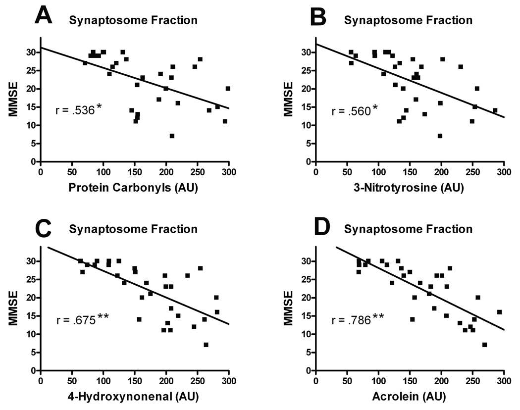
We investigated oxidative stress in human postmortem frontal cortexfrom individuals characterized as mild cognitive impairment (n= 8), mild/moderate Alzheimer disease (n = 4), and late-stage Alzheimer disease (n = 9). Samples from subjects with no cognitive impairment (n = 10) that were age- and postmortem interval-matched with these cases were used as controls. The short postmortem intervalbrain samples were processed for postmitochondrial supernatant, nonsynaptic mitochondria, and synaptosome fractions. Samples were analyzed for several antioxidants (glutathione, glutathione peroxidase, glutathione reductase, glutathione-S-transferase, glucose-6-phosphate dehydrogenase, superoxide dismutase, catalase) and the oxidative marker, thiobarbituric acid reactive substances. The tissue was also analyzed for possible changes in protein damage using neurochemical markers for protein carbonyls, 3-nitrotyrosine, 4-hydroxynonenal, andacrolein. All 3 neuropil fractions (postmitochondrial supernatant, mitochondrial, and synaptosomal) demonstrated significant disease-dependent increases in oxidative markers. The highest changes were observed in the synaptosomal fraction. Both mitochondrial and synaptosomal fractions had significant declines in antioxidants (glutathione, glutathione peroxidase, glutathione-S-transferase, and superoxide dismutase). Levels of oxidative markers significantly correlated with Mini-Mental Status Examination scores. Oxidative stress was more localized to the synapses, with levels increasing in a disease-dependent fashion. These correlations implicate an involvement of oxidative stress in Alzheimer disease-related synaptic loss.
Figures


Similar articles
-
Alteration in glutathione content and associated enzyme activities in the synaptic terminals but not in the non-synaptic mitochondria from the frontal cortex of Parkinson's disease brains.Neurochem Res. 2013 Jan;38(1):186-200. doi: 10.1007/s11064-012-0907-x. Epub 2012 Oct 16. Neurochem Res. 2013. PMID: 23070472
-
Association between frontal cortex oxidative damage and beta-amyloid as a function of age in Down syndrome.Biochim Biophys Acta. 2012 Feb;1822(2):130-8. doi: 10.1016/j.bbadis.2011.10.001. Epub 2011 Oct 8. Biochim Biophys Acta. 2012. PMID: 22009041 Free PMC article.
-
Early time course of oxidative stress in hippocampal synaptosomes and cognitive loss following impaired insulin signaling in rats: Development of sporadic Alzheimer's disease.Brain Res. 2023 Jan 1;1798:148134. doi: 10.1016/j.brainres.2022.148134. Epub 2022 Oct 31. Brain Res. 2023. PMID: 36328067
-
Markers of oxidative damage to lipids, nucleic acids and proteins and antioxidant enzymes activities in Alzheimer's disease brain: A meta-analysis in human pathological specimens.Free Radic Biol Med. 2018 Feb 1;115:351-360. doi: 10.1016/j.freeradbiomed.2017.12.016. Epub 2017 Dec 15. Free Radic Biol Med. 2018. PMID: 29253591 Free PMC article.
-
Antioxidant intervention attenuates oxidative stress in children and teenagers with Down syndrome.Res Dev Disabil. 2014 Jun;35(6):1228-36. doi: 10.1016/j.ridd.2014.03.013. Epub 2014 Mar 28. Res Dev Disabil. 2014. PMID: 24685938
Cited by
-
Oxidative stress and hippocampal synaptic protein levels in elderly cognitively intact individuals with Alzheimer's disease pathology.Neurobiol Aging. 2016 Jun;42:1-12. doi: 10.1016/j.neurobiolaging.2016.02.030. Epub 2016 Mar 8. Neurobiol Aging. 2016. PMID: 27143416 Free PMC article.
-
The synergy of β amyloid 1-42 and oxidative stress in the development of Alzheimer's disease-like neurodegeneration of hippocampal cells.Sci Rep. 2022 Oct 25;12(1):17883. doi: 10.1038/s41598-022-22761-5. Sci Rep. 2022. PMID: 36284177 Free PMC article.
-
The Role of Iron Metabolism, Lipid Metabolism, and Redox Homeostasis in Alzheimer's Disease: from the Perspective of Ferroptosis.Mol Neurobiol. 2023 May;60(5):2832-2850. doi: 10.1007/s12035-023-03245-7. Epub 2023 Feb 3. Mol Neurobiol. 2023. PMID: 36735178 Review.
-
A Perspective on Nrf2 Signaling Pathway for Neuroinflammation: A Potential Therapeutic Target in Alzheimer's and Parkinson's Diseases.Front Cell Neurosci. 2022 Jan 21;15:787258. doi: 10.3389/fncel.2021.787258. eCollection 2021. Front Cell Neurosci. 2022. PMID: 35126058 Free PMC article. Review.
-
Common defects of mitochondria and iron in neurodegeneration and diabetes (MIND): a paradigm worth exploring.Biochem Pharmacol. 2014 Apr 15;88(4):573-83. doi: 10.1016/j.bcp.2013.11.022. Epub 2013 Dec 19. Biochem Pharmacol. 2014. PMID: 24361914 Free PMC article. Review.
References
-
- Halliwell B. Reactive oxygen species and the central nervous system. J Neurochem. 1992;59:1609–1623. - PubMed
-
- Halliwell B. Role of free radicals in the neurodegenerative diseases: Therapeutic implications for antioxidant treatment. Drugs Aging. 2001;18:685–716. - PubMed
-
- Faraci FM. Reactive oxygen species: Influence on cerebral vascular tone. J Appl Physiol. 2006;100:739–743. - PubMed
-
- Mantha AK, Moorthy K, Cowsik SM, et al. Neuroprotective role of neurokinin B (NKB) on beta-amyloid (25–35) induced toxicity in aging rat brain synaptosomes: Involvement in oxidative stress and excitotoxicity. Biogerontology. 2006;7:1–17. - PubMed
-
- Markesbery WR, Lovell MA. DNA oxidation in Alzheimer's disease. Antioxid Redox Signal. 2006;8:2039–2045. - PubMed
Publication types
MeSH terms
Substances
Grants and funding
LinkOut - more resources
Full Text Sources
Medical