. 2010 Feb;69(2):139-54.
doi: 10.1097/NEN.0b013e3181cb5bee.

Pericontusion axon sprouting is spatially and temporally consistent with a growth-permissive environment after traumatic brain injury

Affiliations

Pericontusion axon sprouting is spatially and temporally consistent with a growth-permissive environment after traumatic brain injury

Neil G Harris et al. J Neuropathol Exp Neurol. 2010 Feb.

Abstract

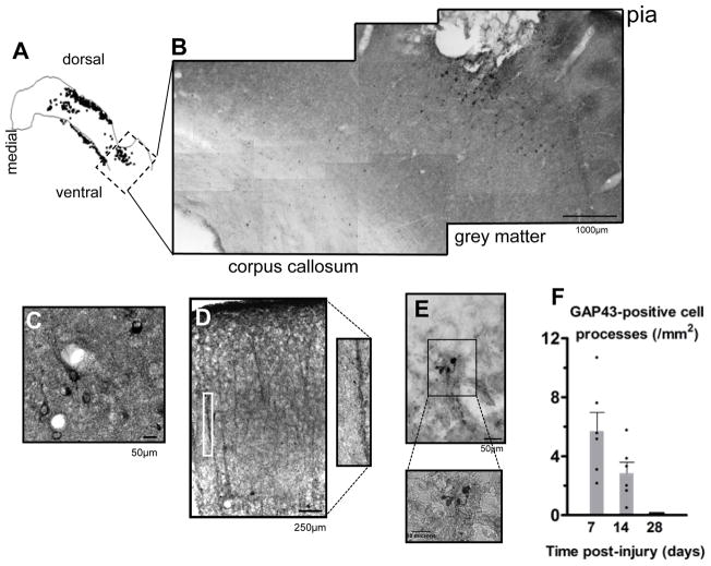
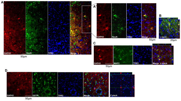
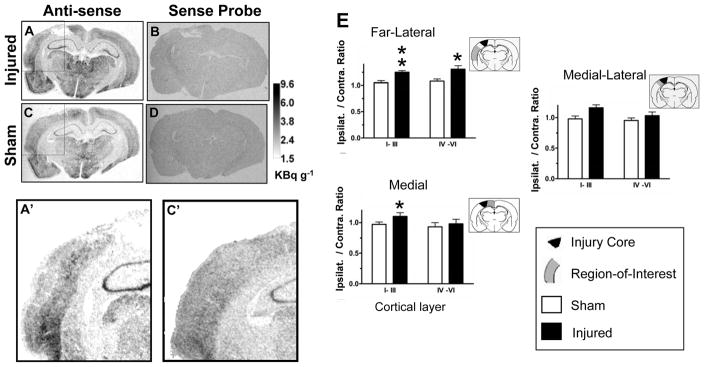
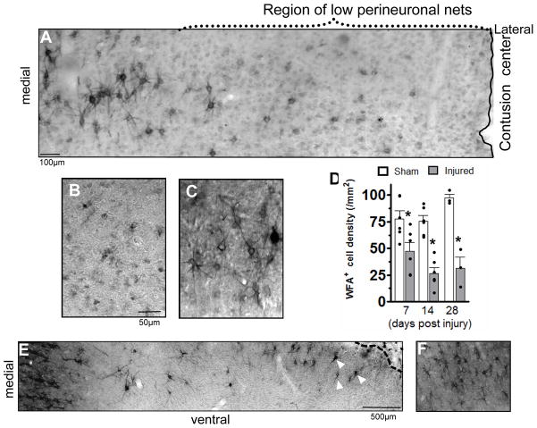
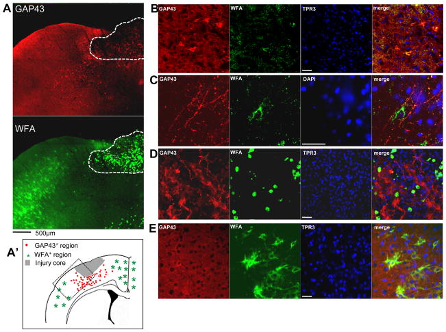
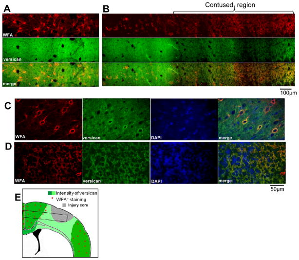
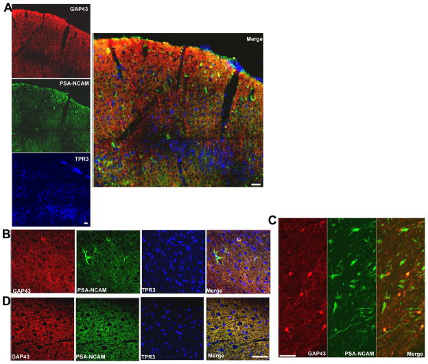
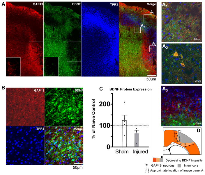
We previously reported that pericontusional extracellular chondroitin sulfate proteoglycans (CSPGs) are profoundly reduced for 3 weeks after experimental traumatic brain injury, indicating a potential growth-permissive window for plasticity. Here, we investigate the extracellular environment of sprouting neurons after controlled cortical impact injury in adult rats to determine the spatial and temporal arrangement of inhibitory and growth-promoting molecules in relation to growth-associated protein 43-positive (GAP43+) neurons. Spontaneous cortical sprouting was maximal in pericontused regions at 7 and 14 days after injury but absent by 28 days. Perineuronal nets containing CSPGs were reduced at 7 days after injury in the pericontused region (p < 0.05), which was commensurate with a reduction in extracellular CSPGs. Sprouting was restricted to the perineuronal nets and CSPG-deficient regions at 7 days, indicating that the pericontused region is temporarily and spatially permissive to new growth. At this time point,GAP43+ neurons were associated with brain regions containing cells positive for polysialic acid neural cell adhesion molecule but not with fibronectin-positive cells. Brain-derived neurotrophic factor was reduced in the immediate pericontused region at 7 days. Along with prior Western blot evidence, these data suggest that a lowered intrinsic growth stimulus, together with a later return of growth-inhibitory CSPGs, may contribute to the ultimate disappearance of sprouting neurons after traumatic brain injury.


Figures

Figure 1
Figure 1
Pericontusional sprouting. Immunostained cortical GAP43+ cells and processes were quantified at 7 to 28 days after controlled cortical impact injury (3–5 coronal sections/rat, >600 μm apart in sensorimotor and parietal cortex). (A–E) Injury resulted in the appearance of GAP43+ cellular profiles at 7 days for distances up to 1 to 2 mm from the injury site, shown in overview (A), montage (B) and at higher power from a different animal (C –E). No staining was present in homotypic regions of the contralateral or in the sham-injured sensorimotor cortex. Both perikarya and neuronal processes were stained throughout the pericontusion cortical gray and white matter (C, D, inset-D) as were numerous “blebs” (i.e. presumed neuronal growth cones) on the contusion edge (E, inset E). (F) Pericontusional sprouting was maximal at 7 and 14 days after injury and was not present by 28 days; the difference between 7 and 14 days was not significant (p = 0.09).
Figure 2
Figure 2
GAP43+ cell phenotype. (A–C) Representative confocal montages of layer V sensorimotor cerebral cortex adjacent to the injury site 7 days after injury illustrate neuronal fiber and perikarya- GAP43+ profiles (A, GAP43 and merge). The pial surface is at the top of the image; the injury site is the left. Perikarya express the neuronal marker NeuN (A, inset A′ and z stack in B) and MAP2 (C). No GAP43+ profiles display the oligodendrocyte marker glutathione S-Transferase pi (GSTPi) in gray or white matter (D). Confocal data trends are based on an analysis of 3 injured brains. TPR3 = nucleic acid marker TOPRO-3.
Figure 3
Figure 3
GAP43 mRNA: (A–D) In situ hybridization for GAP43 mRNA from sham and injured rats 7 days after injury in sensorimotor/parietal cortex showed regional hyperintensity with the GAP43 mRNA anti-sense probe (A, C) but uniform intensity with the sense probe (B, D). Region of interest (ROI) analysis showed increases in cortical GAP43 anti-sense probe intensity in injured cortex compared to sham (A, C- inset of A, C), which was significant in both far lateral regions and more medial ROIs (E). Data are plotted as ipsilateral/contralateral optical density ROI; **p < 0.01, *p < 0.05.
Figure 4
Figure 4
Perineuronal Nets: Numbers of Wisteria floribunda-positive (WFA+) cells 7 days after injury were markedly reduced for up to 1 to 2 mm from the contusion edge, shown in layer V of the injured sensorimotor cortex (A, B), compared to sham-injured rats (C); this was significant at 7, 14 and 28 days (D, *p < 0.05). The number of perineuronal nets was reduced at 28 days (D, E) compared to sham (D, F), but some WFA+ cells (white arrowheads, E) are present closer to the contusion edge in layer V of the cortex (dashed line, E).
Figure 5
Figure 5
Neuronal sprouting and perineuronal nets. (A) Representative confocal montage of pericontused, ipsilateral sensorimotor cortex 7 days after injury showing that the majority of GAP43+ cells occur within regions devoid of Wisteria floribunda-positive (WFA+) cells. The hatched region shows the cystic cavity of the core injured region that contains ovoid, dead or infiltrating cells completely filled intracellularly with very intense WFA+ staining. (A′) Summary drawing based on observations from at least 3 injured brains showing the presence of GAP43+ neurons around the contusion site within a region containing no or few WFA+ cells (dotted box region represents approximate position of panel A). (B–E) Higher power figures at 7 days post-injury (different section from that in A) showing pericontusional GAP43+ perikarya (B) and GAP43+ fibers from cortical layer V containing very few cells double-labeled with WFA. (D) GAP43+ fibers and numerous blebs are present within the cystic contusion cavity, even within a field of highly intense ovoid WFA+ cells; none are double-labeled. (E) No GAP43+ sprouting neurons are observed contralaterally; normal-appearing WFA+ cells are stained only around the cell body and proximal fiber segments. Scale bar = 50 μm.
Figure 6
Figure 6
Perineuronal nets and chondroitin sulphate proteoglycans (CSPGs). (A, B) Representative confocal montages of contralateral (A) and ipsilateral (B) sensorimotor layer IV–V cortex 7 days after injury showing that the growth-inhibitory CSPG versican is markedly reduced within the pericontusional region sparsely populated with Wisteria floribunda-positive (WFA+) cells. (C, D) Higher power images from different sections acquired with different confocal acquisition settings to highlight the perineuronal nets from the high extracellular versican of contralateral cortex showing normal appearing WFA+ perineuronal nets, many of which are versican+ (C). In the pericontusional ipsilateral cortex containing low amounts of extracellular versican, there are no WFA+ or versican+ perineuronal nets (D). (E) Stylized summary diagram of changes in versican and WFA+ cells in injured cortex based on observations from at least 3 injured brains (dotted box corresponds to the approximate location of panels A and B).
Figure 7
Figure 7
Poly-sialic acid neural cell adhesion molecule (PSA-NCAM) and GAP43. Representative confocal montages of the sensorimotor cortical region 7 days after injury within a pericontused region anterior to the core injured tissue (in A the pial surface is shown at the top of the image). GAP43+ processes occur within a region containing numerous PSA-NCAM+ cells (A, merge). Most of the GAP43+ processes are not PSA-NCAM+ (A, Merge), but there are occasional double-labeled processes within the cortical gray and white matter underlying the contusion (B, C, respectively). The significant enhancement in the number of ipsilateral PSA-NCAM+ cells (A) does not extend to the contralateral cortex (D). Scale bars = 50 μm.
Figure 8
Figure 8
Neuronal sprouting and brain-derived neurotrophic factor (BDNF). (A, B) Representative confocal montages of the pericontused cortical region 7 days after injury (A, pial surface is at top, contusion core to right) and contralateral cortex (B). Intensity of BDNF staining was reduced along the border of the contusion (A, insets and merge) compared to more remote ipsilateral tissue and the contralateral cortex (B). GAP43+ neurons were also BDNF+ within the pericontusional ipsilateral cortical tissue (A, A2) but most of the GAP43+ fibers at the contusion edge were BDNF- (A, A3), as were most of the GAP43+ fibers in intact tissue (A1). (C) Western blot analysis graph shows mean BDNF protein concentration and individual data (black symbols) in ipsilateral injured and sham-injured cortex as a percent of uninjured controls 7 days after injury (p = 0.06). (D) Stylized summary diagram of the spatial relationship in ipsilateral cortex between GAP43+ processes and BDNF immunostaining intensity 7 days after injury.

Similar articles

Cited by

References

    1. Hsu JC, Stein SA, Xu X. Temporal and spatial distribution of growth-associated molecules and astroglial cells in the rat corticospinal tract during development. J Neurosc Res. 2005;80:330–40. - PubMed
    1. Greenough WT, Larson JR, Withers GS. Effects of unilateral and bilateral training in a reaching task on dendritic branching of neurons in the rat motor-sensory forelimb cortex. Behav Neural Biol. 1985;44:301–14. - PubMed
    1. Darian-Smith C, Gilbert CD. Topographic reorganization in the striate cortex of the adult cat and monkey is cortically mediated. J Neurosci. 1995;15:1631–47. - PMC - PubMed
    1. Kleim JA, Lussnig E, Schwarz ER, et al. Synaptogenesis and Fos expression in the motor cortex of the adult rat after motor skill learning. J Neurosci. 1996;16:4529–35. - PMC - PubMed
    1. Gogolla N, Galimberti I, Caroni P. Structural plasticity of axon terminals in the adult. Curr Opin Neurobiol. 2007;17:516–24. - PubMed

Publication types

MeSH terms