

Inflammation driven by overexpression of the hypoglycosylated abnormal mucin 1 (MUC1) links inflammatory bowel disease and pancreatitis
- PMID: 20084048
- PMCID: PMC2859977
- DOI: 10.1097/MPA.0b013e3181bd6501
Inflammation driven by overexpression of the hypoglycosylated abnormal mucin 1 (MUC1) links inflammatory bowel disease and pancreatitis
Abstract
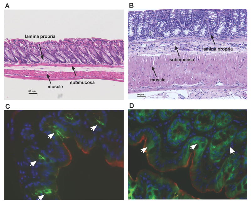
Objective: Pancreatitis occurs as an extraintestinal complication of inflammatory bowel disease (IBD), but the cause is poorly understood. Mucin 1 (MUC1) is overexpressed in an abnormal, hypoglycosylated form on the colonic epithelium in human IBD where it contributes to inflammation. MUC1 is also expressed on pancreatic ductal epithelia. We tested the possibility that in IBD, MUC1 expression on pancreatic ducts is also abnormal leading to inflammation and pancreatitis.
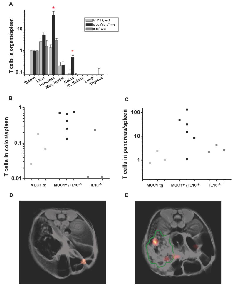
Methods: We used MUC1/interleukin-10 mice that develop IBD. We imaged abnormal MUC1 expression in these mice by adoptively transferring T cells from T cell receptor transgenic mice specific for abnormal MUC1. Cells were labeled with a novel perfluorocarbon tracer reagent and quantified and visualized in vivo using high-throughput F nuclear magnetic resonance spectroscopy and magnetic resonance imaging.
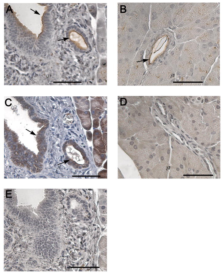
Results: MUC1-specific T cells migrated to the colon in mice with IBD and also to the pancreas. Immunohistochemistry confirmed increased expression on the pancreatic ducts of the abnormal MUC1 seen in the colon and the presence of cellular infiltrate.
Conclusions: Migration of MUC1-specific T cells to the colon and the pancreas in diseased mice suggests that pancreatitis is an extraintestinal site of IBD, characterized by proinflammatory abnormal expression of MUC1. Therapies directed against abnormal MUC1 have the potential of targeting the disease in both sites.
Figures
References
-
- Xavier RJ, Podolsky DK. Unravelling the pathogenesis of inflammatory bowel disease. Nature. 2007;448(7152):427–34. - PubMed
-
- Su CG, Judge TA, Lichtenstein GR. Extraintestinal manifestations of inflammatory bowel disease. Gastroenterol Clin North Am. 2002;31(1):307–27. - PubMed
-
- Gurian LE, Keeffe EB. Pancreatic insufficiency associated with ulcerative colitis and pericholangitis. Gastroenterology. 1982;82(3):581–5. - PubMed
Publication types
MeSH terms
Substances
Grants and funding
LinkOut - more resources
Full Text Sources
Medical
Research Materials
Miscellaneous
