. 2010 Jan 15;6(1):e1000809.
doi: 10.1371/journal.pgen.1000809.

Kidney development in the absence of Gdnf and Spry1 requires Fgf10

Affiliations

Kidney development in the absence of Gdnf and Spry1 requires Fgf10

Odyssé Michos et al. PLoS Genet. .

Abstract

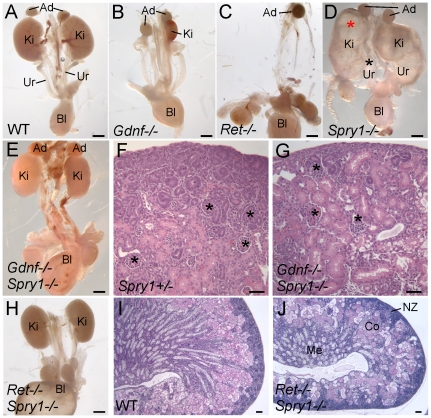
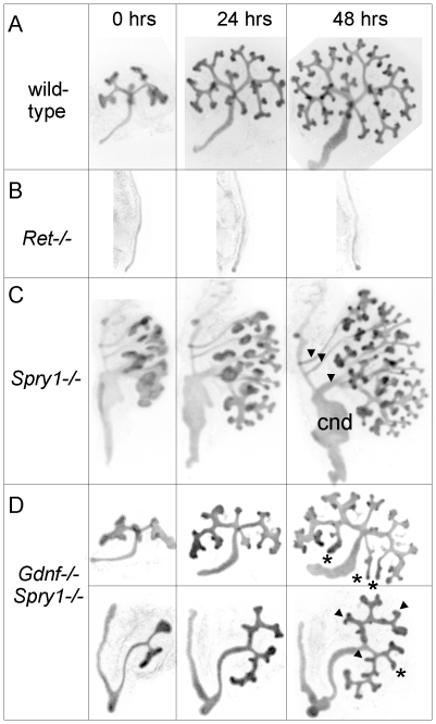
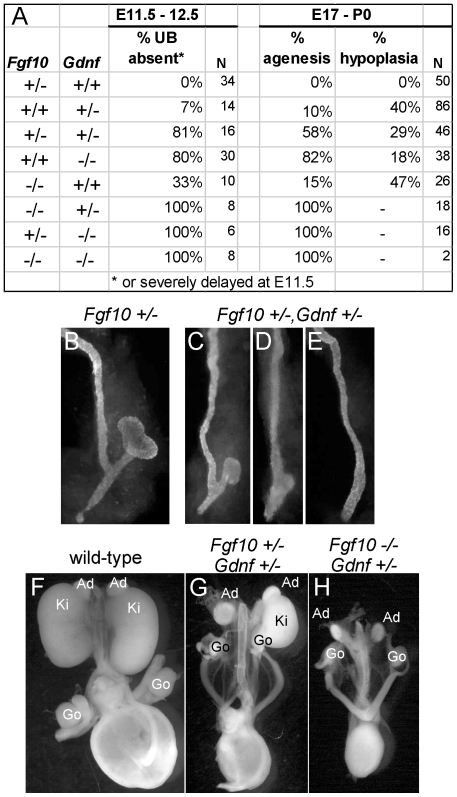
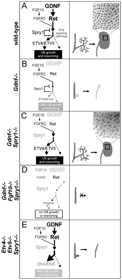
GDNF signaling through the Ret receptor tyrosine kinase (RTK) is required for ureteric bud (UB) branching morphogenesis during kidney development in mice and humans. Furthermore, many other mutant genes that cause renal agenesis exert their effects via the GDNF/RET pathway. Therefore, RET signaling is believed to play a central role in renal organogenesis. Here, we re-examine the extent to which the functions of Gdnf and Ret are unique, by seeking conditions in which a kidney can develop in their absence. We find that in the absence of the negative regulator Spry1, Gdnf, and Ret are no longer required for extensive kidney development. Gdnf-/-;Spry1-/- or Ret-/-;Spry1-/- double mutants develop large kidneys with normal ureters, highly branched collecting ducts, extensive nephrogenesis, and normal histoarchitecture. However, despite extensive branching, the UB displays alterations in branch spacing, angle, and frequency. UB branching in the absence of Gdnf and Spry1 requires Fgf10 (which normally plays a minor role), as removal of even one copy of Fgf10 in Gdnf-/-;Spry1-/- mutants causes a complete failure of ureter and kidney development. In contrast to Gdnf or Ret mutations, renal agenesis caused by concomitant lack of the transcription factors ETV4 and ETV5 is not rescued by removing Spry1, consistent with their role downstream of both RET and FGFRs. This shows that, for many aspects of renal development, the balance between positive signaling by RTKs and negative regulation of this signaling by SPRY1 is more critical than the specific role of GDNF. Other signals, including FGF10, can perform many of the functions of GDNF, when SPRY1 is absent. But GDNF/RET signaling has an apparently unique function in determining normal branching pattern. In contrast to GDNF or FGF10, Etv4 and Etv5 represent a critical node in the RTK signaling network that cannot by bypassed by reducing the negative regulation of upstream signals.


Conflict of interest statement

The authors have declared that no competing interests exist.

Figures

Figure 1
Figure 1. Loss of Spry1 rescues kidney development in Gdnf−/− or Ret−/− mice.
(A–E,H) excretory systems dissected from newborn mice of the indicated genotypes shown in whole mount. Note that the ureter in the Spry1−/− mutant is greatly expanded (black asterisk) and the kidneys are cystic (red asterisk). (F,G) H&E stained sections showing histology of cortex and nephrogenic zone in control (Spry1+/−) and Gdnf−/−;Spry1−/− kidneys, revealing a normal overall organization with well-differentiated glomeruli (*) (I,J) PAS-stained sections of wild-type and Ret−/−;Spry1−/− double mutant kidneys, showing normal overall organization with renal cortex, medulla, and outer nephrogenic zone. Abbreviations: Ad, adrenal gland; Bl, bladder; Co, cortex; Ki, kidney; Me, medulla, NZ, nephrogenic zone; Ur, ureter. Scale bars 1 mm in A-E and H, 100 µm in F,G,I,J.
Figure 2
Figure 2. Numerous nephron and normal nephron–UB connections are observed in double mutant kidneys.
(A,B) Podocalyxin staining of nascent glomeruli in wild-type (A) and Ret−/−;Spry1−/− (B) kidneys at P0, showing numerous, cortically located nephrons in the double mutant, as in the wild-type. (C–H) Six optical sections at different Z-levels of a Gdnf−/−;Spry1−/− E15.5 kidney carrying Hoxb7/myrVenus. The sites where the UB connects to nephrons are visible as “holes” in the myrVenus-labeled UB, as the connecting tubule expresses little or no myrVenus. The connections of three nephrons (1, 2, 3) can be followed at different levels of the image stack. (I) Volume rendering of a wild-type kidney, with nephron connection sites indicated by the pink dots. (J) Volume rendering of double mutant kidneys shown in (C–H), showing normal number and positions of nephron connections per UB tip.
Figure 3
Figure 3. Extensive but irregular UB branching in Gdnf−/−; Spry1−/− and Ret−/−; Spry1−/− double mutant kidneys.
(A–D) Newborn stage kidneys, all carrying the Hoxb7/myrVenus transgene to label the UB branches. Each panel shows a high magnification view of the kidney surface, revealing the shape and organization of branching UB tips; insets show the entire kidney in whole mount. Wild-type kidneys (A) have evenly spaced UB tips with a regular branching pattern, whereas Gdnf−/−;Spry1−/− (B) and Ret−/−;Spry1−/− (C) double mutant kidneys have highly irregular branching. Spry1−/− kidneys (D) have regularly branched, but swollen UB tips. (E–P) 3D volume rendering of E15.5 kidneys. (E–H) Whole kidneys from embryos of the indicated genotypes, carrying Hoxb7/myrVenus. (I–P) Higher power views of two representative surface regions of each genotype. The 3D images were generated from confocal Z-stacks, using Volocity (E–H) or ImageJ (I–P). The yellow dashed lines indicate an interpretation of the branching patterns. While most UB branches in the wild-type (I,M) and Spry1−/− (L,P) kidneys show a reiterative pattern of terminal bifurcation, with branches forming at right angles to their predecessors, most UB branches in the double mutants (J,K,N,O) fail to conform to this pattern, and instead display a variety of abnormal shapes and branching patterns.
Figure 4
Figure 4. Abnormal branching of double mutant kidneys in organ culture.
Kidneys of wild-type (A) and mutant genotypes (B–D), carrying Hoxb7/myrVenus, were excised at E12.5, cultured in vitro, and photographed at the indicated times. The Ret−/− Wolffian duct (B) failed to develop a ureter or kidney, while the Spry1−/− kidney (C) has multiple ureters (arrowheads), swollen UB tips and an enlarged common nephric duct (cnd). (D), in two examples of Gdnf−/−;Spry1−/− mutant kidneys, UB branching is retarded at E12.5, and subsequent branching in culture displays abnormal patterns (asterisks and arrowheads – see text) compared to wild-type.
Figure 5
Figure 5. Differential gene expression in tip and trunk domains is retained in Gdnf−/−;Spry1−/− and Ret−/−;Spry1−/− double mutant kidneys.
Whole mount in situ hybridization for the UB tip markers Ret, Wnt11 and Etv4 and the trunk marker Wnt7b, in wild-type (A,C,E,G) and double mutant E12.5 kidneys (B,F and H, Gdnf−/−;Spry1−/−, D, Ret−/−;Spry1−/−). Solid arrows indicate UB tips and open arrows indicate trunks. Scale bars 100 µm.
Figure 6
Figure 6. Fgf10 expression and function in early ureter and kidney development.
(A,B) In situ hybridization in transverse sections of E10.5 wild type embryos reveals that Fgf10 and Gdnf are expressed in metanephric mesenchyme (arrows). (C,D) Whole-mount in situ hybridization at E11.0 (dorsal view) shows that Fgf10 and Gdnf are expressed in metanephric mesenchyme (MM) surrounding the UB epithelium. The schematic diagram illustrates Fgf10 expression, with purple indicating where the hybridization signal was detected. (E–G) Visualization of Hoxb7/myrVenus shows (E) normal UB branching in an Fgf10+/− kidney, (F) reduced branching in an Fgf10−/− kidney, and (G) rescue of UB branching in an Fgf10−/− kidney when Spry1 dosage is reduced (Spry1+/−). Scale bars, 100 µm. (H–J) Induction of ectopic budding from the Wolffian duct by FGF10. Dissected E10.5 urogenital regions were cultured with control PBS-soaked beads (H) or beads soaked in FGF10 (I,J) placed between the two Wolffian ducts (dotted yellow circles). FGF10 induces multiple ectopic UB outgrowths (marked by asterisks) in both control Gdnf+/− (I) and Gdnf−/− (J) samples. Open arrowhead in H, Wolffian duct; arrows in H-I, normal ureteric buds.
Figure 7
Figure 7. Fgf10 and Gdnf cooperate to support UB outgrowth and kidney development.
(A) Frequency of the failure of UB outgrowth at E11.5–12.5, and renal agenesis or hypoplasia at E17.5-P0. (B) Normal T-stage UB in an Fgf10+/− embryo at E11.5. (C–E) Three examples of UB formation or lack thereof in Fgf10+/−;Gdnf+/− E11.5 embryos. In (C), the UB is slightly retarded, in (D), the UB is severely delayed, and in (E) the UB is absent. (F–H), normal kidneys in wild-type and renal agenesis or hypoplasia in compound Fgf10/Gdnf mutants at P0. The wild-type in (F) has two normal kidneys, the double heterozygote in (G) has renal agenesis on one side and a hypoplastic kidney on the other, and the Fgf10−/−;Gdnf+/− example in (H) has bilateral agenesis. Ad, adrenal; Ki, kidney; Go, gonad. n = number of (potential) kidneys.
Figure 8
Figure 8. Fgf10 is required for ureter and kidney development in the absence of Gdnf and Spry1.
(A) Frequency of absence of the UB at E12.5 and renal agenesis at P0, in Gdnf−/−;Spry1−/− mice with 0, 1, or 2 Fgf10 null alleles. (B) Example of normally branched wild-type UB at E12.5. (C) Gdnf−/−;Spry1−/− UB with moderately reduced branching at E12.5. (D) Absence of the UB in a Gdnf−/−;Spry1−/−;Fgf10+/− embryo at E12.5. n = number of (potential) kidneys.
Figure 9
Figure 9. Loss of Spry1 does not rescue kidney development in Etv4−/−;Etv5−/− mice.
(A) Kidneys in a wild-type mouse at P0. (B) Etv4−/−;Etv5−/− mouse with two ureters but no kidneys. (C) Etv4−/−;Etv5−/−;Spry1−/− mouse with one ureter and no kidneys. Ki, kidney; Ad, adrenal gland; Ur, ureter; Go, gonad.
Figure 10
Figure 10. Model: GDNF and FGF10 cooperate to promote ureteric bud branching morphogenesis, via Etv4 and Etv5, while Sprouty1 regulates signaling downstream of both RET and FGFR2.
(A) In wild-type, GDNF/RET signaling plays a major role and FGF10/FGFR2 a minor role in promoting UB outgrowth and branching morphogenesis. The response to these signals is modulated by SPRY1, leading to a normal kidney at birth (right panel). The transcription factors ETV4 and ETV5 are downstream effectors of GDNF and FGF10 signaling. (B) In the absence of GDNF, there is presumably less SPRY1 produced (indicated by smaller text), but FGF10 is insufficient to overcome negative regulation by SPRY1, causing reduced downstream signaling to induce UB budding and branching (indicated by thinner arrows), one manifestation of which is a severe reduction in Etv4/Etv5 expression . Consequently, renal agenesis or severe hypodysplasia is observed. (C) When GDNF and SPRY1 are both absent, the lack of negative regulation of signaling by FGFR2 allows for Etv4/Etv5 expression, UB branching, and kidney development; however, the pattern of UB branching is altered, suggesting a unique role of GDNF in this process. (D) When FGF10 and GDNF are both absent, there is too little RTK signaling, even in the absence of negative regulation by SPRY1, to allow UB outgrowth from the Wolffian duct, resulting in renal agenesis (whether Etv4/Etv5 would be expressed is not known, as there is no ureter or kidney to analyze). (E) Renal agenesis in Etv4−/−;Etv5−/− mice is not rescued by loss of Spry1, showing that increased RTK signaling is insufficient for kidney development in the absence of Etv4 and Etv5 (dashed arrow). The observation that ureters develop in Etv4;Etv5;Spry1 triple mutants suggests that UB outgrowth, but not later branching, can occur independently of Etv4/Etv5. Insets in a and c show the pattern of branching UB tips in stage P0 wild-type and double mutant kidneys.

Similar articles

Cited by

References

    1. Moore MW, Klein RD, Farinas I, Sauer H, Armanini M, et al. Renal and neuronal abnormalities in mice lacking GDNF. Nature. 1996;382:76–79. - PubMed
    1. Pichel JG, Shen L, Sheng HZ, Granholm AC, Drago J, et al. Defects in enteric innervation and kidney development in mice lacking GDNF. Nature. 1996;382:73–76. - PubMed
    1. Sanchez MP, Silos-Santiago I, Frisen J, He B, Lira SA, et al. Renal agenesis and the absence of enteric neurons in mice lacking GDNF. Nature. 1996;382:70–73. - PubMed
    1. Schuchardt A, D'Agati V, Larsson-Blomberg L, Costantini F, Pachnis V. Defects in the kidney and enteric nervous system of mice lacking the tyrosine kinase receptor Ret. Nature. 1994;367:380–383. - PubMed
    1. Costantini F, Shakya R. GDNF/Ret signaling and the development of the kidney. Bioessays. 2006;28:117–127. - PubMed

Publication types

MeSH terms