. 2010 Jan 15;6(1):e1000816.
doi: 10.1371/journal.pgen.1000816.

Genetic dissection of differential signaling threshold requirements for the Wnt/beta-catenin pathway in vivo

Affiliations

Genetic dissection of differential signaling threshold requirements for the Wnt/beta-catenin pathway in vivo

Michael Buchert et al. PLoS Genet. .

Abstract

Contributions of null and hypomorphic alleles of Apc in mice produce both developmental and pathophysiological phenotypes. To ascribe the resulting genotype-to-phenotype relationship unambiguously to the Wnt/beta-catenin pathway, we challenged the allele combinations by genetically restricting intracellular beta-catenin expression in the corresponding compound mutant mice. Subsequent evaluation of the extent of resulting Tcf4-reporter activity in mouse embryo fibroblasts enabled genetic measurement of Wnt/beta-catenin signaling in the form of an allelic series of mouse mutants. Different permissive Wnt signaling thresholds appear to be required for the embryonic development of head structures, adult intestinal polyposis, hepatocellular carcinomas, liver zonation, and the development of natural killer cells. Furthermore, we identify a homozygous Apc allele combination with Wnt/beta-catenin signaling capacity similar to that in the germline of the Apc(min) mice, where somatic Apc loss-of-heterozygosity triggers intestinal polyposis, to distinguish whether co-morbidities in Apc(min) mice arise independently of intestinal tumorigenesis. Together, the present genotype-phenotype analysis suggests tissue-specific response levels for the Wnt/beta-catenin pathway that regulate both physiological and pathophysiological conditions.


Conflict of interest statement

The authors have declared that no competing interests exist.

Figures

Figure 1
Figure 1. Apc protein expression levels and Wnt/β-catenin pathway activation in primary mouse embryonic fibroblasts (MEFs).
(A) Immunoblot analysis of MEF lysates prepared from mice of the indicated genotype were used to detect full-length (Apc) and mutant (Apcmin) protein using β-actin as loading control. (B) Tcf4 reporter activity in response to Wnt3a in the presence or absence of the antagonists Dkk1 or Sfrp5. Activity was assessed following transient transfection of the pSuperTopFlash reporter plasmid. Where indicated, cells were cotransfected with an expression plasmid encoding Sfrp5, and stimulated with recombinant human Dkk1 and conditioned medium from cells expressing Wnt3a. Cultures were harvested 48h later and assayed for luciferase activity using the dual luciferase system and reporter activity in (min/fl) MEF exposed to a submaximal Wnt3a stimulation was arbitrarily set to 100. At least two independent experiments were performed in triplicates for each genotype. The insert shows relative Tcf4 reporter activation in the absence of stimulation with Wnt3a ligand. Mean ± SD. Genotypes are as follows: wild-type (+/+); Apc+/fl (+/fl); Apcfl/fl (fl/fl); Apcmin/+ (min/+); Apcmin/fl (min/fl). All MEFs were derived from mice on a mixed genetic 129Sv x C57BL/6 background.
Figure 2
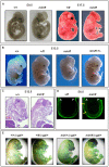
Figure 2. Excessive Wnt/β-catenin signaling results in anterior head defects during embryonic development.
(A) Whole mounts of E9.5 (+/+) and (min/fl) mutant embryos and E17.5 (+/fl) and (min/fl) mutant embryos. (B) Whole mounts of E13.5 wild-type and mutant embryos of the indicated genotypes. Genetic ablation of one allele of β-catenin in (min/fl) “headless” mutant rescues normal head morphology in (min/fl; +/−) mice. (C) Histological cross sections of E12 wild-type (+/+) and mutant (min/fl) embryos. (D) Confocal cross section of E3.5–5.5 (+/fl) and (min/fl) embryos stained for β-catenin protein. The arrowheads demarcate the outer layer (anterior visceral endoderm) of the embryo which shows increased and expanded β-catenin expression in the (min/fl) mutant. A, Anterior; P, Posterior. (E) Whole mount in vivo X-gal staining of E16 embryos to monitor canonical Wnt/β-catenin dependent activity in compound mutant mice harboring the corresponding BAT::gal reporter transgene. Genotypes are as follows: wild-type (+/+); Apc+/fl (+/fl); Apcfl/fl (fl/fl); Apcmin/+ (min/+); Apcmin/fl (min/fl); Apcmin/fl;Ctnnb1+/− (min/fl; +/−). All mice were on a mixed genetic 129Sv x C57BL/6 background.
Figure 3
Figure 3. Intestinal tumor burden and impaired survival in Apc compound mutant mice.
(A) Multiplicity of large (>2mm) tumors in individual moribund mice of the indicated genotypes. Insert shows overall tumor incidence in cohorts of mice of the indicated genotypes. (B) Multiplicity of large (>2 mm) tumors in the small and large intestine of individual moribund (min/+) and compound (min/fl; +/−) mice. (C) Representative allele-specific nucleotide sequence of DNA extracted from an intestinal tumor of a (min/fl; +/−) mouse. Samples were scored as having lost the wild-type allele when the ratio between the peak intensities (boxed area) was ≤0.6 . (D) Survival curve of mice of the indicated genotypes. Livers and the intestines of (min/fl; +/−) mice were analyzed for macroscopic evidence of tumors before being allocated to cohorts with lesions confined to the indicated organ only. Genotypes are as follows: wild-type (+/+); Apcmin/+ (min/+); Apcfl/fl (fl/fl); Apcmin/fl;Ctnnb1+/− (min/fl; +/−). All mice were on a mixed genetic 129Sv x C57BL/6 background.
Figure 4
Figure 4. Liver phenotype in Apc mutant mice.
(A) Hepatocellular carcinoma (HCC) of moribund Apcfl/fl and representative haematoxilin-eosin stained cross section with the dotted line indicating the boundary between normal (N) and tumoral (T) tissue. Representative allele-specific nucleotide sequence of DNA extracted from a liver tumor of a (min/fl; +/−) mouse demonstrating allelic balance between of the Apcmin and the floxed wt allele (boxed area). (B) Incidence of HCC in mice of the indicated genotypes. (C) Western blot and qPCR analysis of full-length Apc protein and Apc mRNA in normal (N) and tumoral (T) liver tissue of Apcfl/fl and Apc+/+ mice. Cell lysates of HeLa cells transfected with a plasmid encoding full-length wild-type Apc serves as an antibody specificity control. The abundance of de-phosphorylated, active (De-PO4) and total β-catenin protein in the same tissue extracts are shown with β-actin serving as a loading control. kDa, protein size marker in kilo Daltons. (D) Comparative qPCR analysis of representative Wnt target gene expression between normal and tumoral liver tissue collected from moribund Apcfl/fl mice (right panel). A comparable analysis was also performed on liver tissue from healthy 5mo old Apcfl/fl and wild-type mice (left panel). Mean ± SD with n≥3 mice per group. * P<0.05. (E) Bisulfite sequencing of the CpG island within the Axin2 promoter from adjacent normal (N) and tumor liver tissue (T1, T2, T3) from Apcfl/fl mice. Each vertical line refers to a CpG dinucleotide at the indicated position relative to the transcriptional start site. Following bisulfite-treatment, DNA was subcloned and sequenced. Horizontal lines represent individual sequences with open and full circles denoting unmethylated and methylated CpG residues, respectively. (F) Boxplot diagram comparing liver tumor multiplicity in +/fl mice (n = 9) and fl/fl mice (n = 12) 6 to 8 months after treatment with DEN. p = 0.0003 (Mann-Whitney). Genotypes are as follows: wild-type (+/+); Apc+/fl (+/fl); Apcfl/fl (fl/fl); Apcfl/fl;Ctnnb1+/− (fl/fl; +/−); Apcmin/fl;Ctnnb1+/− (min/fl; +/−). All mice were on a mixed 129Sv x C57BL/6 background.
Figure 5
Figure 5. Liver zonation is affected in hypomorphic Apcfl/fl mutant mice.
(A) Immunohistochemical expression analysis for β-catenin, glutamine synthetase (GS), carbamoylphosphate synthetase (CPS) and Cyp2E1 was performed on livers of age-matched wild-type (+/+) and (fl/fl) mice. Arrows point to nuclear β-catenin staining. (B) Hepatocytes with nuclear β-catenin staining were expressed as a percentage of total hepatocytes scored in age-matched wild-type (+/+; n = 3) and (fl/fl; n = 4) mice. p = 0.025 (Mann-Whitney). (C) Semi-quantitative RT-PCR analysis from liver of 5mo old healthy (+/+) and (fl/fl) mice for assessment of expression of Apc, CtnnB1, along with the perivenous markers Glt1 (encoding a transporter of glutamate), RHBG (encoding the ammonium transporter), and the periportal markers Arg1 (encoding arginase1) and Glut2 (encoding glutaminase 2). Gapdh serves a as an RT-PCR amplification control.
Figure 6
Figure 6. In vitro assessment of Wnt signaling in MEFs from Apc hypomorphic mice.
(A) Western blot analysis of MEF cell lysates, prepared from (fl/fl) and (min/fl) mice, 48 h after infection with Cre-GFP expressing adenovirus (AdCre-GFP) at different concentrations. Note the gradual reduction of full-length Apc protein (wt) with increasing amount of AdCre-GFP administration and simultaneous accumulation of the truncated Apc580Δ protein encoded by the recombined Apcfl allele. The truncated Apc protein encoded by the Apcmin allele is indicated (min). (B) Tcf4 reporter activity in MEFs of the indicated genotype in response to a submaximally active concentration of Wnt3a-conditioned medium. Activity was assessed following transient transfection with pSuperTopFlash. Cells were harvested 48 h later and assayed for luciferase activity using the dual luciferase system. The activity of the Tcf4 reporter in (min/fl) MEF exposed to a submaximal Wnt3a stimulation was arbitrarily set to 100 and analysis was performed in triplicate cultures. Horizontal lines indicate the signaling threshold predicted for the indicated phenotypes to occur in mice of the corresponding genotypes. The question mark refers to the position of the “intestinal threshold” line relative to reporter activity in min/fl MEFs, since the corresponding adult min/fl mice can not be generated. At least two independent experiments were performed in triplicates for each genotype. Mean ± SD. Note: Histograms refer to situation before LOH, intestinal polyposis threshold to situation after LOH, where applicable. Genotypes are as follows: wild-type (+/+); Apcmin/+ (min/+); Apcfl/fl (fl/fl); Apcmin/fl (min/fl); Apcmin/Δ580 (min/Δ); Apcfl/fl;Ctnnb1+/− (fl/fl;ctnnb1+/−); Apcmin/fl;Ctnnb1+/− (min/fl;ctnnb1+/−); Apcmin/Δ580;Ctnnb1+/− (min/Δ;ctnnb1+/−). All MEFs were derived from mice on a mixed genetic 129Sv x C57BL/6 background.
Figure 7
Figure 7. Lymphodepletion in Apc hypomorphic mice.
(A) Flow cytometry analysis of CD4+/CD8+ stained splenocytes from 17 week old mice of the indicated genotypes. Representative results from one individual mouse are shown with the percentages contribution to each quadrant shown as Mean ± SD from at least 3 mice. (B) Flow cytometry analysis of CD3+/DX5+ stained splenocytes from 17 week old mice of the indicated genotypes. The NK cell population (DX5+;CD3) are within the top left gate, and the bottom gates contain CD3+ T cells. Representative results from one individual mouse are shown with the percentages contribution to each quadrant shown as Mean ± SD from at least 3 mice. Genotypes are as follows: wild-type (+/+); Apcmin/+ (min/+); Apcfl/fl (fl/fl); Apcfl/fl;Ctnnb1+/− (fl/fl; +/−). All mice were on a mixed genetic 129Sv x C57BL/6 background.

Similar articles

Cited by

References

    1. Klaus A, Birchmeier W. Wnt signalling and its impact on development and cancer. Nat Rev Cancer. 2008;8:387–398. - PubMed
    1. Clevers H. Wnt/beta-catenin signaling in development and disease. Cell. 2006;127:469–480. - PubMed
    1. Mukhopadhyay M, Shtrom S, Rodriguez-Esteban C, Chen L, Tsukui T, et al. Dickkopf1 is required for embryonic head induction and limb morphogenesis in the mouse. Dev Cell. 2001;1:423–434. - PubMed
    1. Satoh W, Gotoh T, Tsunematsu Y, Aizawa S, Shimono A. Sfrp1 and Sfrp2 regulate anteroposterior axis elongation and somite segmentation during mouse embryogenesis. Development. 2006;133:989–999. - PubMed
    1. Popperl H, Schmidt C, Wilson V, Hume CR, Dodd J, et al. Misexpression of Cwnt8C in the mouse induces an ectopic embryonic axis and causes a truncation of the anterior neuroectoderm. Development. 1997;124:2997–3005. - PubMed

Publication types

MeSH terms

LinkOut - more resources