. 2010 Jan 13;5(1):e8678.
doi: 10.1371/journal.pone.0008678.

Two regulators of Vibrio parahaemolyticus play important roles in enterotoxicity by controlling the expression of genes in the Vp-PAI region

Affiliations

Two regulators of Vibrio parahaemolyticus play important roles in enterotoxicity by controlling the expression of genes in the Vp-PAI region

Toshio Kodama et al. PLoS One. .

Abstract

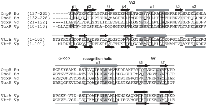
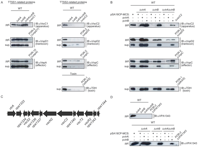
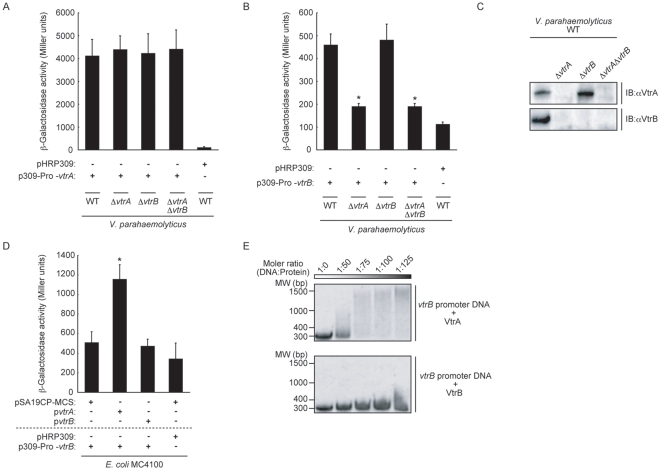
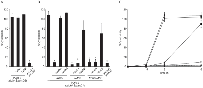
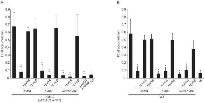
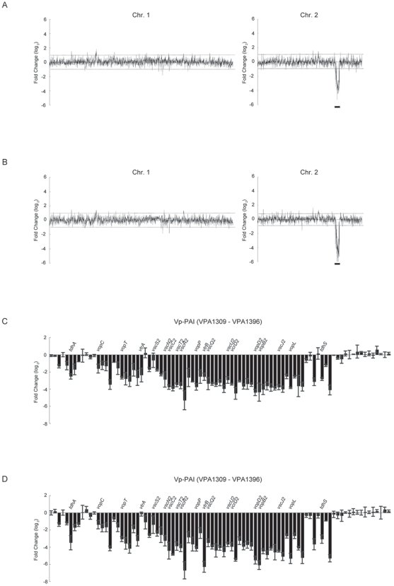
Vibrio parahaemolyticus is an important pathogen causing food-borne disease worldwide. An 80-kb pathogenicity island (Vp-PAI), which contains two tdh (thermostable direct hemolysin) genes and a set of genes for the type III secretion system (T3SS2), is closely related to the pathogenicity of this bacterium. However, the regulatory mechanisms of Vp-PAI's gene expression are poorly understood. Here we report that two novel ToxR-like transcriptional regulatory proteins (VtrA and VtrB) regulate the expression of the genes encoded within the Vp-PAI region, including those for TDH and T3SS2-related proteins. Expression of vtrB was under control of the VtrA, as vector-expressed vtrB was able to recover a functional protein secretory capacity for T3SS2, independent of VtrA. Moreover, these regulatory proteins were essential for T3SS2-dependent biological activities, such as in vitro cytotoxicity and in vivo enterotoxicity. Enterotoxic activities of vtrA and/or vtrB deletion strains derived from the wild-type strain were almost absent, showing fluid accumulation similar to non-infected control. Whole genome transcriptional profiling of vtrA or vtrB deletion strains revealed that the expression levels of over 60 genes were downregulated significantly in these deletion mutant strains and that such genes were almost exclusively located in the Vp-PAI region. These results strongly suggest that VtrA and VtrB are master regulators for virulence gene expression in the Vp-PAI and play critical roles in the pathogenicity of this bacterium.


Conflict of interest statement

Competing Interests: The authors have declared that no competing interests exist.

Figures

Figure 1
Figure 1. VPA1332 (VtrA) and VPA1348 (VtrB) have a winged-helix-turn-helix DNA-binding domain of OmpR family.
Multiple sequence alignment and secondary structure assignments of DNA-binding and trans-activation domains of OmpR, PhoB, ToxR, VtrA, and VtrB proteins are shown. The amino acids that form the hydrophobic cores are highlighted with boxes. Highly conserved amino acids are highlighted with gray boxes.
Figure 2
Figure 2. VtrA and VtrB regulate the expression levels of T3SS2-related proteins and TDH.
A. Loss of vtrA and vtrB diminished the expression of T3SS2-related proteins and TDH. Western blot analysis of bacterial pellets (ppt.) and secreted proteins (sup.) from isogenic mutants of wild-type (WT) V. parahaemolyticus. Lane 1, wild-type V. parahaemolyticus (WT); lane 2, vtrA deletion strain (WTΔvtrA); lane 3, vtrB deletion strain (WTΔvtrB); lane 4, vtrA and vtrB double deletion strain (WTΔvtrAΔvtrB). Samples from indicated strains were loaded in lane 5 to confirm the specificity of each antibody. Blots were probed with anti-VscC1, anti-VopD1, anti-VepA, anti-VscC2, anti-VopD2, anti-VopC, and anti-TDH polyclonal antibodies. B. Vector-induced vtrB could restore the secretory capacity of T3SS2 independent of vtrA. Western blot analyses of bacterial pellets (ppt.) and secreted proteins (sup.) from indicated strains are shown. Blots were probed with anti-VscC2, anti-VopD2, anti-VopC, and anti-TDH polyclonal antibodies. C. Genetic organization of the DNA region containing vscC2 and vpa1343 of V. parahaemolyticus RIMD2210633. D. VPA1343 protein expression was strictly regulated by VtrB. Western blot analysis of bacterial pellets (ppt.) from isogenic mutants of wild-type (WT) V. parahaemolyticus (upper panel) and their complemented strains (lower panel). Blots were probed with anti-VPA1343 polyclonal antibodies.
Figure 3
Figure 3. VtrB expression is under the control of VtrA.
A. Neither vtrA nor vtrB was involved in the transcription of vtrA. V. parahaemolyticus strains carrying the vtrA-lacZ transcriptional fusion vector were assayed for β-galactosidase activity. The bars show the average of three separate experiments, and the standard deviations are indicated by error bars. B. Transcription of vtrB was decreased in vtrA deletion strains. V. parahaemolyticus strains carrying the vtrB-lacZ transcriptional fusion vector were assayed for β-galactosidase activity. The bars show the average of three separate experiments, and the standard deviations are indicated by error bars. C. Deletion of vtrA caused a decrease in the production of VtrB. Immunoblot analysis of VtrA and VtrB protein expression in vtrA and vtrB mutant strains are shown. Lane 1, wild-type V. parahaemolyticus (WT); lane 2, vtrA mutant strain (WTΔvtrA); lane 3, vtrB mutant strain (WTΔvtrB); lane 4, vtrA and vtrB double mutant strain (WTΔvtrAΔvtrB). Blots were probed with anti-VtrA (upper panel) and anti-VtrB (lower panel) polyclonal antibodies. D. Effects of vtrA and vtrB expression on vtrB transcription in E. coli. E. coli MC4100 carrying vtrB-lacZ transcriptional fusion vector were assayed for β-galactosidase activity. The bars show the average of three separate experiments, and the standard deviations are indicated by error bars. E. Binding of purified VtrA DNA binding domain to the upstream region of vtrB is shown by an electrophoretic mobility shift assay. Each lane contains the same amount of upstream region of vtrB (30 nM) and various concentrations (0, 1.5, 2.25, 3.0, 4 µM) of VtrA DNA binding domain (upper panel) or VtrB DNA binding domain (lower panel). The molecular ratios are indicated in the top line.
Figure 4
Figure 4. VtrA and VtrB are not necessary for T3SS1-dependent cytotoxicity but necessary for T3SS2-dependent cytotoxicity.
A. vtrA and vtrB are not necessary for T3SS1-dependent cytotoxicity. Caco-2 cells were infected for 6 h with isogenic strains of POR-3 (ΔtdhASΔvcrD2). Bar 1: POR-3 (ΔtdhASΔvcrD2); bar 2: POR-3ΔvtrA; bar 3: POR-3ΔvtrB; bar 4: ΔvcrD1ΔvcrD2 (ΔtdhASΔvcrD1ΔvcrD2). Cytotoxicity was evaluated by the amount of LDH released. Error bars represent standard deviations for results from triplicate experiments. B. vtrA and vtrB are essential for T3SS2-dependent cytotoxicity. Caco-2 cells were infected for 6 h with isogenic mutant strains of POR-2 (ΔtdhASΔvcrD1). Bar 1: POR-2 (ΔtdhASΔvcrD1); bar 2: POR-2ΔvtrA (ΔtdhASΔvcrD1ΔvtrA); bar 3: POR-2ΔvtrA expressing vtrA (POR-2ΔvtrA+pvtrA); bar 4: POR-2ΔvtrA expressing vtrB (POR-2ΔvtrA+pvtrB); bar 5: POR-2ΔvtrB (ΔtdhASΔvcrD1ΔvtrB); bar 6; POR-2ΔvtrB expressing vtrA (POR-2ΔvtrB+pvtrA); bar 7: POR-2ΔvtrB expressing vtrB (POR-2ΔvtrB+pvtrB); bar 8: POR-2ΔvtrAΔvtrB (ΔtdhASΔvcrD1ΔvtrAΔvtrB); bar 9: POR-2ΔvtrAΔvtrB expressing vtrA (POR-2ΔvtrAΔvtrB+pvtrA); bar 10: POR-2ΔvtrAΔvtrB expressing vtrB (POR-2ΔvtrAΔvtrB+pvtrB); bar 11: ΔvcrD1ΔvcrD2 (ΔtdhASΔvcrD1ΔvcrD2). Cytotoxicity was evaluated by the amount of LDH released. Error bars represent standard deviations for results from triplicate experiments. Asterisks indicate significant differences from the results obtained with the parent strain (*P<0.05). C. Overexpressing of vtrA and vtrB promoted T3SS2-dependent cytotoxicity. Caco-2 cells were infected for 1.5–6 h with V. parahaemolyticus. Cytotoxicity was evaluated by the amount of LDH released. POR-2 (ΔtdhASΔvcrD1) with control vector (pSA19CP-MCS) (filled squares, solid line), POR-2 expressing vtrA (filled circles, solid line), POR-2 expressing vtrB (filled triangles, solid line), ΔvcrD1ΔvcrD2 (ΔtdhASΔvcrD1ΔvcrD2) with control vector (pSA19CP-MCS) (open squares, dashed line), ΔvcrD1ΔvcrD2 expressing vtrA (open circles, dashed line), and ΔvcrD1ΔvcrD2 expressing vtrB (open triangles, dashed line). Error bars represent standard deviations for results from triplicate experiments. Asterisks indicate significant differences from the results obtained with the parent strain (*P<0.05).
Figure 5
Figure 5. VtrA and VtrB have a critical role in V. parahaemolyticus-induced enterotoxicity.
A. VtrA and VtrB are essential for T3SS2-dependent enterotoxicity. The enterotoxic activity levels of isogenic mutants of POR-2 (ΔtdhASΔvcrD1) and complemented strains in rabbit ileal loops were examined. Bar 1, POR-2 (ΔtdhASΔvcrD1); bar 2, POR-2ΔvtrA (ΔtdhASΔvcrD1ΔvtrA); bar 3, POR-2ΔvtrA expressing vtrA (POR-2ΔvtrA+pvtrA); bar 4, POR-2ΔvtrA expressing vtrB (POR-2ΔvtrA+pvtrB); bar 5, POR-2ΔvtrB (ΔtdhASΔvcrD1ΔvtrB); bar 6, POR-2ΔvtrB expressing vtrA (POR-2ΔvtrB+pvtrA); bar 7, POR-2ΔvtrB expressing vtrB (POR-2ΔvtrB+pvtrB); bar 8, POR-2ΔvtrAΔvtrB (ΔtdhASΔvcrD1ΔvtrAΔvtrB); bar 9, POR-2ΔvtrAΔvtrB expressing vtrA (POR-2ΔvtrAΔvtrB+pvtrA); bar 10, POR-2ΔvtrAΔvtrB expressing vtrB (POR-2ΔvtrAΔvtrB+pvtrB); bar 11, ΔvcrD1ΔvcrD2 (ΔtdhASΔvcrD1ΔvcrD2); bar 12, non-infected (NI) control. Results were measured as the amount of accumulated fluid (in milliliters) per length (in centimeters) of ligated rabbit small intestine. Error bars represent standard deviations for results from triplicate experiments. Asterisks indicate significant differences from the results obtained with the parental strain (P<0.05). B. VtrA and VtrB are essential for V. parahaemolyticus-induced enterotoxicity. The enterotoxic activity of isogenic mutants of wild-type V. parahaemolyticus (WT) and complemented strains in rabbit ileal loops were examined. Bar 1, wild-type (WT); bar 2, WTΔvtrA; bar 3, WTΔvtrA expressing vtrA (WTΔvtrA+pvtrA); bar 4, WTΔvtrA expressing vtrB (WTΔvtrA+pvtrB); bar 5, WTΔvtrB; bar 6, WTΔvtrB expressing vtrA (WTΔvtrB+pvtrA); bar 7, WTΔvtrB expressing vtrB (WTΔvtrB+pvtrB); bar 8, WTΔvtrAΔvtrB; bar 9, WTΔvtrAΔvtrB expressing vtrA (WTΔvtrAΔvtrB+pvtrA); bar 10, WTΔvtrAΔvtrB expressing vtrB (WTΔvtrAΔvtrB+pvtrB); bar 11, NI control. Error bars represent standard deviations for results from triplicate experiments. Asterisks indicate significant differences from the results obtained with the parental strain (P<0.05).
Figure 6
Figure 6. Whole-genome transcriptional profiling of vtrA and vtrB deletion strain.
Genome-wide transcript analysis of the VtrA and VtrB regulons is shown. Gene expression was determined by comparing cDNA generated from WTΔvtrA (A) or WTΔvtrB (B) in exponential phase grown in LB medium with 0.5% NaCl with that from the WT strain. The Vp-PAI region is indicated by a bold line. Effect of the vtrA (C) or vtrB (D) deletion on expression of genes located within Vp-PAI (vpa1309-vpa1396). Representative gene functions are indicated at the top.

Similar articles

Cited by

References

    1. Blake PA, Weaver RE, Hollis DG. Diseases of humans (other than cholera) caused by vibrios. Annu Rev Microbiol. 1980;34:341–367. - PubMed
    1. Morris JG, Jr, Black RE. Cholera and other vibrioses in the United States. N Engl J Med. 1985;312:343–350. - PubMed
    1. Hlady WG, Klontz KC. The epidemiology of Vibrio infections in Florida, 1981–1993. J Infect Dis. 1996;173:1176–1183. - PubMed
    1. Daniels NA, MacKinnon L, Bishop R, Altekruse S, Ray B, et al. Vibrio parahaemolyticus infections in the United States, 1973–1998. J Infect Dis. 2000;181:1661–1666. - PubMed
    1. Miyamoto Y, Kato T, Obara Y, Akiyama S, Takizawa K, et al. In vitro hemolytic characteristic of Vibrio parahaemolyticus: its close correlation with human pathogenicity. J Bacteriol. 1969;100:1147–1149. - PMC - PubMed

Publication types

MeSH terms

Substances