. 2010 Jan 13;5(1):e8686.
doi: 10.1371/journal.pone.0008686.

Genetic and molecular analyses of PEG10 reveal new aspects of genomic organization, transcription and translation

Affiliations

Genetic and molecular analyses of PEG10 reveal new aspects of genomic organization, transcription and translation

Heike Lux et al. PLoS One. .

Abstract

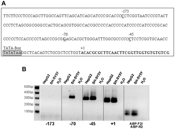
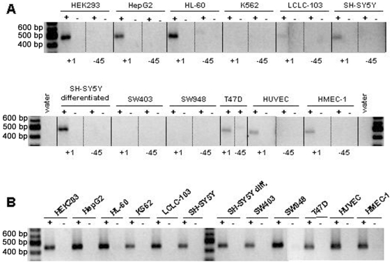
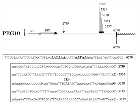
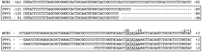
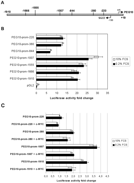
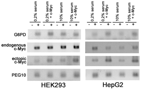
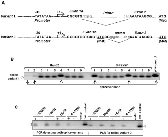
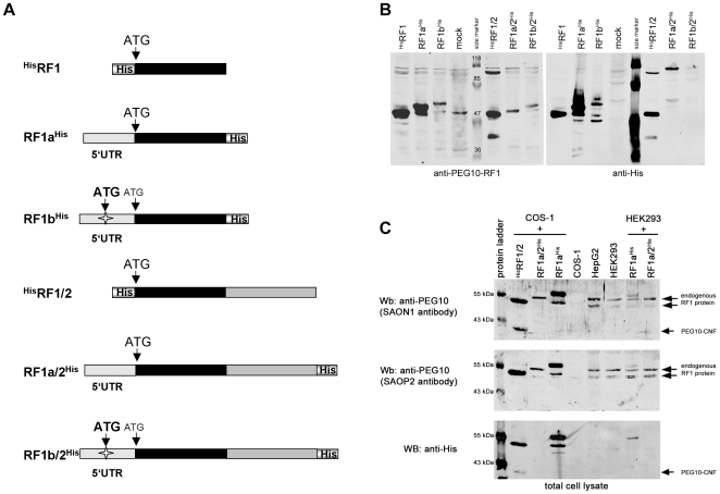
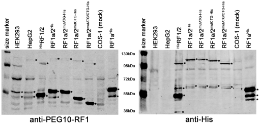
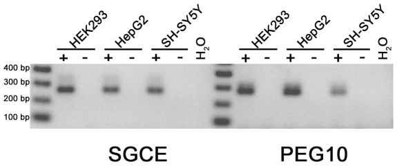
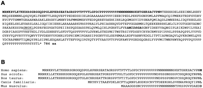
The paternally expressed gene PEG10 is a retrotransposon derived gene adapted through mammalian evolution located on human chromosome 7q21. PEG10 codes for at least two proteins, PEG10-RF1 and PEG10-RF1/2, by -1 frameshift translation. Overexpression or reinduced PEG10 expression was seen in malignancies, like hepatocellular carcinoma or B-cell acute and chronic lymphocytic leukemia. PEG10 was also shown to promote adipocyte differentiation. Experimental evidence suggests that the PEG10-RF1 protein is an inhibitor of apoptosis and mediates cell proliferation. Here we present new data on the genomic organization of PEG10 by identifying the major transcription start site, a new splice variant and report the cloning and analysis of 1.9 kb of the PEG10 promoter. Furthermore, we show for the first time that PEG10 translation is initiated at a non-AUG start codon upstream of the previously predicted AUG codon as well as at the AUG codon. The finding that PEG10 translation is initiated at different sides adds a new aspect to the already interesting feature of PEG10's -1 frameshift translation mechanism. It is now important to unravel the cellular functions of the PEG10 protein variants and how they are related to normal or pathological conditions. The generated promoter-reporter constructs can be used for future studies to investigate how PEG10 expression is regulated. In summary, our study provides new data on the genomic organization as well as expression and translation of PEG10, a prerequisite in order to study and understand the role of PEG10 in cancer, embryonic development and normal cell homeostasis.


Conflict of interest statement

Competing Interests: The authors have declared that no competing interests exist.

Figures

Figure 1
Figure 1. The PEG10 major transcription start site and alternative start sites.
A. Upstream and downstream sequence around the major transcription start site (mTSS) numbered +1. The positions −45, −70 and −173 represent binding sites of primers used to analyse whether alternative TSSs exist located further upstream. In addition, at positions −79 and −112, a T/G and T/C polymorphism, respectively, was identified (data not shown). B. Total RNA of the indicated cell lines were reversely transcribed with a PEG10-specific oligonucleotide (KIAA-3′INNER) located in exon 2. The cDNA was then used in a PCR reaction with the PEG10 exon 2-specific reverse primer ABP-R2 (located upstream of KIAA-3′INNER) and forward primers located at the indicated positions in regard of the mTSS (see A.). The ABP-F2 oligonucleotide is located in exon 1 next to the exon1|intron border.
Figure 2
Figure 2. Analysis of PEG10-specific transcription and transcription start sites.
PEG10 expression and transcription start sites were analysed by semi-quantitative RT-PCR. A. Total RNA of the indicated cell lines was reverse transcribed (RT) with a mixture of oligo-dT/hexamer oligonucleotides. The cDNAs were used in PCR reactions with an identical PEG10-specific reverse primer (KIAA-3′INNER) but variable forward primers specific for transcription start sites at position +1 or −45, respectively, resulting in approximately 500 bp products. B. PCRs for the GAPDH gene served as a positive control for the RT reaction and the use of equal amounts of template RNA. +, PCR with cDNA; −, PCR with total RNA.
Figure 3
Figure 3. PEG10 polyadenylation.
Shown is a schematic view of the 6570 bp long PEG10 transcript with a poly-A tail at its 3′-end. Shown is also the PEG10 mRNA 3′-end sequence with the two underlined canonical polyadenylation signal motifs. The arrows and appendant numbers mark the nucleotide positions of putative alternative polyadenylation sites according to identified PEG10 transcripts. The sequences preceding the polyadenylated nucleotides do not contain a canonical polyadenylation signal motif.
Figure 4
Figure 4. PEG10 promoter variants.
The sequence named NCBI −162 to +2 corresponds to nucleotides 19518795 to 19519959 of reference sequence NT_007933|Hs7_8090 and represents the PEG10 promoter sequence in regard to the PEG10 mTSS. Analyses of this promoter region revealed three different PEG10 promoter variants (PPV) named PPV1, 2 and 3. The variants are characterised by 31, 37 and 71 nucleotide deletions as compared to the reference sequence.
Figure 5
Figure 5. Analysis of PEG10 promoter fragments by promoter-reporter luciferase assays.
A. Shown is a schematic view of 1.9 kb of the PEG10 promoter variant 1 (PPV1). The numbers indicate the 5′-end and length in bp of the evaluated promoter regions in (B.) and (C.) in relation to the transcription start site +1. B. Functional analysis of the PEG10 promoter. PEG10 promoter regions of different length, as indicated, were cloned upstream of the luciferase gene into the reporter plasmid pGL2-Basic. Equal molar amounts of the different promoter constructs were transfected into HEK293 cells. Luciferase activity was measured 48h after transfection and an over-night cultivation with either 10% FCS or 0.2% FCS. The data represent the mean of two independent experiments performed in triplicates plus standard deviation and normalized for transfection efficiency. C. Ectopic c-MYC expression inhibits PEG10 promoter activity. HEK293 cells were co-transfected with a c-myc expression construct and equal molar amounts of the indicated promoter constructs. Luciferase assays were performed as described for (B.). The activity of the PEG10-prom−220 reporter in the presence of 10% FCS was set to one and used as a reference point to calculate the relative activity changes. The data represent the mean of two independent experiments performed in triplicates plus standard deviation and normalized for transfection efficiency.
Figure 6
Figure 6. No obvious evidence for enhanced PEG10 expression due to c-MYC.
HEK293 and HepG2 cells were transfected with a c-MYC expression construct and then cultered over night in the presence of 0.2% or 10% FCS in parallel with non-transfected cells. Subsequently, total RNA was isolated, reverse transcribed and used for sqRT-PCR with primers specific for G6PD, PEG10, c-MYC (endogenous) and ectopic c-MYC. For further details see Results.
Figure 7
Figure 7. PEG10 transcript analysis reveals the existence of two splice variants.
A. Shown is the sequence around the splice junctions for the two PEG10 splice variants. A predicted ATG start codon in exon 2 is underlined. The exon 1b transcript represents a rare splice event, which leads to a new ATG start codon (underlined) in exon 1b. B. In order to estimate the frequency of PEG10 alternative splicing PEG10-specific PCR was done with cell line HepG2 and SH-SY5Y cDNA using forward and reverse primers located in exon 1 and exon 2 respectively. PCR products were cloned and transformed into bacteria. 9 colonies were analysed by colony PCR with the ABP-F2/ABP-R2 primer pair that allows to amplify both splice variants. PCR products were analysed on an agarose gel and variants were distinguished by size: Variant 1 87bp, variant 2 98bp C. PCR with splice variant specific primers shows that all of the tested cell lines express the exon 1a as well as the exon 1b transcript variants. In order to detect both splice variants (left half of Figure) or only variant 2 (right half of Figure), PCRs were performed with the following primer combinations, ABP-F2/ABP-R2 (variant 1 and 2) or ABP-F2b/ABP-R2 (variant 2 specific) respectively. +, PCR with cDNA; -, PCR with total RNA.
Figure 8
Figure 8. Analysis of the PEG10 5′-UTR in protein translation.
A. Schematic presentation of different PEG10 expression constructs. HisRF1 and HisRF1/2 represent constructs without the 5′-UTR and the PEG10 coding sequence starts at the in previous reports predicted ATG start codon. The star in the 5′-UTR of the RF1bHis and RF1b/2His constructs represent the location of a putative new ATG start codon. B. HEK293 cells were mock-transfected or with the indicated PEG10 expression constructs. 48 h after transfection cells were lysed and aliquots of lysates were subjected to SDS-PAGE (8% gel) under reducing conditions and Western blots (Wb). PEG10 proteins were detected with PEG10-RF1-specific antibodies (SAON1/SAOP2) or an anti-His antibody. C. COS-1 and HEK293 cells were mock-transfected or with the indicated PEG10 expression constructs. 48 h after transfection cells were lysed and aliquots of lysates were subjected to SDS-PAGE (8% gel) under reducing conditions and Western blot. Lysates of non-transfected HepG2 cells were analysed in parallel. PEG10 proteins were detected with PEG10-RF1-specific antibodies (SAOP2 and SAON1) or an anti-His antibody. The size of the RF1a/2His expressed PEG10-RF1 protein corresponds to the HepG2 and HEK293 (mock) endogenous PEG10-RF1 protein.
Figure 9
Figure 9. Endogenous PEG10 proteins use non-AUG translation intiation.
In order to test whether PEG10 uses a putative non-AUG start codon (CUG) different PEG10 expression constructs (PEG10-RF1a/2mutATG-His, PEG10-RF1a/2mutCTG-His and PEG10-RF1a/2mutATG/CTG-His) mutated at the putative translation initiation sites were transfected into COS-1 cells and cell lysates were analysed by SDS-PAGE (8% gel) and Western blot with PEG10-RF1-specific antibodies and an anti-His antibody. Lysates of non-transfected HEK293 and HepG2 cells were analysed in parallel for endogenous PEG10 proteins. Proteins considered to be PEG10-specific are marked by asterisks.
Figure 10
Figure 10. The head-to-head orientated PEG10 and SGCE gene are co-expressed in different cell lines.
PCR with the cDNA of HEK293, HepG2 and non-differentiated SH-SY5Y cells and gene-specific primers demonstrates that PEG10 as well as SGCE are co-expressed. +, PCR with cDNA; −, PCR with total RNA.
Figure 11
Figure 11. Putative amino acid sequence of the PEG10-RF1b/2 protein and comparison of the N-terminal region with orthologous proteins.
A. Shown is the amino acid sequence for the translated PEG10-RF1b/2 protein starting from the putative ATG start codon in exon 1b. The N-terminal part of the human PEG10-RF1b/2 up to the methionine coded by the previously predicted ATG start codon in exon 2 is in bold. The amino acid sequence around the −1 frameshift site is underlined. The aspartate protease motif AMIDSGA is in italic and bold. B. A comparison of the N-terminal part of PEG10 orthologous proteins up to the position corresponding to the human methionine is depicted.

Similar articles

Cited by

References

    1. Finishing the euchromatic sequence of the human genome. Nature. 2004;431:931–945. - PubMed
    1. Lux A, Beil C, Majety M, Barron S, Gallione CJ, et al. Human retroviral gag- and gag-pol-like proteins interact with the transforming growth factor-beta receptor activin receptor-like kinase 1. J Biol Chem. 2005;280:8482–8493. - PubMed
    1. Shigemoto K, Brennan J, Walls E, Watson CJ, Stott D, et al. Identification and characterisation of a developmentally regulated mammalian gene that utilises -1 programmed ribosomal frameshifting. Nucleic Acids Res. 2001;29:4079–4088. - PMC - PubMed
    1. Farabaugh PJ. Programmed translational frameshifting. Microbiol Rev. 1996;60:103–134. - PMC - PubMed
    1. Wills NM, Moore B, Hammer A, Gesteland RF, Atkins JF. A functional -1 ribosomal frameshift signal in the human paraneoplastic Ma3 gene. J Biol Chem. 2006;281:7082–7088. - PubMed

Publication types

LinkOut - more resources