

Prostatic acid phosphatase is expressed in peptidergic and nonpeptidergic nociceptive neurons of mice and rats
- PMID: 20084276
- PMCID: PMC2800773
- DOI: 10.1371/journal.pone.0008674
Prostatic acid phosphatase is expressed in peptidergic and nonpeptidergic nociceptive neurons of mice and rats
Abstract
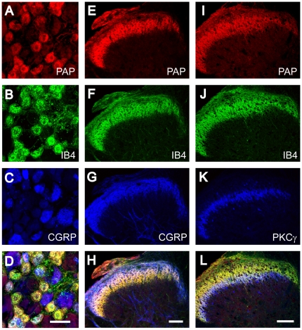
Thiamine monophosphatase (TMPase, also known as Fluoride-resistant acid phosphatase or FRAP) is a classic histochemical marker of small- to medium-diameter dorsal root ganglia (DRG) neurons and has primarily been studied in the rat. Previously, we found that TMPase was molecularly identical to Prostatic acid phosphatase (PAP) using mice. In addition, PAP was expressed in a majority of nonpeptidergic, isolectin B4-binding (IB4+) nociceptive neurons and a subset of peptidergic, calcitonin gene-related peptide-containing (CGRP+) nociceptive neurons. At the time, we were unable to determine if PAP was present in rat DRG neurons because the antibody we used did not cross-react with PAP in rat tissues. In our present study, we generated a chicken polyclonal antibody against the secretory isoform of mouse PAP. This antibody detects mouse, rat and human PAP protein on western blots. Additionally, this antibody detects PAP in mouse and rat small- to medium-diameter DRG neurons and axon terminals in lamina II of spinal cord. In the rat, 92.5% of all PAP+ cells bind the nonpeptidergic marker IB4 and 31.8% of all PAP+ cells contain the peptidergic marker CGRP. Although PAP is found in peptidergic and nonpeptidergic neurons of mice and rats, the percentage of PAP+ neurons that express these markers differs between species. Moreover, PAP+ axon terminals in the rat partially overlap with Protein kinase Cgamma (PKCgamma+) interneurons in dorsal spinal cord whereas PAP+ axon terminals in the mouse terminate dorsal to PKCgamma+ interneurons. Collectively, our studies highlight similarities and differences in PAP localization within nociceptive neurons of mice and rats.
Conflict of interest statement
Figures


Similar articles
-
Critical evaluation of the colocalization between calcitonin gene-related peptide, substance P, transient receptor potential vanilloid subfamily type 1 immunoreactivities, and isolectin B4 binding in primary afferent neurons of the rat and mouse.J Pain. 2007 Mar;8(3):263-72. doi: 10.1016/j.jpain.2006.09.005. Epub 2006 Nov 16. J Pain. 2007. PMID: 17113352 Free PMC article.
-
Co-localization of nociceptive markers in the lumbar dorsal root ganglion and spinal cord of dromedary camel.J Comp Neurol. 2021 Dec;529(17):3710-3725. doi: 10.1002/cne.25240. Epub 2021 Sep 24. J Comp Neurol. 2021. PMID: 34468017
-
Non-peptidergic primary afferents are presynaptic to neurokinin-1 receptor immunoreactive lamina I projection neurons in rat spinal cord.Mol Pain. 2012 Sep 10;8:64. doi: 10.1186/1744-8069-8-64. Mol Pain. 2012. PMID: 22963197 Free PMC article.
-
Transgenic Mouse Models for the Tracing of “Pain” Pathways.In: Kruger L, Light AR, editors. Translational Pain Research: From Mouse to Man. Boca Raton (FL): CRC Press/Taylor & Francis; 2010. Chapter 7. In: Kruger L, Light AR, editors. Translational Pain Research: From Mouse to Man. Boca Raton (FL): CRC Press/Taylor & Francis; 2010. Chapter 7. PMID: 21882471 Free Books & Documents. Review.
-
Itch Modulation by VGLUT2-Dependent Glutamate Release from Somatic Sensory Neurons.In: Carstens E, Akiyama T, editors. Itch: Mechanisms and Treatment. Boca Raton (FL): CRC Press/Taylor & Francis; 2014. Chapter 21. In: Carstens E, Akiyama T, editors. Itch: Mechanisms and Treatment. Boca Raton (FL): CRC Press/Taylor & Francis; 2014. Chapter 21. PMID: 24830022 Free Books & Documents. Review.
Cited by
-
EXPRESS: NGF-trkA signaling modulates the analgesic effects of prostatic acid phosphatase in resiniferatoxin-induced neuropathy.Mol Pain. 2016 Jun 15;12:1744806916656846. doi: 10.1177/1744806916656846. Print 2016. Mol Pain. 2016. PMID: 27306411 Free PMC article.
-
Neuronal adenosine release, and not astrocytic ATP release, mediates feedback inhibition of excitatory activity.Proc Natl Acad Sci U S A. 2012 Apr 17;109(16):6265-70. doi: 10.1073/pnas.1120997109. Epub 2012 Mar 15. Proc Natl Acad Sci U S A. 2012. PMID: 22421436 Free PMC article.
-
Downregulation of adenosine and adenosine A1 receptor contributes to neuropathic pain in resiniferatoxin neuropathy.Pain. 2018 Aug;159(8):1580-1591. doi: 10.1097/j.pain.0000000000001246. Pain. 2018. PMID: 29672450 Free PMC article.
-
Expression of green fluorescent protein defines a specific population of lamina II excitatory interneurons in the GRP::eGFP mouse.Sci Rep. 2020 Aug 6;10(1):13176. doi: 10.1038/s41598-020-69711-7. Sci Rep. 2020. PMID: 32764601 Free PMC article.
-
CGRPα-expressing sensory neurons respond to stimuli that evoke sensations of pain and itch.PLoS One. 2012;7(5):e36355. doi: 10.1371/journal.pone.0036355. Epub 2012 May 1. PLoS One. 2012. PMID: 22563493 Free PMC article.
References
-
- Colmant HJ. Aktivitatsschwankungen der sauren Phosphatase im Ruckenmark und den Spinalganglien der Ratte nach Durchschneidung des Nervus ischiadicus. Arch Psychiat Nervnekr. 1959;199:60–71. - PubMed
-
- Dodd J, Jahr CE, Hamilton PN, Heath MJ, Matthew WD, et al. Cytochemical and physiological properties of sensory and dorsal horn neurons that transmit cutaneous sensation. Cold Spring Harb Symp Quant Biol. 1983;48 Pt 2:685–695. - PubMed
-
- Silverman JD, Kruger L. Acid phosphatase as a selective marker for a class of small sensory ganglion cells in several mammals: spinal cord distribution, histochemical properties, and relation to fluoride-resistant acid phosphatase (FRAP) of rodents. Somatosens Res. 1988;5:219–246. - PubMed
-
- Sanyal S, Rustioni A. Phosphatases in the substantia gelatinosa and motoneurones: a comparative histochemical study. Brain Res. 1974;76:161–166. - PubMed
-
- Knyihar E, Gerebtzoff MA. Localisation ultrastructurale de l' isoenzyme fluororesistant de la phosphatase acide dans la moelle epiniere de rat. Bull Assoc Anat. 1970;149:768–790. - PubMed
Publication types
MeSH terms
Substances
Grants and funding
LinkOut - more resources
Full Text Sources
Molecular Biology Databases
Research Materials
Miscellaneous
