

Promoter recognition by a complex of Spx and the C-terminal domain of the RNA polymerase alpha subunit
- PMID: 20084284
- PMCID: PMC2801614
- DOI: 10.1371/journal.pone.0008664
Promoter recognition by a complex of Spx and the C-terminal domain of the RNA polymerase alpha subunit
Abstract
Background: Spx, an ArsC (arsenate reductase) family member, is a global transcriptional regulator of the microbial stress response and is highly conserved amongst Gram-positive bacteria. Bacillus subtilis Spx protein exerts positive and negative control of transcription through its interaction with the C-terminal domain of the RNA polymerase (RNAP) alpha subunit (alphaCTD). Spx activates trxA (thioredoxin) and trxB (thioredoxin reductase) in response to thiol stress, and bears an N-terminal C10XXC13 redox disulfide center that is oxidized in active Spx.
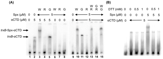
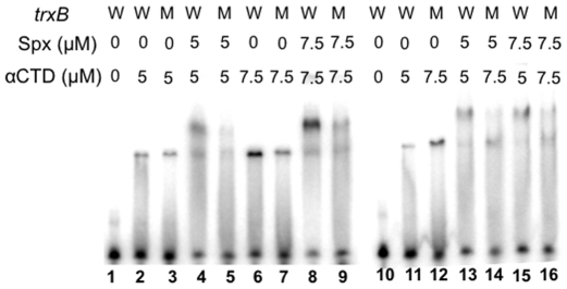
Methodology/principal findings: The structure of mutant Spx(C10S) showed a change in the conformation of helix alpha4. Amino acid substitutions R60E and K62E within and adjacent to helix alpha4 conferred defects in Spx-activated transcription but not Spx-dependent repression. Electrophoretic mobility-shift assays showed alphaCTD interaction with trxB promoter DNA, but addition of Spx generated a supershifted complex that was disrupted in the presence of reductant (DTT). Interaction of alphaCTD/Spx complex with promoter DNA required the cis-acting elements -45AGCA-42 and -34AGCG-31 of the trxB promoter. The Spx(G52R) mutant, defective in alphaCTD binding, did not interact with the alphaCTD-trxB complex. Spx(R60E) not only failed to complex with alphaCTD-trxB, but also disrupted alphaCTD-trxB DNA interaction.
Conclusions/significance: The results show that Spx and alphaCTD form a complex that recognizes the promoter DNA of an Spx-controlled gene. A conformational change during oxidation of Spx to the disulfide form likely alters the structure of Spx alpha helix alpha4, which contains residues that function in transcriptional activation and alphaCTD/Spx-promoter interaction. The results suggest that one of these residues, R60 of the alpha4 region of oxidized Spx, functions in alphaCTD/Spx-promoter contact but not in alphaCTD interaction.
Conflict of interest statement
Figures


Similar articles
-
Exploring the Amino Acid Residue Requirements of the RNA Polymerase (RNAP) α Subunit C-Terminal Domain for Productive Interaction between Spx and RNAP of Bacillus subtilis.J Bacteriol. 2017 Jun 27;199(14):e00124-17. doi: 10.1128/JB.00124-17. Print 2017 Jul 15. J Bacteriol. 2017. PMID: 28484046 Free PMC article.
-
Residue substitutions near the redox center of Bacillus subtilis Spx affect RNA polymerase interaction, redox control, and Spx-DNA contact at a conserved cis-acting element.J Bacteriol. 2013 Sep;195(17):3967-78. doi: 10.1128/JB.00645-13. J Bacteriol. 2013. PMID: 23813734 Free PMC article.
-
Activation of transcription initiation by Spx: formation of transcription complex and identification of a Cis-acting element required for transcriptional activation.Mol Microbiol. 2008 Aug;69(3):765-79. doi: 10.1111/j.1365-2958.2008.06330.x. Mol Microbiol. 2008. PMID: 18687074 Free PMC article.
-
Roles and regulation of Spx family transcription factors in Bacillus subtilis and related species.Adv Microb Physiol. 2019;75:279-323. doi: 10.1016/bs.ampbs.2019.05.003. Epub 2019 Jul 5. Adv Microb Physiol. 2019. PMID: 31655740 Free PMC article. Review.
-
Spx, a versatile regulator of the Bacillus subtilis stress response.Curr Genet. 2019 Aug;65(4):871-876. doi: 10.1007/s00294-019-00950-6. Epub 2019 Mar 4. Curr Genet. 2019. PMID: 30830258 Review.
Cited by
-
Genome-wide identification of genes directly regulated by the pleiotropic transcription factor Spx in Bacillus subtilis.Nucleic Acids Res. 2012 Oct;40(19):9571-83. doi: 10.1093/nar/gks755. Epub 2012 Aug 16. Nucleic Acids Res. 2012. PMID: 22904090 Free PMC article.
-
Genetic regulation of the intercellular adhesion locus in staphylococci.Front Cell Infect Microbiol. 2012 Mar 26;2:38. doi: 10.3389/fcimb.2012.00038. eCollection 2012. Front Cell Infect Microbiol. 2012. PMID: 23061050 Free PMC article. Review.
-
Exploring the Amino Acid Residue Requirements of the RNA Polymerase (RNAP) α Subunit C-Terminal Domain for Productive Interaction between Spx and RNAP of Bacillus subtilis.J Bacteriol. 2017 Jun 27;199(14):e00124-17. doi: 10.1128/JB.00124-17. Print 2017 Jul 15. J Bacteriol. 2017. PMID: 28484046 Free PMC article.
-
Phase Transition of the Bacterium upon Invasion of a Host Cell as a Mechanism of Adaptation: a Mycoplasma gallisepticum Model.Sci Rep. 2016 Oct 24;6:35959. doi: 10.1038/srep35959. Sci Rep. 2016. PMID: 27775027 Free PMC article.
-
Determination of the Gene Regulatory Network of a Genome-Reduced Bacterium Highlights Alternative Regulation Independent of Transcription Factors.Cell Syst. 2019 Aug 28;9(2):143-158.e13. doi: 10.1016/j.cels.2019.07.001. Epub 2019 Aug 21. Cell Syst. 2019. PMID: 31445891 Free PMC article.
References
Publication types
MeSH terms
Substances
Grants and funding
LinkOut - more resources
Full Text Sources
