. 2010 Apr;67(8):1343-51.
doi: 10.1007/s00018-009-0256-3. Epub 2010 Jan 20.

Cross-reactivity of autoantibodies from patients with epidermolysis bullosa acquisita with murine collagen VII

Affiliations

Cross-reactivity of autoantibodies from patients with epidermolysis bullosa acquisita with murine collagen VII

Kinga Csorba et al. Cell Mol Life Sci. 2010 Apr.

Abstract

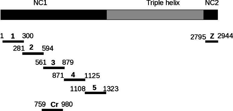
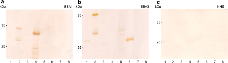
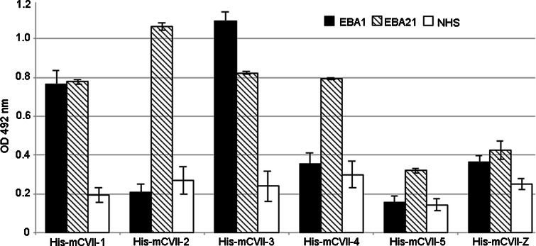
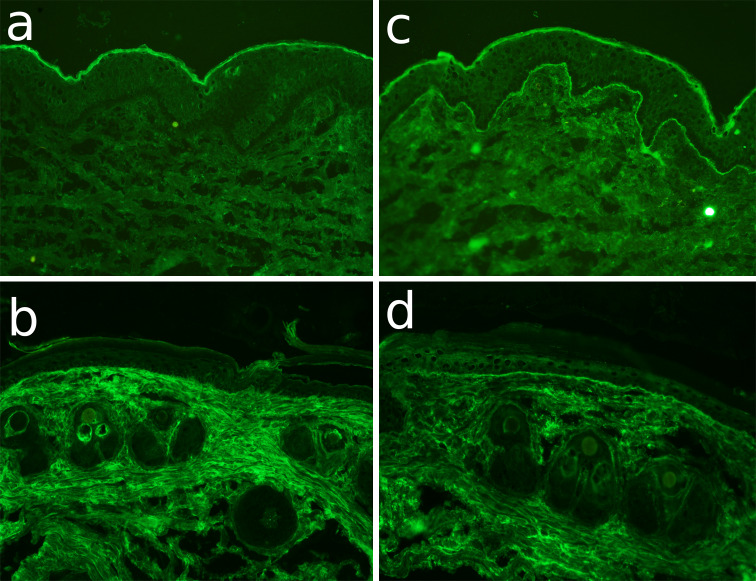
The pathomechanism of antibody-mediated tissue damage in autoimmune diseases can be best studied in experimental models by passively transferring specific autoantibodies into animals. The reproduction of the disease in animals depends on several factors, including the cross-reactivity of patient autoantibodies with the animal tissue. Here, we show that autoantibodies from patients with epidermolysis bullosa acquisita (EBA), a subepidermal autoimmune blistering disease, recognize multiple epitopes on murine collagen VII. Indirect immunofluorescence microscopy revealed that EBA patients' IgG cross-reacts with mouse skin. Overlapping, recombinant fragments of murine collagen VII were used to characterize the reactivity of EBA sera and to map the epitopes on the murine antigen by ELISA and immunoblotting. The patients' autoantibody binding to murine collagen VII triggered pathogenic events as demonstrated by a complement fixing and an ex vivo granulocyte-dependent dermal-epidermal separation assay. These findings should greatly facilitate the development of improved disease models and novel therapeutic strategies.


Figures

Fig. 1
Fig. 1
Cross-reactivity of EBA patients sera with murine skin by immunofluorescence microscopy. Cryosections of ad murine and e, f human skin were incubated with a serum from a rabbit immunized with GST-mCVIICr (SA6310), b,e serum from EBA3, c,f serum from EBA1, and d normal human serum (NHS). After washing in PBS, sections were incubated with a anti-rabbit IgG and bf anti-human IgG both labeled with AlexaFluor 488 (magnifications, all ×200)
Fig. 2
Fig. 2
Recombinant fragments of collagen VII generated in this study. Collagen VII is composed of three identical α chains, each consisting of a central triple helical collagenous domain, flanked by a large amino-terminal noncollagenous domain (NC1) and a smaller carboxy-terminal noncollagenous domain (NC2). Fragments of murine collagen VII cDNA corresponding to the NC1 and NC2 domains were cloned into pGEX-6P-1 and pQE41 and expressed in E. coli. Amino acid residue numbers are shown above the fragments
Fig. 3
Fig. 3
Immunoblot analysis of the EBA sera reactivity with mCVII. His-tagged recombinant mCVII fragments 1, 2, 3, 4, 5 and Z (lanes 2–8) as well as His-DHFR (lane 1) were separated by 12% SDS-PAGE and electrophoretically transferred to nitrocellulose. The membranes were immunoblotted with a serum from patients EBA1, b EBA3, and c normal human serum (NHS). Migration positions of molecular weight markers (kDa) are shown on the left
Fig. 4
Fig. 4
Analysis of the reactivity of EBA sera with murine collagen VII by ELISA. Levels of serum IgG autoantibodies were measured in triplicate by ELISA using recombinant fragments of murine collagen VII as described in “Materials and methods”. OD readings of serum samples from patients EBA1, EBA21 and from a healthy donor are given as mean ± SD
Fig. 5
Fig. 5
EBA patient autoantibodies fix complement to the dermal–epidermal junction of murine skin sections. Frozen sections of human (a,c) and murine (b,d) skin were incubated with serum from a healthy donor (a,b) and from EBA1 patient (c,d). After washing in phosphate-buffered saline, the sections were further incubated with fresh human serum as a source of complement. C3 deposits were visualized by incubation with a monoclonal antibody specific to human C3 followed by an AlexaFluor-488-conjugated anti-mouse IgG antibody. While normal human serum (a,b) did not fix complement in the cryosections, the EBA serum bound complement at the dermal–epidermal junction of both human (c) and murine (d) skin sections
Fig. 6
Fig. 6
EBA patient autoantibodies induce dermal–epidermal separation in sections of murine skin. Frozen sections of human (a,c) and murine (b,d) skin were incubated with serum from a healthy donor (a,b) and from EBA1 patient (c,d). After washing in phosphate-buffered saline, the sections were further incubated with fresh human serum as a source of leucocytes. In contrast to sections incubated with serum from a healthy donor (a,b), sections treated with serum from the EBA patient (c,d) developed dermal–epidermal separation after incubation with normal human granulocytes

Similar articles

Cited by

References

    1. Nishie W, Sawamura D, Goto M, Ito K, Shibaki A, McMillan J, Sakai K, Nakamura H, Olasz E, Yancey K, Akiyama M, Shimizu H. Humanization of autoantigen. Nat Med. 2007;13:378–383. doi: 10.1038/nm1496. - DOI - PubMed
    1. Toyka KV, Brachman DB, Pestronk A, Kao I. Myasthenia gravis: passive transfer from man to mouse. Science. 1975;190:397–399. doi: 10.1126/science.1179220. - DOI - PubMed
    1. Liu Z, Diaz LA, Troy JL, Taylor AF, Emery DJ, Fairley JA, Giudice GJ. A passive transfer model of the organ-specific autoimmune disease, bullous pemphigoid, using antibodies generated against the hemidesmosomal antigen, BP180. J Clin Invest. 1993;92:2480–2488. doi: 10.1172/JCI116856. - DOI - PMC - PubMed
    1. Lazarova Z, Yee C, Darling T, Briggaman RA, Yancey KB. Passive transfer of anti-laminin 5 antibodies induces subepidermal blisters in neonatal mice. J Clin Invest. 1996;98:1509–1518. doi: 10.1172/JCI118942. - DOI - PMC - PubMed
    1. Sitaru C, Mihai S, Otto C, Chiriac MT, Hausser I, Dotterweich B, Saito H, Rose C, Ishiko A, Zillikens D. Induction of dermal–epidermal separation in mice by passive transfer of antibodies specific to type VII collagen. J Clin Invest. 2005;115:870–878. - PMC - PubMed

Publication types

MeSH terms

LinkOut - more resources