

Bmi-1 gene is upregulated in early-stage hepatocellular carcinoma and correlates with ATP-binding cassette transporter B1 expression
- PMID: 20085590
- PMCID: PMC11158551
- DOI: 10.1111/j.1349-7006.2009.01431.x
Bmi-1 gene is upregulated in early-stage hepatocellular carcinoma and correlates with ATP-binding cassette transporter B1 expression
Abstract
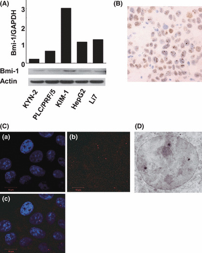
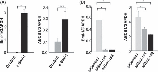
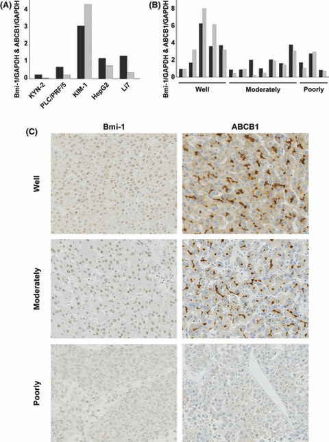
Overexpression of "stemness gene"Bmi-1 has been identified in some solid tumors. We investigated Bmi-1 expression in hepatocellular carcinoma (HCC) and ATP-binding cassette transporter B1 (ABCB1) as a new potential target for Bmi-1. Bmi-1 was highly expressed in HCC cell lines and the most well differentiated cell line, KIM-1, showed the highest expression. Immunohistochemical, immunocytochemical, and immunoelectron microscopic analysis showed the Bmi-1 protein as having a high intensity of small dots within the nucleus which reflected concentrated sites of Bmi-1 repressive activity. Clear "dot-pattern" staining was observed in 24 of 37 (65%) well differentiated HCC (including 13 of 21 early nodules [62%]), in 32 of 71 (45%) moderately differentiated HCC, and 7 of 14 (50%) poorly differentiated HCC. A similar expression was not observed in non-cancerous background regions. High Bmi-1 expression was observed in the early and well differentiated HCC. Furthermore, overexpression and suppression of Bmi-1 was followed by a respective increase and decrease in ABCB1 expression. As with Bmi-1, high ABCB1 expression was also observed in the early and well differentiated HCC. A strong correlation between ABCB1 and Bmi-1 mRNA expression was seen in HCC cell lines and clinical samples (Pearson's correlation coefficient 0.95 and 0.90, respectively). The Bmi-1 gene is upregulated in HCC, and in particular is highly expressed in early and well differentiated HCC. The fact that this expression correlated with that of ABCB1 suggests a new regulation target for Bmi-1, and gives new insight into early hepatocarcinogenesis mechanisms and potential targets for future HCC treatment.
Figures

Similar articles
-
Molecular pathology in early hepatocarcinogenesis.Oncology. 2010;78(2):157-60. doi: 10.1159/000312658. Epub 2010 Apr 13. Oncology. 2010. PMID: 20389138 Review.
-
The overexpression of polycomb group proteins Bmi1 and EZH2 is associated with the progression and aggressive biological behavior of hepatocellular carcinoma.Lab Invest. 2008 Aug;88(8):873-82. doi: 10.1038/labinvest.2008.52. Epub 2008 Jun 30. Lab Invest. 2008. PMID: 18591938
-
Two stem cell markers, ATP-binding cassette, G2 subfamily (ABCG2) and BMI-1, predict the transformation of oral leukoplakia to cancer: a long-term follow-up study.Cancer. 2012 Mar 15;118(6):1693-700. doi: 10.1002/cncr.26483. Epub 2011 Aug 25. Cancer. 2012. PMID: 22009787
-
The polycomb gene product BMI1 contributes to the maintenance of tumor-initiating side population cells in hepatocellular carcinoma.Cancer Res. 2008 Oct 1;68(19):7742-9. doi: 10.1158/0008-5472.CAN-07-5882. Cancer Res. 2008. PMID: 18829528
-
Roles of BMI1 in the Initiation, Progression, and Treatment of Hepatocellular Carcinoma.Technol Cancer Res Treat. 2022 Jan-Dec;21:15330338211070689. doi: 10.1177/15330338211070689. Technol Cancer Res Treat. 2022. PMID: 35072573 Free PMC article. Review.
Cited by
-
Epigenetic regulation in hepatocellular carcinoma requires long noncoding RNAs.Biomed Res Int. 2015;2015:473942. doi: 10.1155/2015/473942. Epub 2015 Mar 10. Biomed Res Int. 2015. PMID: 25861629 Free PMC article. Review.
-
Early hepatocellular carcinoma with high-grade atypia in small vaguely nodular lesions.Cancer Sci. 2016 Apr;107(4):543-50. doi: 10.1111/cas.12893. Epub 2016 Mar 18. Cancer Sci. 2016. PMID: 26797961 Free PMC article.
-
Celecoxib Prevents Doxorubicin-Induced Multidrug Resistance in Canine and Mouse Lymphoma Cell Lines.Cancers (Basel). 2020 Apr 29;12(5):1117. doi: 10.3390/cancers12051117. Cancers (Basel). 2020. PMID: 32365663 Free PMC article.
-
miR-200c inhibits melanoma progression and drug resistance through down-regulation of BMI-1.Am J Pathol. 2012 Nov;181(5):1823-35. doi: 10.1016/j.ajpath.2012.07.009. Epub 2012 Sep 13. Am J Pathol. 2012. PMID: 22982443 Free PMC article.
-
Gankyrin induces STAT3 activation in tumor microenvironment and sorafenib resistance in hepatocellular carcinoma.Cancer Sci. 2017 Oct;108(10):1996-2003. doi: 10.1111/cas.13341. Epub 2017 Sep 2. Cancer Sci. 2017. PMID: 28777492 Free PMC article.
References
-
- Parkin DM, Bray F, Ferlay J, Pisani P. Global cancer statistics, 2002. CA Cancer J Clin 2005; 55: 74–108. - PubMed
-
- Sakamoto M, Hirohashi S, Shimosato Y. Early stages of multistep hepatocarcinogenesis: adenomatous hyperplasia and early hepatocellular carcinoma. Hum Pathol 1991; 22: 172–8. - PubMed
-
- Reya T, Morrison SJ, Clarke MF, Weissman IL. Stem cells, cancer, and cancer stem cells. Nature 2001; 414: 105–11. - PubMed
-
- Pardal R, Clarke MF, Morrison SJ. Applying the principles of stem‐cell biology to cancer. Nat Rev Cancer 2003; 3: 895–902. - PubMed
-
- Van Lohuizen M, Verbeek S, Scheijen B, Wientjens E, Van Der Gulden H, Berns A. Identification of cooperating oncogenes in E mu‐myc transgenic mice by provirus tagging. Cell 1991; 65: 737–52. - PubMed
Publication types
MeSH terms
Substances
LinkOut - more resources
Full Text Sources
Medical
Research Materials
