Acute mucosal pathogenesis of feline immunodeficiency virus is independent of viral dose in vaginally infected cats
- PMID: 20085648
- PMCID: PMC2835650
- DOI: 10.1186/1742-4690-7-2
Acute mucosal pathogenesis of feline immunodeficiency virus is independent of viral dose in vaginally infected cats
Abstract
Background: The mucosal pathogenesis of HIV has been shown to be an important feature of infection and disease progression. HIV-1 infection causes depletion of intestinal lamina propria CD4+ T cells (LPL), therefore, intestinal CD4+ T cell preservation may be a useful correlate of protection in evaluating vaccine candidates. Vaccine studies employing the cat/FIV and macaque/SIV models frequently use high doses of parenterally administered challenge virus to ensure high plasma viremia in control animals. However, it is unclear if loss of mucosal T cells would occur regardless of initial viral inoculum dose. The objective of this study was to determine the acute effect of viral dose on mucosal leukocytes and associated innate and adaptive immune responses.
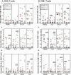
Results: Cats were vaginally inoculated with a high, middle or low dose of cell-associated and cell-free FIV. PBMC, serum and plasma were assessed every two weeks with tissues assessed eight weeks following infection. We found that irrespective of mucosally administered viral dose, FIV infection was induced in all cats. However, viremia was present in only half of the cats, and viral dose was unrelated to the development of viremia. Importantly, regardless of viral dose, all cats experienced significant losses of intestinal CD4+ LPL and CD8+ intraepithelial lymphocytes (IEL). Innate immune responses by CD56+CD3- NK cells correlated with aviremia and apparent occult infection but did not protect mucosal T cells. CD4+ and CD8+ T cells in viremic cats were more likely to produce cytokines in response to Gag stimulation, whereas aviremic cats T cells tended to produce cytokines in response to Env stimulation. However, while cell-mediated immune responses in aviremic cats may have helped reduce viral replication, they could not be correlated to the levels of viremia. Robust production of anti-FIV antibodies was positively correlated with the magnitude of viremia.
Conclusions: Our results indicate that mucosal immune pathogenesis could be used as a rapid indicator of vaccine success or failure when combined with a physiologically relevant low dose mucosal challenge. We also show that innate immune responses may play an important role in controlling viral replication following acute mucosal infection, which has not been previously identified.
Figures


Similar articles
-
Domestic cats infected with lion or puma lentivirus develop anti-feline immunodeficiency virus immune responses.J Acquir Immune Defic Syndr. 2003 Sep 1;34(1):20-31. doi: 10.1097/00126334-200309010-00003. J Acquir Immune Defic Syndr. 2003. PMID: 14501789
-
Prior virus exposure alters the long-term landscape of viral replication during feline lentiviral infection.Viruses. 2011 Oct;3(10):1891-908. doi: 10.3390/v3101891. Epub 2011 Oct 13. Viruses. 2011. PMID: 22069521 Free PMC article.
-
FIV infection induces unique changes in phenotype and cellularity in the medial iliac lymph node and intestinal IEL.AIDS Res Hum Retroviruses. 2007 May;23(5):720-8. doi: 10.1089/aid.2006.0025. AIDS Res Hum Retroviruses. 2007. PMID: 17530999
-
Mucosal infection and vaccination against feline immunodeficiency virus.J Biotechnol. 1999 Aug 20;73(2-3):213-21. doi: 10.1016/s0168-1656(99)00139-x. J Biotechnol. 1999. PMID: 10486930 Review.
-
Feline immunodeficiency virus (FIV) neutralization: a review.Viruses. 2011 Oct;3(10):1870-90. doi: 10.3390/v3101870. Epub 2011 Oct 13. Viruses. 2011. PMID: 22069520 Free PMC article. Review.
Cited by
-
Feline Leukemia Virus Frequently Spills Over from Domestic Cats to North American Pumas.J Virol. 2022 Dec 14;96(23):e0120122. doi: 10.1128/jvi.01201-22. Epub 2022 Nov 14. J Virol. 2022. PMID: 36374109 Free PMC article.
-
Suppression of NK cells and regulatory T lymphocytes in cats naturally infected with feline infectious peritonitis virus.Vet Microbiol. 2013 May 31;164(1-2):46-59. doi: 10.1016/j.vetmic.2013.01.042. Epub 2013 Feb 8. Vet Microbiol. 2013. PMID: 23434014 Free PMC article.
-
A novel test for determination of wild felid-domestic cat hybridization.Forensic Sci Int Genet. 2020 Jan;44:102160. doi: 10.1016/j.fsigen.2019.102160. Epub 2019 Oct 22. Forensic Sci Int Genet. 2020. PMID: 31683165 Free PMC article.
-
Prior mucosal exposure to heterologous cells alters the pathogenesis of cell-associated mucosal feline immunodeficiency virus challenge.Retrovirology. 2010 May 28;7:49. doi: 10.1186/1742-4690-7-49. Retrovirology. 2010. PMID: 20507636 Free PMC article.
-
Recent developments in human immunodeficiency virus-1 latency research.J Gen Virol. 2013 May;94(Pt 5):917-932. doi: 10.1099/vir.0.049296-0. Epub 2013 Jan 30. J Gen Virol. 2013. PMID: 23364195 Free PMC article. Review.
References
-
- HIV vaccine failure prompts Merck to halt trial. Nature. 2007;449:390. - PubMed
-
- Vaccine trial discontinued. AIDS Patient Care STDS. 2007;21:778–779. - PubMed
-
- Priddy FH, Brown D, Kublin J, Monahan K, Wright DP, Lalezari J, Santiago S, Marmor M, Lally M, Novak RM, Brown SJ, Kulkarni P, Dubey SA, Kierstead LS, Casimiro DR, Mogg R, DiNubile MJ, Shiver JW, Leavitt RY, Robertson MN, Mehrotra DV, Quirk E. Merck V520-016 Study Group. Safety and immunogenicity of a replication-incompetent adenovirus type 5 HIV-1 clade B gag/pol/nef vaccine in healthy adults. Clin Infect Dis. 2008;46:1769–1781. doi: 10.1086/587993. - DOI - PubMed
-
- Buchbinder SP, Mehrotra DV, Duerr A, Fitzgerald DW, Mogg R, Li D, Gilbert PB, Lama JR, Marmor M, Del Rio C, McElrath MJ, Casimiro DR, Gottesdiener KM, Chodakewitz JA, Corey L, Robertson MN. Step Study Protocol Team. Efficacy assessment of a cell-mediated immunity HIV-1 vaccine (the Step Study): a double-blind, randomised, placebo-controlled, test-of-concept trial. Lancet. 2008;372:1881–1893. doi: 10.1016/S0140-6736(08)61591-3. - DOI - PMC - PubMed
Publication types
MeSH terms
Substances
Grants and funding
LinkOut - more resources
Full Text Sources
Research Materials
Miscellaneous

