. 2010 Jan 8;140(1):99-110.
doi: 10.1016/j.cell.2009.12.022.

A region of the human HOXD cluster that confers polycomb-group responsiveness

Affiliations

A region of the human HOXD cluster that confers polycomb-group responsiveness

Caroline J Woo et al. Cell. .

Abstract

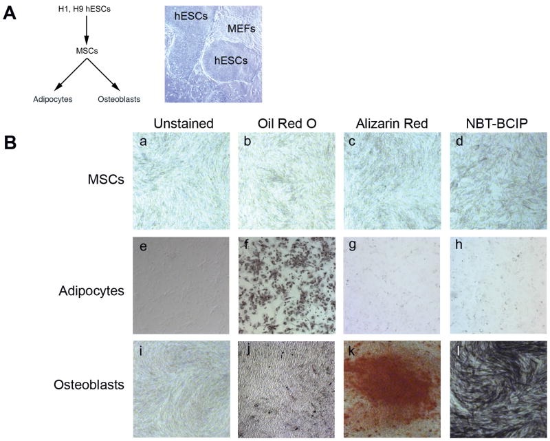
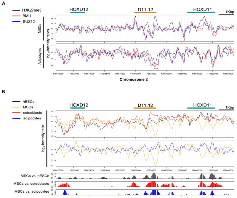
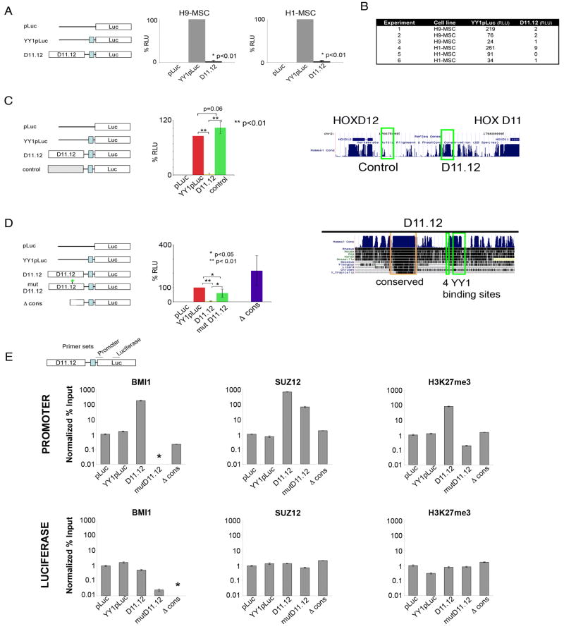
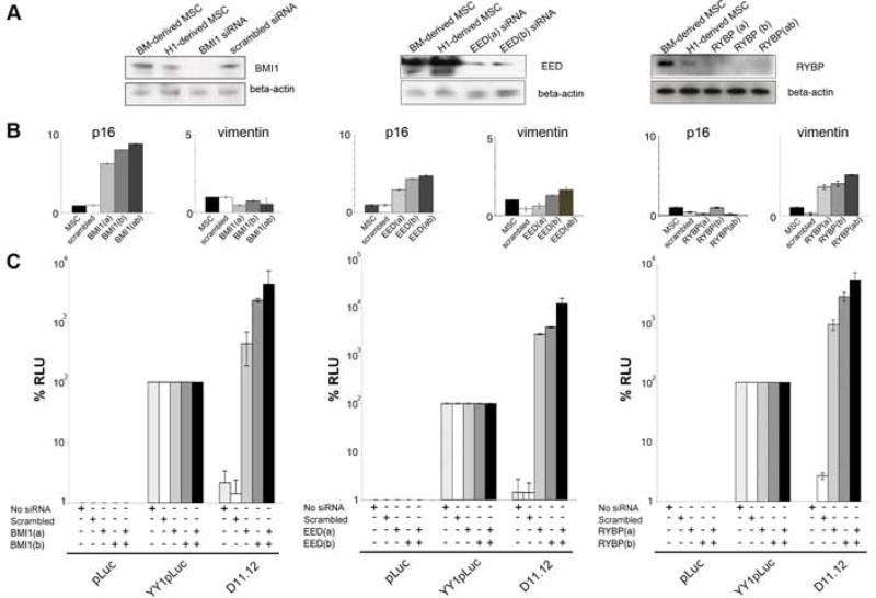
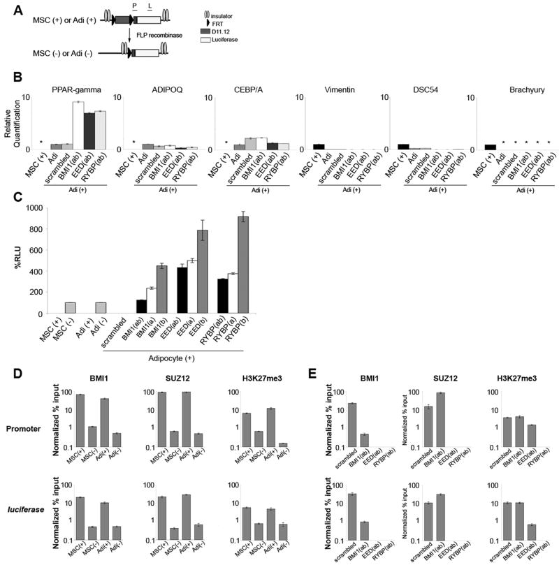
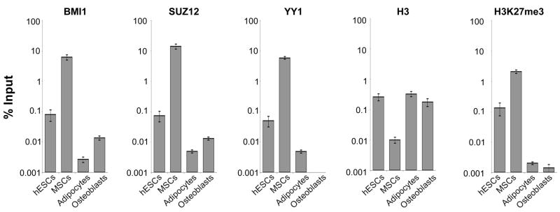
Polycomb group (PcG) proteins are essential for accurate axial body patterning during embryonic development. PcG-mediated repression is conserved in metazoans and is targeted in Drosophila by Polycomb response elements (PREs). However, targeting sequences in humans have not been described. While analyzing chromatin architecture in the context of human embryonic stem cell (hESC) differentiation, we discovered a 1.8kb region between HOXD11 and HOXD12 (D11.12) that is associated with PcG proteins, becomes nuclease hypersensitive, and then shows alteration in nuclease sensitivity as hESCs differentiate. The D11.12 element repressed luciferase expression from a reporter construct and full repression required a highly conserved region and YY1 binding sites. Furthermore, repression was dependent on the PcG proteins BMI1 and EED and a YY1-interacting partner, RYBP. We conclude that D11.12 is a Polycomb-dependent regulatory region with similarities to Drosophila PREs, indicating conservation in the mechanisms that target PcG function in mammals and flies.


Figures

Figure 1
Figure 1. Lineage commitment from pluripotent cells to differentiated cell types
(A) Differentiation schematic; H9 hESCs cultured with MEFs (B) MSCs (panels a–d: unstained, treated with Oil Red O, Alizarin Red, and NBT-BCIP); Adipocytes, day 14 (panels e–h: unstained, treated with Oil Red O, Alizarin Red, and NBT-BCIP); and Osteoblasts, day 28 (panels i–l: unstained, treated with Oil Red O, Alizarin Red, and NBT-BCIP). See Figure S1 for qRT-PCR results.
Figure 2
Figure 2. ChIP-chip and MNase hypersensitivity results
(A) Summary of location analyses across HOXD13 to HOXD10 for H3K27me3, BMI1, and SUZ12 in MSCs. Log2 ratios were normalized and sliding windows of 20 bp were used to plot the values. (B) Nucleosomal mapping of MNase-sensitive sites. Top graphs: plots of normalized log2 ratios from MNase-digested chromatin for hESCs (black), MSCs (orange), osteoblasts (red), and adipocytes (blue). X-axis represents 21kb along chromosome 2 along the HOXD cluster. The bracket points to a large region of difference between HOXD11 and HOXD12. Bottom graphs: Plots of the statistically significant differences of the log2 ratios of the MNase profiles between the cells. Figure S2 displays data across the HOX clusters. Figure S3 displays MNase sensitivity data of D11.12.
Figure 3
Figure 3. D11.12 represses luciferase activity in MSCs
(A) Constructs are displayed on the left: the firefly luciferase construct (pLuc), with an upstream YY1 enhancer (YY1pLuc), and with a 1.8kb region between HOXD11 and HOXD12 upstream of YY1pLuc (D11.12). Luciferase measurements of the co-transfected firefly luciferase and renilla luciferase constructs (n=3, each cell line). Data are represented as mean +/−SEM. (B) Chart of the individual lucifease results, presented as RLU (n=6). (C) Luciferase activity of a control region located between HOXD11 and HOXD12 (left panel). The UCSC Genome Browser map shows the locations of the control region and D11.12 and the level of mammalian conservation of sequence (right panel). (D) Luciferase activity of D11.12 having mutated YY1 binding sites (mutD11.12) or deletion of the conserved region (Δ cons) (left panel). The UCSC Genome Browser map depicts the degree of conservation across 10 other vertebrate species (right panel) with the conserved region (orange) and the 4 YY1 binding sites (green). (E) ChIP-qPCR of BMI1, SUZ12, and H3K27me3. ChIP results are displayed as ratios of the % input(IP)/% input (histone H3). These templates had H3 levels that were consistently 5–10 fold lower in the luciferase gene than for the promoter, perhaps indicative of differences in nucleosome occupancy across these transiently transfected templates. (*) indicates values lower than 0.01% input. Data are represented as mean +/−SEM.
Figure 4
Figure 4. The repressive activity of D11.12 is dependent on BMI1, EED, and RYBP
Western blots of BMI1, EED, and RYBP knockdown in MSCs. Western blots of beta-actin were used to demonstrate equal loading of samples. (B) qRT-PCR results p16/Arf and vimentin in the 3 different BMI1 knockdown cells (Bmi1(a), Bmi1(b), Bmi1(ab), EED knockdown cells (Eed(a), Eed(b), Eed(ab)), and RYBP knockdown cells (RYBP(a), RYBP(b), RYBP(ab)). (C) Luciferase activity of pLuc, YY1pLuc, and D11.12 in the scrambled, BMI1, SUZ12, and RYBP knockdown cells. Data are represented as mean +/−SEM. See Figure S4 for HOXD11-D13 expression levels in the knockdown cells.
Figure 5
Figure 5. D11.12 repression is maintained when MSCs are differentiated into adipocytes
(A) D11.12 construct in the stable cell lines. FRT sites flank D11.12 and insulators flank the D11.12 construct. (B) qRT-PCR from adipocytes (Adi(+)) derived from MSCs carrying the stably integrated D11.12 luciferase construct and from BMI1, SUZ12, and RYBP knockdown Adi(+) cells shows PPAR-gamma, AdipoQ, CEBP-alpha, vimentin, DSC54, and brachyury were similarly expressed in the adipocytes and Adi(+) cells. (*) were not detectable. (C) Luciferase results from MSC(+), MSC(−), Adi(+) and Adi(−) cells normalized by beta-galactosidase measurements. Adi(+) cells infected with scrambled, BMI1, SUZ12, and RYBP lentiviruses were assayed in parallel. Data are represented as mean +/−SEM (D) Bar graphs represent results from ChIP-qPCR experiments for MSC(+), MSC(−), Adi(+), and Adi(−) cells for BMI1, SUZ12, H3, and H3K27me3 at the promoter and the luciferase gene. ChIP results represented as Normalized % Input were normalized for the % input of histone H3. (E) ChIP-qPCR results for Bmi1, Suz12, and H3K27me3 from Adi(+) cells and lentiviral control, BMI1, SUZ12, and RYBP knockdown derivatives. Data are represented as mean +/−SEM.
Figure 6
Figure 6. PcG proteins are enriched at the endogenous D11.12 in different cell types
ChIP-qPCR results of BMI1, SUZ12, YY1, H3, and H3K27me3 at the endogenous D11.12 region in MSCs. Results are represented as % input and were not normalized against histone H3. Data are represented as mean +/−SEM.

References

    1. Atchison L, Ghias A, Wilkinson F, Bonini N, Atchison ML. Transcription factor YY1 functions as a PcG protein in vivo. Embo J. 2003;22:1347–1358. - PMC - PubMed
    1. Barberi T, Willis LM, Socci ND, Studer L. Derivation of multipotent mesenchymal precursors from human embryonic stem cells. PLoS Med. 2005;2:e161. - PMC - PubMed
    1. Beckers J, Duboule D. Genetic analysis of a conserved sequence in the HoxD complex: regulatory redundancy or limitations of the transgenic approach? Dev Dyn. 1998;213:1–11. - PubMed
    1. Bejarano F, Gonzalez I, Vidal M, Busturia A. The Drosophila RYBP gene functions as a Polycomb-dependent transcriptional repressor. Mech Dev. 2005;122:1118–1129. - PubMed
    1. Beuchle D, Struhl G, Muller J. Polycomb group proteins and heritable silencing of Drosophila Hox genes. Development. 2001;128:993–1004. - PubMed

Publication types

MeSH terms

Associated data