

High resolution melting analysis for gene scanning
- PMID: 20085814
- PMCID: PMC2836412
- DOI: 10.1016/j.ymeth.2010.01.013
High resolution melting analysis for gene scanning
Abstract
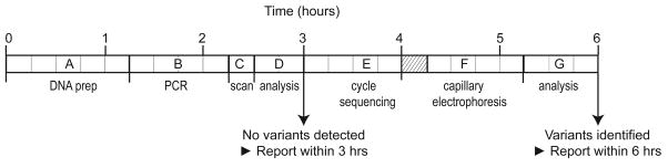
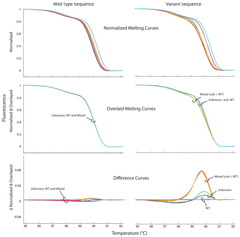
High resolution melting is a new method of genotyping and variant scanning that can be seamlessly appended to PCR amplification. Limitations of genotyping by amplicon melting can be addressed by unlabeled probe or snapback primer analysis, all performed without labeled probes. High resolution melting can also be used to scan for rare sequence variants in large genes with multiple exons and is the focus of this article. With the simple addition of a heteroduplex-detecting dye before PCR, high resolution melting is performed without any additions, processing or separation steps. Heterozygous variants are identified by atypical melting curves of a different shape compared to wild-type homozygotes. Homozygous or hemizygous variants are detected by prior mixing with wild-type DNA. Design, optimization, and performance considerations for high resolution scanning assays are presented for rapid turnaround of gene scanning. Design concerns include primer selection and predicting melting profiles in silico. Optimization includes temperature gradient selection of the annealing temperature, random population screening for common variants, and batch preparation of primer plates with robotically deposited and dried primer pairs. Performance includes rapid DNA preparation, PCR, and scanning by high resolution melting that require, in total, only 3h when no variants are present. When variants are detected, they can be identified in an additional 3h by rapid cycle sequencing and capillary electrophoresis. For each step in the protocol, a general overview of principles is provided, followed by an in depth analysis of one example, scanning of CYBB, the gene that is mutated in X-linked chronic granulomatous disease.
Copyright 2010 Elsevier Inc. All rights reserved.
Figures
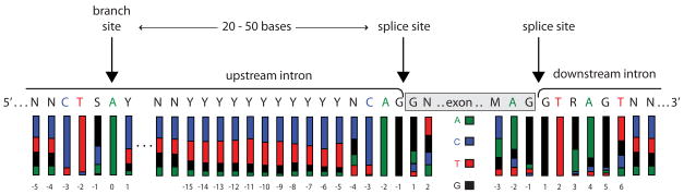
 , C
, C , T
, T , G■). Primers should be designed to avoid consensus bases where variation is likely to effect splicing.
References
Publication types
MeSH terms
Substances
Grants and funding
LinkOut - more resources
Full Text Sources
Other Literature Sources
Miscellaneous
