. 2010 Mar 30;399(1):31-38.
doi: 10.1016/j.virol.2009.12.018. Epub 2010 Jan 20.

Elevated levels of macrophage migration inhibitory factor (MIF) in the plasma of HIV-1-infected patients and in HIV-1-infected cell cultures: a relevant role on viral replication

Affiliations

Elevated levels of macrophage migration inhibitory factor (MIF) in the plasma of HIV-1-infected patients and in HIV-1-infected cell cultures: a relevant role on viral replication

Eduardo G Regis et al. Virology. .

Abstract

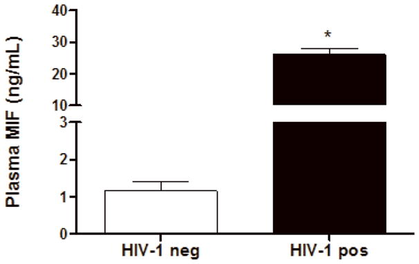
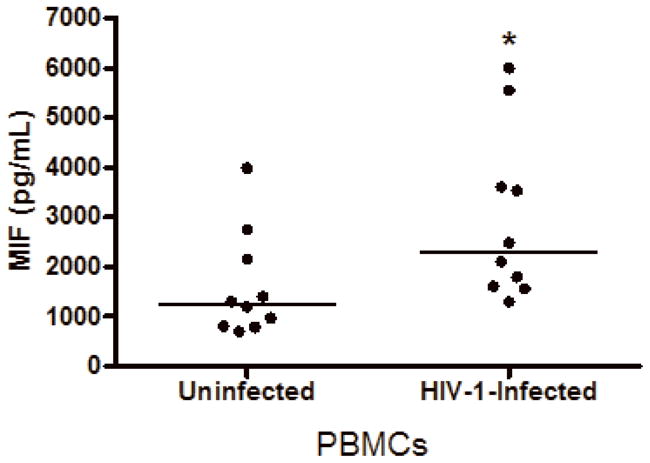
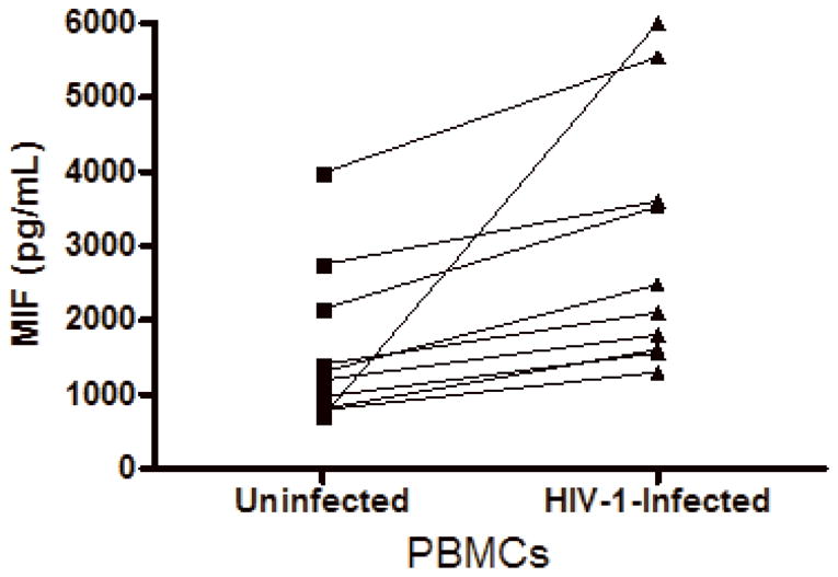
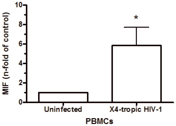
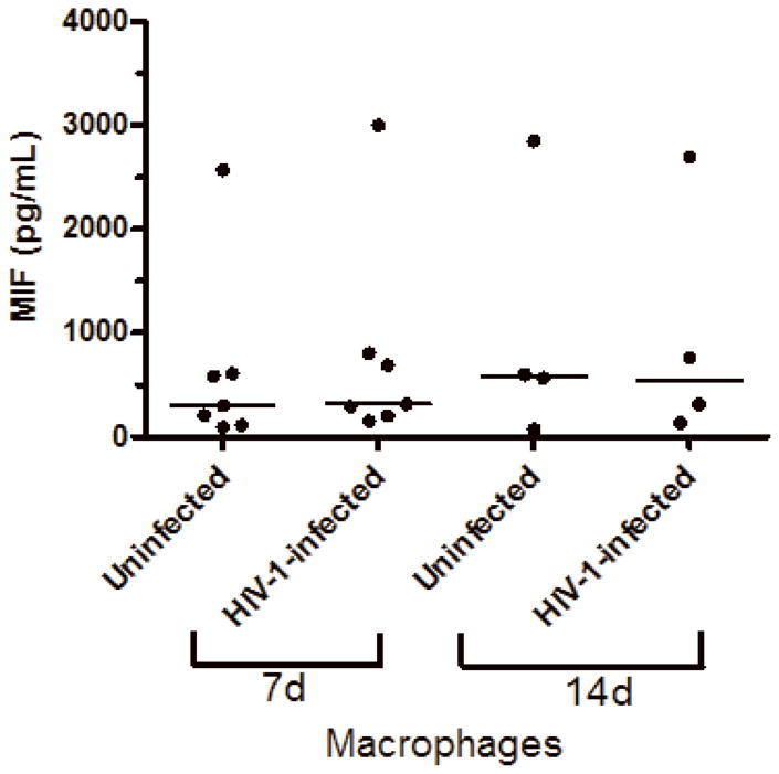
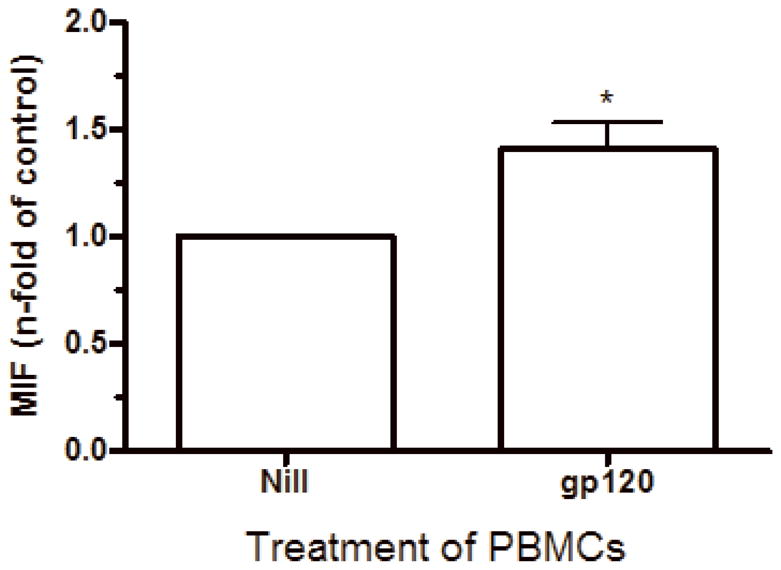
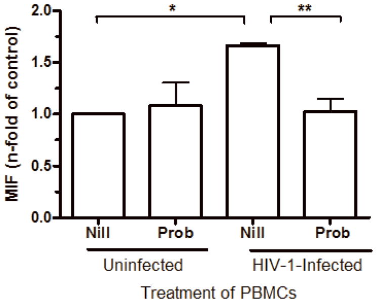
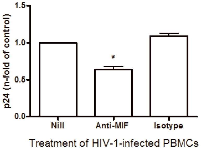
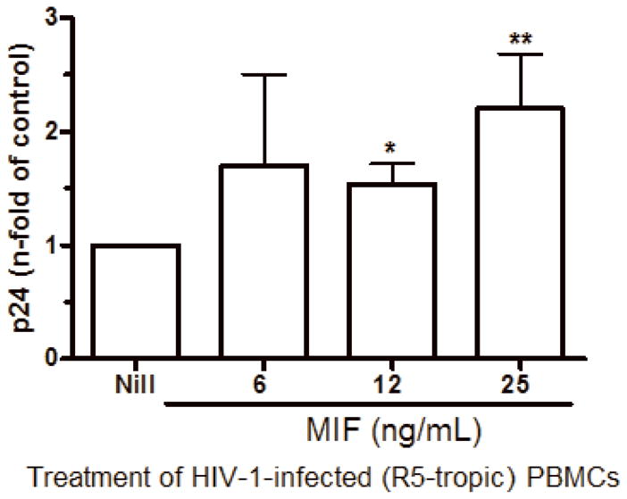
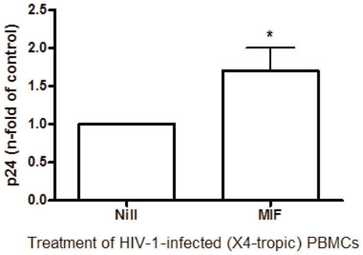
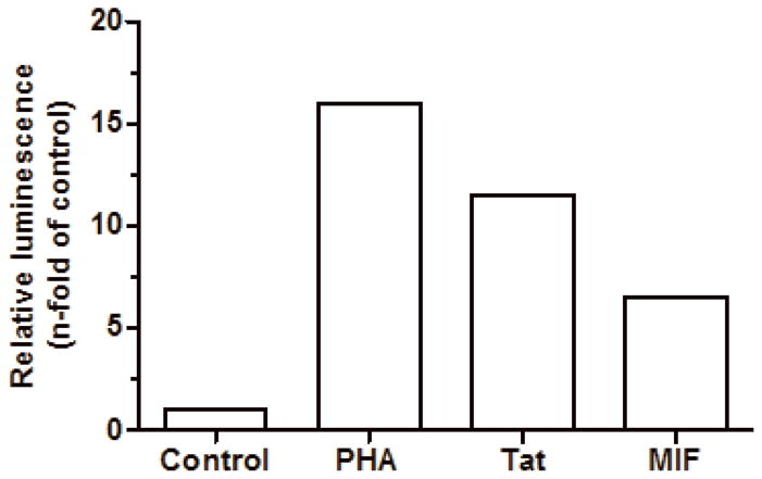
The cytokine macrophage migration inhibitory factor (MIF) is involved in the pathogenesis of inflammatory and infectious diseases, however its role in HIV-1 infection is unknown. Here we show that HIV-1-infected patients present elevated plasma levels of MIF, that HIV-1-infected peripheral blood mononuclear cells (PBMCs) release a greater amount of MIF, and that the HIV-1 envelope glycoprotein gp120 induces MIF secretion from uninfected PBMCs. The HIV-1 replication in PBMCs declines when these cells are treated with anti-MIF antibodies, and exposure of HIV-1-infected cells to the ABC-transporter inhibitor probenecid results in inhibition of MIF secretion. The addition of recombinant MIF (rhMIF) to HIV-1-infected PBMCs enhances viral replication of CCR5- or CXCR4-tropic HIV-1 isolates. Using a T CD4(+) cell lineage containing an HIV long terminal repeats (LTR)-Luciferase construct, we detected that rhMIF promotes transcription from HIV-1 LTR. Our results show that HIV-1 induces MIF secretion and suggest that MIF influences the HIV-1 biology through activation of HIV-1 LTR.


Figures

Figure 1
Figure 1
HIV-1-infected patients present higher plasma levels of MIF relative to uninfected individuals. Plasmas from HIV-1-infected individuals (HIV-1 pos), and from healthy volunteers (control plasmas; HIV-1 neg) were assessed for MIF concentrations by ELISA. Bars represent means ± SEM of 10 healthy volunteers (HIV-1 neg) or 30 HIV-1-infected patients (HIV-1 pos). * p<0.0001
Figure 2
Figure 2
HIV-1 infection and HIV-1 gp120 induce MIF release by PBMCs. (A) PBMCs were infected by an R5-tropic HIV-1 isolate (Ba-L), and after 7 days MIF contents were evaluated in the culture supernatants by ELISA (* p<0.04). Individual values and medians are represented for each group. (B) Lines show individual MIF release in the uninfected and in HIV-1-infected PBMC depicted in figure 2-A. (C) MIF contents in culture supernatants of PBMCs infected by an X4-tropic HIV-1 isolate (Tybe) (* p<0.04). Mean of MIF values by cells cultured only with medium (Nill): 757.8 pg/mL. (D) Macrophages were infected by the HIV-1 isolate Ba-L and MIF release was assessed by ELISA at 7 (7d) and 14 (14d) days after infection. Individual values and medians are represented for each group. (E) Uninfected PBMCs were exposed to recombinant HIV-1 envelope protein gp120 (5 μg/mL) derived from the HIV-1 isolate Ba-L, and MIF secretion was evaluated in culture supernatants by ELISA after 16 hours (* p<0.006). Mean of MIF values by cells cultured only with medium (Nill): 427 pg/mL. In (C) and (E) bars show means ± SEM for 3 and 5 different donors, respectively, and all experiments were done in triplicates per donor.
Figure 2
Figure 2
HIV-1 infection and HIV-1 gp120 induce MIF release by PBMCs. (A) PBMCs were infected by an R5-tropic HIV-1 isolate (Ba-L), and after 7 days MIF contents were evaluated in the culture supernatants by ELISA (* p<0.04). Individual values and medians are represented for each group. (B) Lines show individual MIF release in the uninfected and in HIV-1-infected PBMC depicted in figure 2-A. (C) MIF contents in culture supernatants of PBMCs infected by an X4-tropic HIV-1 isolate (Tybe) (* p<0.04). Mean of MIF values by cells cultured only with medium (Nill): 757.8 pg/mL. (D) Macrophages were infected by the HIV-1 isolate Ba-L and MIF release was assessed by ELISA at 7 (7d) and 14 (14d) days after infection. Individual values and medians are represented for each group. (E) Uninfected PBMCs were exposed to recombinant HIV-1 envelope protein gp120 (5 μg/mL) derived from the HIV-1 isolate Ba-L, and MIF secretion was evaluated in culture supernatants by ELISA after 16 hours (* p<0.006). Mean of MIF values by cells cultured only with medium (Nill): 427 pg/mL. In (C) and (E) bars show means ± SEM for 3 and 5 different donors, respectively, and all experiments were done in triplicates per donor.
Figure 3
Figure 3
MIF release upon HIV-1 infection is dependent of ABC transporters. PBMCs were treated with probenecid (Prob; 10 μM, 45 minutes), then infected or not by an R5-tropic HIV-1 isolate (BaL), and probenecid was re-applied. MIF contents were measured by ELISA in culture supernatants 7 days after infection (* p<0.0002; ** p<0.004). Mean of MIF values by HIV-1-infected cells cultured only with medium (Nill): 1.6 ng/mL. Data represent means ± SEM for 3 different donors done in triplicates.
Figure 4
Figure 4
MIF neutralization diminishes HIV-1 replication. PBMCs were infected by an R5-tropic HIV-1 isolate (BaL) and treated with anti- human MIF polyclonal antibodies (20 μg/mL) or isotype control. Viral replication was measured after 7 days in culture supernatants by ELISA (* p<0.0001). Mean of p24 values by HIV-1-infected cells cultured only with medium (Nill): 147 ng/mL. Data represent means ± SEM for 5 different donors, done in triplicates.
Figure 5
Figure 5
MIF addition exacerbates HIV-1 replication. (A) PBMCs were infected by an R5-tropic HIV-1 isolate (Ba-L), and treated with rhMIF at indicated concentrations. HIV-1 replication was measured after 7 days by a p24 Ag ELISA in culture supernatants (12 ng/mL, * p<0.009; 25 ng/mL, ** p<0.03, both relative to untreated cells). Mean of p24 values by HIV-1-infected cells cultured only with medium (Nill): 123 ng/mL; (B) PBMCs were infected by an X4-tropic HIV-1 (Tybe) and exposed to rhMIF (25 ng/mL). Viral replication was measured 7 days after infection, as described (* p<0.04). Mean of p24 values by HIV-1-infected cells cultured only with medium (Nill): 73 ng/mL. Data represent means ± SEM for 3 (6 ng/ml), 5 (12 ng/mL) and 9 (25 ng/mL) (A), or 5 (B) different donors, done in triplicates.
Figure 5
Figure 5
MIF addition exacerbates HIV-1 replication. (A) PBMCs were infected by an R5-tropic HIV-1 isolate (Ba-L), and treated with rhMIF at indicated concentrations. HIV-1 replication was measured after 7 days by a p24 Ag ELISA in culture supernatants (12 ng/mL, * p<0.009; 25 ng/mL, ** p<0.03, both relative to untreated cells). Mean of p24 values by HIV-1-infected cells cultured only with medium (Nill): 123 ng/mL; (B) PBMCs were infected by an X4-tropic HIV-1 (Tybe) and exposed to rhMIF (25 ng/mL). Viral replication was measured 7 days after infection, as described (* p<0.04). Mean of p24 values by HIV-1-infected cells cultured only with medium (Nill): 73 ng/mL. Data represent means ± SEM for 3 (6 ng/ml), 5 (12 ng/mL) and 9 (25 ng/mL) (A), or 5 (B) different donors, done in triplicates.
Figure 6
Figure 6
rhMIF promotes HIV-1 transcription from LTR. CD4+ T Jurkat derivative cell lineage (1G5) containing stably transfection of luciferase under control of HIV-1 LTR was left untreated (control) or treated with PHA (2 μg/mL), Tat (100 ng/mL) or MIF (25 ng/mL). After 20hs, cells were washed and lyzed for luminescence evaluation. Bars represent one representative of 3 independent experiments with similar results.

Similar articles

Cited by

References

    1. Aguilar-Cordova E, Chinen J, Donehower L, Lewis DE, Belmont JW. A sensitive reporter cell line for HIV-1 tat activity, HIV-1 inhibitors, and T cell activation effects. AIDS Res Hum Retroviruses. 1994;10(3):295–301. - PubMed
    1. Alfano M, Crotti A, Vicenzi E, Poli G. New players in cytokine control of HIV infection. Curr HIV/AIDS Rep. 2008;5(1):27–32. - PubMed
    1. Amano T, Nishihira J, Miki I. Blockade of macrophage migration inhibitory factor (MIF) prevents the antigen-induced response in a murine model of allergic airway inflammation. Inflamm Res. 2007;56(1):24–31. - PubMed
    1. Amin MA, Haas CS, Zhu K, Mansfield PJ, Kim MJ, Lackowski NP, Koch AE. Migration inhibitory factor up-regulates vascular cell adhesion molecule-1 and intercellular adhesion molecule-1 via Src, PI3 kinase, and NFkappaB. Blood. 2006;107(6):2252–61. - PMC - PubMed
    1. Arjona A, Foellmer HG, Town T, Leng L, McDonald C, Wang T, Wong SJ, Montgomery RR, Fikrig E, Bucala R. Abrogation of macrophage migration inhibitory factor decreases West Nile virus lethality by limiting viral neuroinvasion. J Clin Invest. 2007;117(10):3059–66. - PMC - PubMed

Publication types

MeSH terms

Substances