. 2010 Apr;16(4):241-50.
doi: 10.1093/molehr/gaq003. Epub 2010 Jan 19.

Toward gene therapy of premature ovarian failure: intraovarian injection of adenovirus expressing human FSH receptor restores folliculogenesis in FSHR(-/-) FORKO mice

Affiliations

Toward gene therapy of premature ovarian failure: intraovarian injection of adenovirus expressing human FSH receptor restores folliculogenesis in FSHR(-/-) FORKO mice

M Ghadami et al. Mol Hum Reprod. 2010 Apr.

Abstract

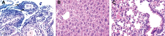
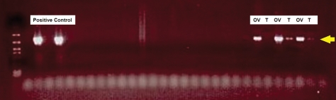
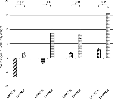
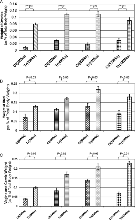
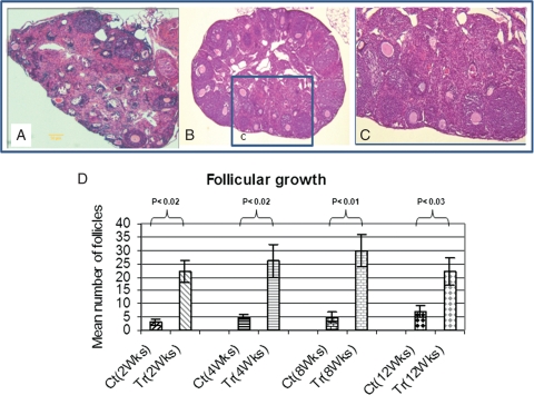
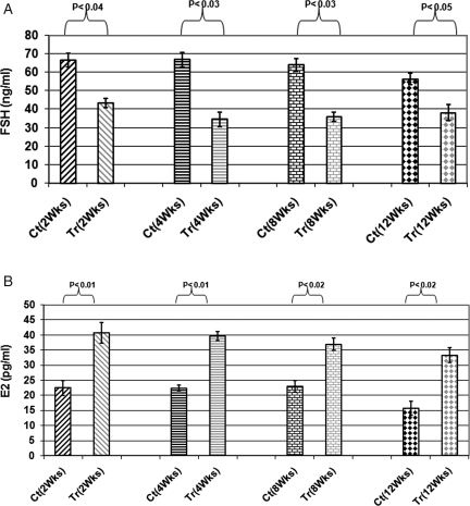
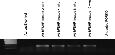
A homozygous missense mutation, C566T, in the follicle stimulation hormone receptor (FSHR) gene has been linked to premature ovarian failure. The disease leads to infertility in a normal karyotype female with an elevated follicle stimulating hormone (FSH) and decreased serum estrogen level. Female mice carrying mutated FSHR gene, called follitropin receptor knockout (FORKO), display similar phenotype and are sterile because of a folliculogenesis block at a primary stage. We investigated the effects of bilateral intra-ovarian injection of an adenovirus expressing a normal copy of human FSHR on the reproductive system of 6-10 weeks female FORKO mice. Ad-LacZ was injected directly into each ovary of the control group. Animals were sacrificed at 2, 4, 8 and 12 weeks post-injection and tissues collected for evaluation. Treated mice showed estrogenic changes in daily vaginal smear whereas control animals remained fixated in the diestrus stage. Histological evaluation showed on average 26 +/- 4 follicles/ovary in treated group with 8 +/- 2 follicles at the antral stage compared with only 5 +/- 2 with zero follicles at antral stage in Ad-LacZ control mice. There was no significant change in serum level of progesterone, however, estrogen level increased 2-3-fold (P < 0.02) and FSH decreased by up to 50% (P < 0.04) in treated animals. FSHR mRNA was detected in the ovaries of the treated group. In conclusion, intra-ovarian injection of an adenovirus expressing human FSHR gene is able to restore FSH responsiveness and reinitiate ovarian folliculogenesis as well as resume estrogen production in female FORKO mice. Ad-LacZ injections indicate the absence of systemic viral dissemination or germ line transmission of adenovirus DNA to offspring.


Figures

Figure 1
Figure 1
(A) Ad-LacZ was effective in infecting both granulosa (gr) cells and stromal (st) cells when delivered by direct intra-ovarian injection in nude mice (108 PFU/ovary), (o) indicates ooplasm; (B) Section of liver and (C) lung from nude mice treated by direct intraovarian injection with Ad-LacZ (103 PFU/ovary). They appear healthy with no LacZ (blue) expression. These photos were taken 4 weeks post-inoculation, H&E, ×400.
Figure 2
Figure 2
For safety and toxicity evaluation of direct intraovarian inoculation of adenovirus vectors, a group of mice were treated by unilateral (left) intraovarian injection of adenovirus. Multiple organ samples were collected to detect adenovirus DNA 1 week post-intra-ovarian injection. The presence of adenovirus was documented by PCR amplification of 714 bp fragment from the essential E4 region of the virus genome documented on 1% agarose gel. Adenoviral specific DNA was detectable only in the injected ovary (OV) and adjacent oviduct (T). Positive controls are shown on the left side. The following organs were run in the middle of the figure and were negative for E4 DNA: uterus, vagina, contralateral ovary, spleen, liver, lung, kidney and brain.
Figure 3
Figure 3
Changes of total body weight in treated (Tr) versus Control (Ct) animals at different time points. Control groups at Weeks 2 and 4 after treatment lost weight although body weight in treated group increased after 2 weeks and continue to increase by 12 weeks of experiment (P < 0.02).
Figure 4
Figure 4
(A) Ovaries weight of treated (Tr) versus control (Ct) animals. Weight considered as percentage of total body weight for each animal: as indicated weight of ovaries significantly increased at all time points of experiments (P < 0.02); (B) Uterus weight of treated (Tr) versus control (Ct) animals: weight considered as percentage of total body weight for each animal: uteri weight of treated animals increased at all time points of experiments up to 12 weeks (P < 0.04); (C) Vagina and Cervix weight of treated (Tr) versus control (Ct) animals: weight considered as percentage of total body weight for each animal. As indicated vagina and cervix weight increased in treated animals compared with control animals (P < 0.03).
Figure 5
Figure 5
Development of the ovary in Ad-LacZ control animal (A) compared with Ad-FSHR treated animal (B). Both the total number of follicles and the number of antral follicles are significantly higher in Ad-FSHR treated than Ad-LacZ control group. Histological evaluation showed on average 26 ± 4 follicles/ovary in treated group with 8 ± 2 follicles at the antral stage compared with only 5 ± 2 with zero follicles at antral stage in Ad-LacZ control mice. Photos were taken at the same magnification (×600); (C) is an area from treated ovary (inset marked—c) shown at 10× higher magnification; (D) Mean number of follicles at different time points of experiments. As shown the total numbers of follicles are significantly increased in treated versus control animals (P < 0.02). The most number of follicle increase observed at 8 weeks of treatment.
Figure 6
Figure 6
(A) Serum FSH of treated (Tr) versus Cotrol (Ct) animals at different time points. Ad-FSHR treated animals have shown 40–50% decrease in serum FSH compared with Ad-LacZ treated animals (P < 0.04); (B) Serum estrogen of treated (Tr) versus control (Ct) animals at different time points. Ad-FSHR treated animals have shown 2–3-fold increased in serum estrogen compared with Ad-LacZ treated at all time points of experiment (P < 0.02).
Figure 7
Figure 7
Expression of FSHR mRNA at different time points of experiment in treated animals and controls. As shown, there is no FSHR mRNA expression in Ad-LacZ treated and untreated controls. But FSHR mRNA was expressed in Ad-FSHR treated animals up to 12 weeks.

Similar articles

Cited by

References

    1. Aittomaki K, Lucerna JL, Pakarinen P, Sistonen P, Tapanainen JS, Gromoll J, Kaskikari R, Sankila EM, Lehvaslaiho H, Engel AR. Mutation in the follicle-stimulating hormone receptor gene causes hereditary hypergonadotropic ovarian failure. Cell. 1995;82:959–968. - PubMed
    1. Al-Hendy A, Lee EJ, Wang HQ, Copland JA. Gene therapy of uterine leiomyomas: adenovirus-mediated expression of dominant negative estrogen receptor inhibits tumor growth in nude mice. Am J Obstet Gynecol. 2004;191:1621–1631. - PubMed
    1. Al-Hendy A, Wang H, Salama SA. Towards Gene Therapy of Ovarian Failure: Intraovarian injected adenovirus successfully transduced granulosa and stromal but not germ cells. J Soc Gynecol Investig. 2005;12 Ref Type: Abstract.
    1. Allen LA, Achermann JC, Pakarinen P, Kotlar TJ, Huhtaniemi IT, Jameson JL, Cheetham TD, Ball SG. A novel loss of function mutation in exon 10 of the FSH receptor gene causing hypergonadotrophic hypogonadism: clinical and molecular characteristics. Hum Reprod. 2003;18:251–256. - PubMed
    1. Balla A, Danilovich N, Yang Y, Sairam MR. Dynamics of ovarian development in the FORKO immature mouse: structural and functional implications for ovarian reserve. Biol Reprod. 2003;69:1281–1293. - PubMed

Publication types