. 2010 Mar 19;285(12):8808-23.
doi: 10.1074/jbc.M109.075028. Epub 2010 Jan 19.

Proteolysis of mutant huntingtin produces an exon 1 fragment that accumulates as an aggregated protein in neuronal nuclei in Huntington disease

Affiliations

Proteolysis of mutant huntingtin produces an exon 1 fragment that accumulates as an aggregated protein in neuronal nuclei in Huntington disease

Christian Landles et al. J Biol Chem. .

Abstract

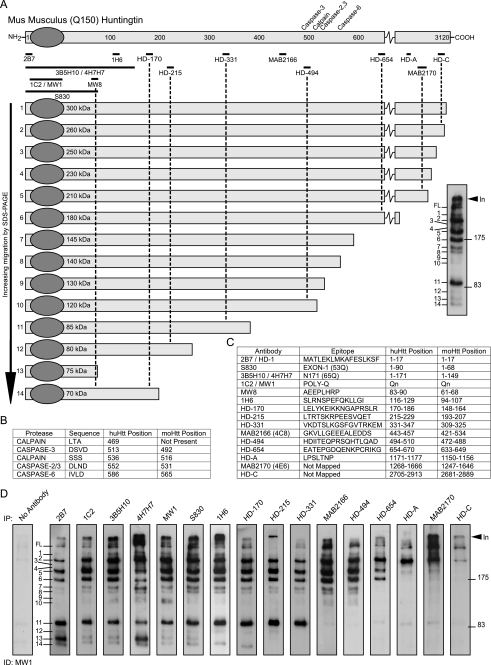
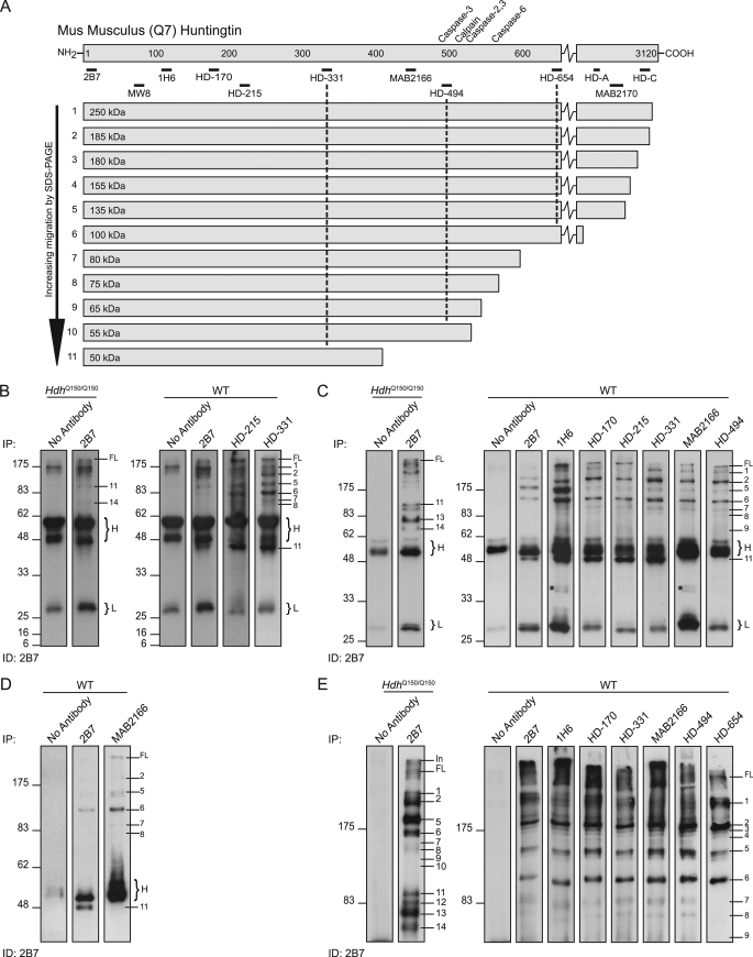
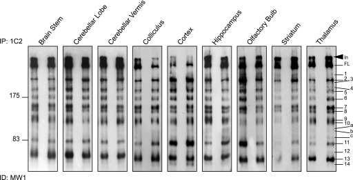
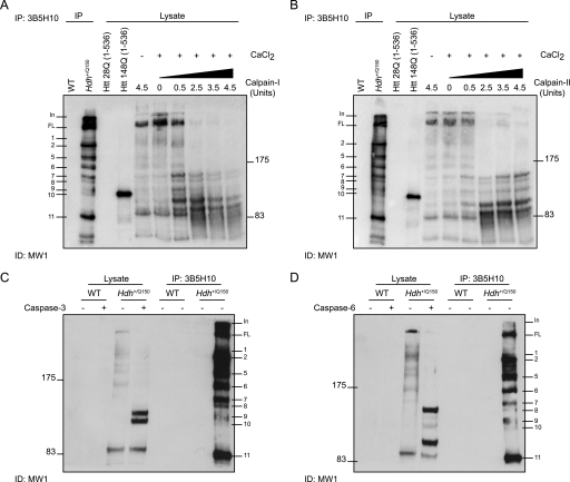
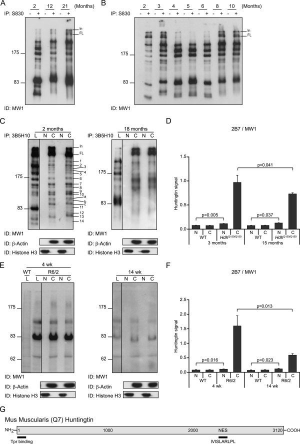
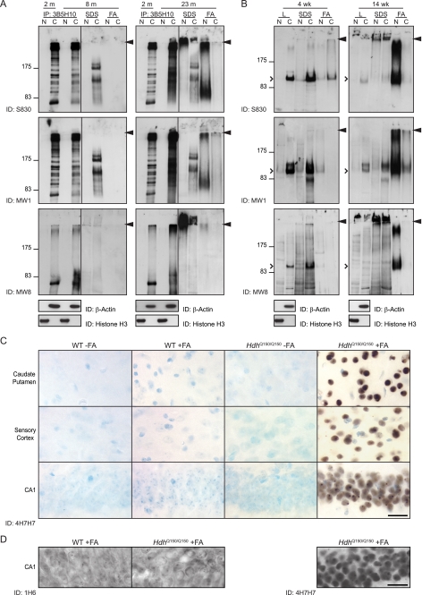
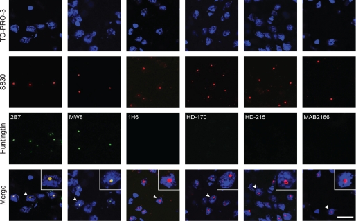
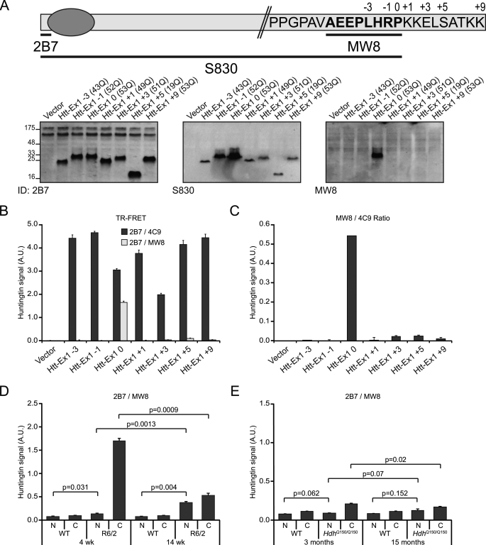
Huntingtin proteolysis has been implicated in the molecular pathogenesis of Huntington disease (HD). Despite an intense effort, the identity of the pathogenic smallest N-terminal fragment has not been determined. Using a panel of anti-huntingtin antibodies, we employed an unbiased approach to generate proteolytic cleavage maps of mutant and wild-type huntingtin in the HdhQ150 knock-in mouse model of HD. We identified 14 prominent N-terminal fragments, which, in addition to the full-length protein, can be readily detected in cytoplasmic but not nuclear fractions. These fragments were detected at all ages and are not a consequence of the pathogenic process. We demonstrated that the smallest fragment is an exon 1 huntingtin protein, known to contain a potent nuclear export signal. Prior to the onset of behavioral phenotypes, the exon 1 protein, and possibly other small fragments, accumulate in neuronal nuclei in the form of a detergent insoluble complex, visualized as diffuse granular nuclear staining in tissue sections. This methodology can be used to validate the inhibition of specific proteases as therapeutic targets for HD by pharmacological or genetic approaches.


Figures

FIGURE 1.
FIGURE 1.
Map of proteolytic cleavage sites that generate the N-terminal mutant Htt fragments present in HdhQ150 knock-in mouse brains. A, schematic representation of mutant mouse Htt protein showing the position of the previously mapped caspase and calpain sites (above) and the antibodies used for immunoprecipitation (below). Htt was immunoprecipitated with each antibody from 2-month Hdh+/Q150 whole brain extracts and immunoprobed with the N-terminal antibody MW1 after fractionation by 8% SDS-PAGE. Fourteen prominent N-terminal fragments can be detected (right panel). The schematic shows the order in which the fragments migrate on the SDS gel, their size (in kilodaltons based on the co-migration of size markers), and their predicted length (based on the location of the antibodies with which they have been immunoprecipitated). B, details of the previously mapped caspase and calpain sites. The position in both human (with 23Q) and mouse Htt (with 7Q) is given. C, details of the antibodies used to generate the protease cleavage map: the epitope that they recognize and its location in both human (23Q) and mouse (7Q) Htt. D, series of SDS-PAGE immunoblots from which the map is derived. Mutant Htt was immunoprecipitated from a single 2-month Hdh+/Q150 brain with the panel of antibodies as indicated and immunodetected with MW1. Results were confirmed using multiple additional 2-month brains. The control immunoprecipitation from a WT brain was routinely performed and showed no signal (supplemental Fig. S3). In = interface between stacking and resolving gel; FL = full-length protein.
FIGURE 2.
FIGURE 2.
Map of proteolytic cleavage sites that generate N-terminal WT Htt fragments. A, schematic representation of WT mouse Htt showing the position of the previously mapped caspase and calpain sites (above) and the antibodies used for immunoprecipitation (below). Htt was immunoprecipitated with each antibody from 2-month WT whole brain extracts and immunoprobed with 2B7. The schematic shows the order in which the fragments migrate on the SDS gel, their size (in kilodaltons based on the co-migration of size markers), and their predicted length (based on the location of the antibodies with which they have been immunoprecipitated). B–E, series of SDS-PAGE immunoblots from which the map is derived. B, right-hand panel: fragments were resolved by 12% SDS-PAGE to identify the smallest N-terminal fragments, however, Fragments 12–14, predicted to be 8–36 kDa in size, could not be detected. Left-hand panel: no antibody and HdhQ150/Q150 controls for comparison with the position of mutant Htt Fragments 11 and 14 indicated for reference. C, right-hand panel: fragments were resolved by 10% SDS-PAGE, and Fragment 11 can be detected with antibodies 2B7 to HD-331, but not MAB2166 and HD-494. Left-hand panel: no antibody and HdhQ150/Q150 controls for comparison with the position of mutant Htt Fragments 11, 13, and 14 indicated for reference. D, fragments were separated by 8% SDS-PAGE to better resolve Fragment 11 from the IgG heavy chain. E, right-hand panel: fragments were resolved by 8% SDS-PAGE. Fragments 7–9 can be detected with antibodies 2B7 to HD-494 and not HD-654. Many more N-terminal fragments of a size between that of Fragment 2 and the full-length protein can be detected on this gel. Left-hand panel: no antibody and HdhQ150/Q150 controls for comparison with the position of mutant Htt fragments indicated for reference. H = IgG heavy chain; L = IgG light chain; *, bands detected by the secondary anti-mouse antibody.
FIGURE 3.
FIGURE 3.
The spatial distribution of N-terminal mutant Htt fragments throughout the mouse brain. N-terminal Htt fragments were immunoprecipitated from the dissected brain regions of two Hdh+/Q150 mice at 2 months of age with antibody 1C2. Immunoprecipitates from both mice were fractionated by 8% SDS-PAGE and immunodetected with MW1. In = interface between stacking and resolving gel; FL = full-length protein.
FIGURE 4.
FIGURE 4.
Identification of Htt fragments generated by calpain or caspase proteolysis. A and B, the lysate from a 2-month-old Hdh+/Q150 mice was digested with increasing concentrations of calpain-I (A) or calpain-II (B) in the presence of CaCl2, fractionated by 8% SDS-PAGE, and immunoprobed with MW1. Control lanes included: (i) WT and Hdh+/Q150 lysates that had been immunoprecipitated with 3B5H10 (the same Hdh+/Q150 brain as digested with calpains), (ii) Htt fragments that terminate at amino acid 536 and contain 23Q or 148Q; and (iii) lysate digested with the maximum concentration (4.5 units) of calpain-I or -II in the absence of CaCl2. C and D, lysates from 2-month-old WT and Hdh+/Q150 mice were digested with or without either caspase-3 (C) or caspase-6 (D) and fractionated by 8% SDS-PAGE and immunoprobed with MW1. Control lanes were lysates that had been immunoprecipitated with 3B5H10 from the same WT and Hdh+/Q150 mice.
FIGURE 5.
FIGURE 5.
Detection of native N-terminal fragments of mutant Htt in the nucleus. A, N-terminal Htt fragments were immunoprecipitated with or without S830 from whole brain lysates of Hdh+/Q150 mice at 2, 12, and 21 months of age and immunodetected with MW1. The three smaller fragments (Fragments 12–14) cannot be detected in the lysates from mice aged 12 and 21 months. The resolution of the larger fragments is less distinct in the older mice. B, N-terminal Htt fragments were immunoprecipitated with or without S830 from Hdh+/Q150 whole brain lysates aged from 2 to 10 months and immunodetected with MW1. Fragments 12–14 are prominent up to and including 6 months of age but have diminished by 8 months. The variability in the migration of specific fragments between ages is a consequence of the difference in the CAG repeat length in the mice used in panels A and B. C, cytoplasmic and nuclear fractions were prepared from whole brains from two Hdh+/Q150 mice at each of 2 and 18 months of age and N-terminal Htt fragments were immunoprecipitated with 3B5H10 and immunodetected with MW1. These were compared with the fragments that were obtained by immunoprecipitation from whole brain lysates. Purity of the cytoplasmic and nuclear preparations was determined by immunoprobing with antibodies to β-actin and histone H3, respectively. D, TR-FRET (2B7-MW1) was performed on cytoplasmic and nuclear preparations from WT and HdhQ150/Q150 brains at 3 months and 15 months of age (n = 3/genotype/age). E, cytoplasmic and nuclear fractions were prepared from whole brains from two R6/2 mice at each of 4 and 14 weeks of age, the R6/2 transprotein was immunoprobed with MW1 and compared with the signal obtained from whole brain lysates. Purity of the cytoplasmic and nuclear preparations was determined by immunoprobing with antibodies to β-actin and histone H3, respectively. F, TR-FRET (2B7-MW1) was performed on cytoplasmic and nuclear preparations from WT and R6/2 brains at 4 and 14 weeks of age (n = 3/genotype/age). G, schematic of the position of putative nuclear export (NES) sites in the mouse Htt protein. N = nuclear, C = cytoplasm, L = lysates. In = interface between stacking and resolving gel; FL = full-length protein; Tpr = translocated promoter region.
FIGURE 6.
FIGURE 6.
Aggregated but not soluble N-terminal mutant Htt fragments are present in the nucleus. A, cytoplasmic and nuclear fractions were prepared from brains from Hdh+/Q150 mice aged 8 and 23 months. After centrifugation, the supernatant was immunoprecipitated with 3B5H10, and the pellet was sequentially solubilized in SDS and formic acid. Samples were fractionated on 8% SDS-PAGE gels alongside N-terminal fragments immunoprecipitated from brain lysates from Hdh+/Q150 mice aged 2 months for comparison. Western blots were probed with S830, MW1, and MW8 as indicated. B, cytoplasmic and nuclear fractions were prepared from the brains of R6/2 mice at 4 and 14 weeks of age. After centrifugation the supernatant was retained, and the pellet was sequentially solubilized in SDS and formic acid. Samples were fractionated by 8% SDS-PAGE, and Western blots were immunoprobed with S830, MW1, and MW8 as indicated. Arrowhead = interface between stacking and resolving gels. N = nuclear, C = cytoplasm; open arrowhead = R6/2 transprotein. C, diffuse nuclear Htt was immunodetected with the polyQ-specific antibody 4H7H7 in sections from HdhQ150/Q150 brains aged 6 months only after prior treatment with formic acid. Staining was absent from formic acid-treated and untreated WT sections. D, diffuse nuclear Htt was immunodetected with the polyQ-specific antibody 4H7H7 in sections from HdhQ150/Q150 brains aged 6 months after prior treatment with formic acid. Staining was absent from formic acid-treated HdhQ150/Q150 and WT sections when immunoprobed with 1H6. FA = formic acid. Scale bars: 20 μm.
FIGURE 7.
FIGURE 7.
Nuclear inclusions are immunodetected by antibodies that recognize epitopes N-terminal to 1H6. The exon 1 Htt antibody, S830, readily detects nuclear inclusions in HdhQ150/Q150 mouse brains aged 15 months. To determine which Htt fragments might be present in the nuclear inclusions, frozen sections were fixed in 4% paraformaldehyde, and immunohistochemistry was performed with antibodies that spanned the Htt protein and confocal microscopy used to look for co-localization with S830. The antibodies 2B7 and MW8 that detect exon 1 Htt epitopes, but not 1H6, HD-170, HD-215, and MAB216, were found to co-localize. Therefore, we could only detect epitopes that are present in the smallest N-terminal fragment (Fragment 13) in the nuclear inclusions. Scale bar: 20 μm.
FIGURE 8.
FIGURE 8.
The smallest N-terminal fragment is an exon 1 protein. A, schematic of the exon-1 Htt protein showing the C-terminal amino acid sequence, the positions of the antigens against which 2B7 and S830 were raised, and the epitope recognized by MW8 (39). The numbers above the amino acid sequence indicate the C termini of the various exon 1 mutant constructs. MW8 only recognizes the exon 1 protein in lysates prepared from the constructs when transiently expressed in COS-1 cells, whereas 2B7 and S830 detect exon 1 as well as all of the deletion and addition mutants. B and C, demonstration of the specificity of MW8 against the C terminus of exon 1. Htt specific signal (Htt levels) measured by TR-FRET (B) from cell lysates expressing the mutated exon 1 constructs using 2B7–4C9 (black bars) or 2B7-MW8 (gray bars). Normalization (C) of the MW8 signal with the 4C9 signal is shown. D and E, 2B7-MW8 TR-FRET of cytoplasmic and nuclear fractions from R6/2 mice aged 4 and 14 weeks (D) and HdhQ150/Q150 mice aged 3 and 15 months (E). For comparison, please see Fig. 5 (D and F), where the same tissue samples were analyzed by 2B7-MW1 TR-FRET.

Similar articles

Cited by

References

    1. Bates G. P., Harper P. S., Jones A. L. (eds) (2002) Huntington's Disease, Oxford University Press, Oxford
    1. Landles C., Bates G. P. (2004) EMBO Rep. 5, 958–963 - PMC - PubMed
    1. Harjes P., Wanker E. E. (2003) Trends Biochem. Sci. 28, 425–433 - PubMed
    1. Mangiarini L., Sathasivam K., Seller M., Cozens B., Harper A., Hetherington C., Lawton M., Trottier Y., Lehrach H., Davies S. W., Bates G. P. (1996) Cell 87, 493–506 - PubMed
    1. Li H., Li S. H., Cheng A. L., Mangiarini L., Bates G. P., Li X. J. (1999) Hum. Mol. Genet. 8, 1227–1236 - PubMed

Publication types