. 2010 Apr 2;285(14):10761-76.
doi: 10.1074/jbc.M109.053348. Epub 2010 Jan 10.

E-cadherin differentially regulates the assembly of Connexin43 and Connexin32 into gap junctions in human squamous carcinoma cells

Affiliations

E-cadherin differentially regulates the assembly of Connexin43 and Connexin32 into gap junctions in human squamous carcinoma cells

Souvik Chakraborty et al. J Biol Chem. .

Abstract

It is as yet unknown how the assembly of connexins (Cx) into gap junctions (GJ) is initiated upon cell-cell contact. We investigated whether the trafficking and assembly of Cx43 and Cx32 into GJs were contingent upon cell-cell adhesion mediated by E-cadherin. We also examined the role of the carboxyl termini of these Cxs in initiating the formation of GJs. Using cadherin and Cx-null cells, and by introducing Cx43 and Cx32, either alone or in combination with E-cadherin, our studies demonstrated that E-cadherin-mediated cell-cell adhesion was neither essential nor sufficient to initiate GJ assembly de novo in A431D human squamous carcinoma cells. However, E-cadherin facilitated the growth and assembly of preformed GJs composed of Cx43, although the growth of cells on Transwell filters was required to initiate the assembly of Cx32. Our results also documented that the carboxyl termini of both Cxs were required in this cell type to initiate the formation of GJs de novo. Our findings also showed that GJ puncta composed of Cx43 co-localized extensively with ZO-1 and actin fibers at cell peripheries and that ZO-1 knockdown attenuated Cx43 assembly. These findings suggest that the assembly of Cx43 and Cx32 into GJs is differentially modulated by E-cadherin-mediated cell-cell adhesion and that direct or indirect cross-talk between carboxyl tails of Cxs and actin cytoskeleton via ZO-1 may regulate GJ assembly and growth.


Figures

FIGURE 1.
FIGURE 1.
Isolation of A431D and A431DE clones expressing equal levels of connexin32 and connexin43. A431D and A431DE cells were infected with LXSN, LXSNCx32S, and LXSNCx43S and selected as described (see under “Materials and Methods”). A, localization of Cx43 and Cx32 by immunocytochemical analyses. A431D-43, A431DE-43, A431D-32, and A431DE-32 cells were immunostained for Cx43 and Cx32. Note that Cx43 assembles into GJs in the absence (−E-cad) and in the presence (+E-cad) of E-cadherin (E-cad), whereas Cx32 remains intracellular and fails to assemble. Note also that Cx43 forms larger GJs in E-cadherin-expressing cells. B, E-cadherin expression recruits α-catenin (α-cat) and β-catenin (β-cat) to cell-cell contact areas. A431D and A431DE cells were immunostained for α- and β-catenins. Note localization of both catenins at cell-cell contact areas in A431DE but not in A431D cells. C and D, expression level of Cx43 (C) and Cx32 (D) and E-cadherin, β-catenin, α-catenin, and occludin in parental (A431D and A431DE) cells and in cells infected with LXSN (A431D-NEO and A431DE-NEO), LXSNCx43S (A431D-43 and A432DE-43), and LXSNCx32S (A431D-32 and A431DE-32). Ten micrograms of total protein was resolved on 12% SDS-PAGE and immunoblotted for Cx32, Cx43, E-cadherin, β-catenin, α-catenin, and occludin. The blots were re-probed with β-actin to verify equal loading. Note that Cx43 (C) and Cx32 (D) are expressed only in clones infected with LXSNCx32S and LXSNCx43S, respectively. Note that expression of E-cadherin, α-catenin, and β-catenin is detected only in A431DE clones. Bar, 15 μm.
FIGURE 2.
FIGURE 2.
Detergent-solubility of connexin32 and connexin43 in A431D and A431DE cells. A, assembly of Cxs into GJs was examined in A431D-43, A431DE-43, A431D-32, and A431DE-32 cells upon in situ extraction with 1% Triton X-100 at 4 °C (see under “Materials and Methods”). Cells were immunostained for Cx32/Cx43 (green) and E-cadherin (red). Note the disappearance of intracellular, but not junctional, Cx43 and E-cadherin (E-cad) upon extraction. Note also the larger size of Cx43 puncta in A431DE-43 cells and their significant co-localization with E-cadherin under control and Triton X-100-extracted conditions. B, total (T), Triton X-100-soluble (S), and Triton X-100-insoluble (I) fractions from A431D-43, A431DE-43, A431D-32, and A431DE-32 were analyzed by Western blot analysis (see under “Materials and Methods”). Note that although Cx43 is detected in the both detergent-soluble and -insoluble fractions, Cx32 is detected only in the soluble fraction, and E-cadherin expression has no detectable effect on detergent solubility of Cx43. The blots were stripped and re-probed with anti-β-actin antibody as a loading control. C, connexin expression does not affect the detergent solubility of E-cadherin. Total, Triton X-100-soluble and -insoluble fractions from A431DE, A431DE-43, and A431DE-32 cells were analyzed by Western blot analysis (see under “Materials and Methods”). Note that similar levels of E-cadherin are detected in the detergent-insoluble fraction in all cells. The blots were stripped and re-probed with anti-β-actin to verify equal loading. Bar, 10 μm.
FIGURE 3.
FIGURE 3.
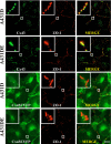
E-cadherin expression does not affect trafficking and subcellular localization of connexin43 and connexin32. A and B, cell-surface biotinylation of Cx43 and Cx32 in A431D and A431DE cells. Confluent A431D-43 and A431DE-43 (A) and A431D-32 and A431DE-32 cells (B) were biotinylated with sulfo-NHS-SS-biotin at 4 °C, and biotinylated Cxs from 100 μg of total protein were detected by streptavidin pulldown followed by Western blotting (see under “Materials and Methods”). As an input, 10 μg of total protein was used and probed for Cx43 or Cx32. Note that in both A431D-43 and A431DE-43 cells (A; labeled as + Biotin) and A431D-32 and A431DE-32 cells (B; labeled as + Biotin), similar levels of Cx43 and Cx32 were detected in the streptavidin Pulldown lanes, respectively. A nonbiotinylated dish (labeled as − Biotin) was used as a negative control. C and D, co-localization of Cx43 and Cx32 with EEA-1 in A431D-43 and A431DE-32 and A431DE-43 and A431DE-32 cells. Note the co-localization of both Cx43 (C) and Cx32 (D, green) with EEA1 (red) in all cells. Note extensive co-localization of Cx32 with EEA1. The boxed region is magnified and shown in the inset. Bar, 15 μm.
FIGURE 4.
FIGURE 4.
Connexin32 assembles into gap junctions in A431D-32 and A431DE-32 cells grown on Transwell filters. A, A431D-32 and A431DE-32 cells, seeded on clear Transwell filters, were grown for 5 days and immunostained for Cx32 (green) and ZO-1 (red) as described under “Materials and Methods.” Note the formation of distinct GJ puncta at the cell-cell contact sites as delineated by ZO-1. Note lack of significant difference in the size and number of Cx32 puncta at the cell-cell contact sites between A431D-32 and A431DE-32 cells. Note also that the GJ puncta remain detergent-resistant (B). Bar, 15 μm.
FIGURE 5.
FIGURE 5.
Carboxyl-terminally truncated untagged and EYFP-tagged chimeras of Cx43 fail to assemble into gap junctions. Polyclonal cultures of A431D and A431DE cells expressing Cx43Δ233, Cx43Δ233-EYFP, Cx43Δ257, Cx43Δ257-EYFP, Cx43Δ363, Cx43Δ363EYFP, Cx43, and Cx43-EYFP were grown on glass coverslips. Cells expressing untagged Cx43 and truncated mutants were immunostained with polyclonal antibody raised against amino acid residues 252–271 of Cx43 (see under “Materials and Methods”). Note that all truncated and EYFP-tagged chimeras of Cx43 remain intracellular, whereas untagged Cx43 is assembled into GJs in cadherin-null (A431D) and E-cadherin-expressing (A431DE) cells. Note that the localization pattern of untagged versus EYFP-tagged full-length and mutant Cx chimeras appears to be different partly due to the brighter diffuse EYFP fluorescence of tagged Cx mutants compared with untagged Cx mutants, which were detected immunocytochemically. All EYFP-tagged and carboxyl-terminally truncated mutants are lost upon in situ extraction (data not shown). Bar, 10 μm.
FIGURE 6.
FIGURE 6.
Carboxyl-terminally truncated untagged and EYFP-tagged chimera of Cx32 fail to assemble into gap junctions. Polyclonal cultures of A431D and A431DE cells expressing Cx32Δ220 and Cx32Δ220-EYFP were grown on Transwell filters and were extracted in situ with 1% Triton X-100 (see under “Materials and Methods”). Note that both Cx32Δ220 and Cx32Δ220-EYFP remain as scattered intracellular puncta or aggregates that are lost upon detergent extraction. Note that the localization pattern of untagged versus EYFP-tagged full-length and mutant Cx chimeras appears to be different partly due to the brighter diffuse EYFP fluorescence of tagged Cx mutants compared with untagged Cx mutants, which were detected immunocytochemically. Bar, 20 μm.
FIGURE 7.
FIGURE 7.
Gap junction assembly-dependent co-localization of connexin43 and ZO-1. Polyclonal cultures of A431D-43 and A431DE-43 cells, seeded on glass coverslips, were fixed, permeabilized, and immunostained for Cx43 (green) and ZO-1 (red), whereas those of Cx43-EYFP were immunostained for ZO-1 (see under “Materials and Methods”). The insets show the magnified images of co-localized Cx43 and ZO-1 puncta. Note that only untagged Cx43 is assembled into GJs and is co-localized with ZO-1. Note also that ZO-1 and Cx43-EYFP do not co-localize significantly. Bar, 10 μm.
FIGURE 8.
FIGURE 8.
Connexin43 and ZO-1 puncta are formed on F-actin at cell-cell contact. A431D-43 (top row) and A431DE-43 (bottom row) cells, seeded on glass coverslips, were immunostained for Cx43 (green) and ZO-1 (red). F-actin (blue) was detected using Alexa Fluor 350-conjugated phalloidin. The magnified images of co-localized Cx43, ZO-1, and F-actin are shown on the right, and the key describing the corresponding images in color is shown in the middle. Note that significant co-localization of Cx43, ZO-1, and F-actin is observed only at puncta and at the regions of cell-cell contact. Bar, 5 μm.
FIGURE 9.
FIGURE 9.
E-cadherin delays gap junction disassembly. A431D-43 and A431DE-43 cells, seeded on glass coverslips, were treated with cytochalasin B at 1 μg/ml (labeled CT-B) for 1–6 h and immunostained for Cx43 and ZO-1. F-actin was detected with Alexa Fluor 350-conjugated phalloidin. Note that co-localized GJ puncta of Cx43 and ZO-1 are disrupted/internalized within 1 h in A431D-43 cells, whereas no detectable disruption/internalization is noticed in A431DE-43 cells within 1 h and even until 6 h (supplemental Fig. S11). Bar, 5 μm. CONT, control.
FIGURE 10.
FIGURE 10.
ZO-1 knockdown disrupts gap junction assembly. A431D-43 and A431DE-43 cells were either mock-transfected with Oligofectamine (labeled MOCK) or transfected with 130 nm ZO-1 SMARTpool siRNA (labeled siRNA) or DY-547-labeled control RISC-free siGLO RNA (labeled SiGLO) as described under “Materials and Methods.” A, expression of ZO-1 and Cx43 was determined by immunoblotting 10 μg of total protein. The blots were re-probed with anti-β-actin antibody to verify equal loading. Note that ZO-1 expression is reduced significantly in cells transfected with ZO-1 SMARTpool siRNAs. B, cells were immunostained for Cx43 and ZO-1 together. Note that ZO-1 staining (red) is significantly reduced in ZO-1 SMART pool siRNA-transfected cells compared with MOCK-transfected cells. Note significant loss of gap junction puncta (green), with a concomitant increase in the intracellular Cx43 staining, in ZO-1 siRNA-transfected cells compared with MOCK-transfected cells. Bar, 10 μm.

Similar articles

Cited by

References

    1. Beyer E. C., Berthoud V. M. (2009) in Connexins: A Guide (Harris A., Locke D. eds) pp. 3–26, Springer, Humana Press, NY
    1. Laird D. W. (2006) Biochem. J. 394, 527–543 - PMC - PubMed
    1. Sosinsky G. E., Nicholson B. J. (2005) Biochim. Biophys. Acta 1711, 99–125 - PubMed
    1. Falk M. M., Baker S. M., Gumpert A. M., Segretain D., Buckheit R. W., 3rd (2009) Mol. Biol. Cell 20, 3342–3352 - PMC - PubMed
    1. Gaietta G., Deerinck T. J., Adams S. R., Bouwer J., Tour O., Laird D. W., Sosinsky G. E., Tsien R. Y., Ellisman M. H. (2002) Science 296, 503–507 - PubMed

Publication types

MeSH terms