. 2010 Mar 19;285(12):9147-60.
doi: 10.1074/jbc.M109.082008. Epub 2010 Jan 10.

Pannexin1 and pannexin3 delivery, cell surface dynamics, and cytoskeletal interactions

Affiliations

Pannexin1 and pannexin3 delivery, cell surface dynamics, and cytoskeletal interactions

Ruchi Bhalla-Gehi et al. J Biol Chem. .

Abstract

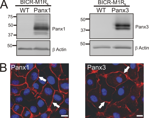
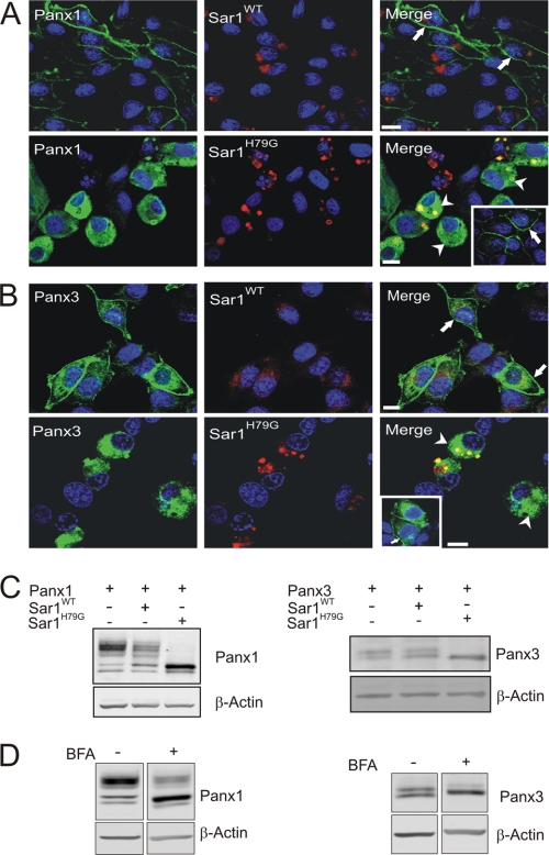
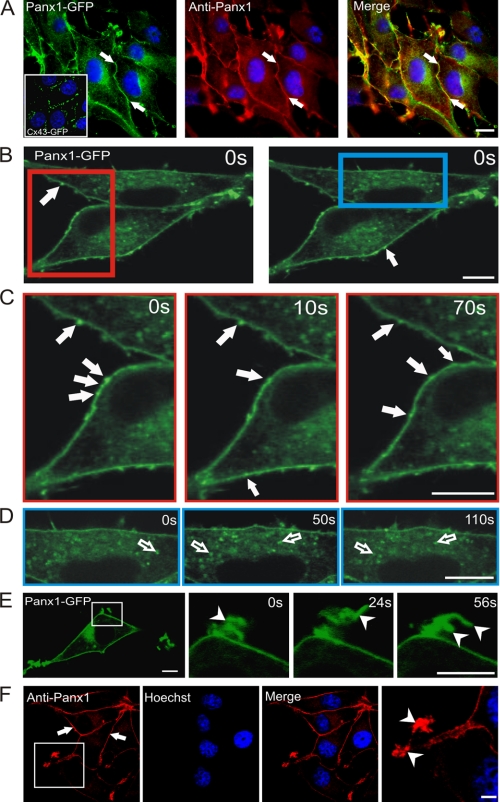
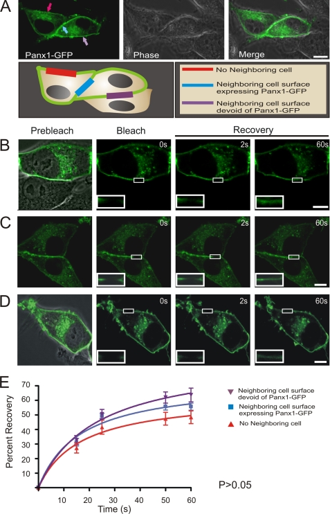
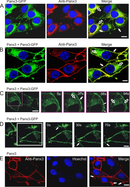
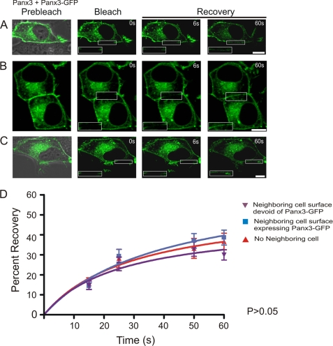
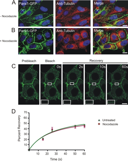
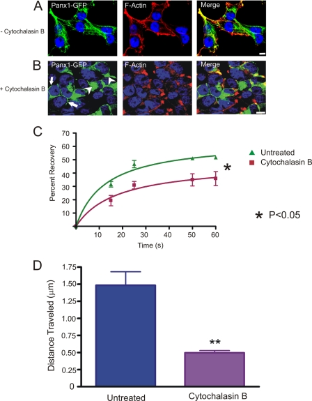
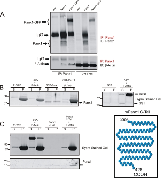
Pannexins (Panx) are a class of integral membrane proteins that have been proposed to exhibit characteristics similar to those of connexin family members. In this study, we utilized Cx43-positive BICR-M1R(k) cells to stably express Panx1, Panx3, or Panx1-green fluorescent protein (GFP) to assess their trafficking, cell surface dynamics, and interplay with the cytoskeletal network. Expression of a Sar1 dominant negative mutant revealed that endoplasmic reticulum to Golgi transport of Panx1 and Panx3 was mediated via COPII-dependent vesicles. Distinct from Cx43-GFP, fluorescence recovery after photobleaching studies revealed that both Panx1-GFP and Panx3-GFP remained highly mobile at the cell surface. Unlike Cx43, Panx1-GFP exhibited no detectable interrelationship with microtubules. Conversely, cytochalasin B-induced disruption of microfilaments caused a severe loss of cell surface Panx1-GFP, a reduction in the recoverable fraction of Panx1-GFP that remained at the cell surface, and a decrease in Panx1-GFP vesicular transport. Furthermore, co-immunoprecipitation and co-sedimentation assays revealed actin as a novel binding partner of Panx1. Collectively, we conclude that although Panx1 and Panx3 share a common endoplasmic reticulum to Golgi secretory pathway to Cx43, their ultimate cell surface residency appears to be independent of cell contacts and the need for intact microtubules. Importantly, Panx1 has an interaction with actin microfilaments that regulates its cell surface localization and mobility.


Figures

FIGURE 1.
FIGURE 1.
Panx1 and Panx3 are capable of trafficking to the cell surface in BICR-M1Rk cells. BICR-M1Rk cells were engineered to stably express Panx1 or Panx3. Immunoblotting with affinity-purified antibodies (anti-Panx1 and anti-Panx3) revealed multiple banding profiles of Panx1 (∼41–48 kDa) and Panx3 (∼41–43 kDa) (A). β-Actin was used as a protein loading control (A). Immunolabeling of Panx1 and Panx3 (B) identified that both pannexins trafficked and localized to the cell surface (arrows). Nuclei are stained with Hoechst 33342 (blue). Bars, 10 μm. Results shown are representative of three independent experiments.
FIGURE 2.
FIGURE 2.
Trafficking of Panx1 and Panx3 was disrupted in the presence of a dominant-negative Sar1 mutant. BICR-M1Rk cells expressing Panx1 or Panx3 together with Sar1WT or Sar1H79G were immunolabeled for Panx1 (A) or Panx3 (B). Both Panx1 and Panx3 were capable of trafficking and localizing to the cell surface in the presence of Sar1WT (A and B, filled arrows). Expression of Sar1H79G resulted in Panx1 and Panx3 being retained in an ER-like compartment (A and B, arrowheads); however, when cells expressed Panx1 or Panx3 without expressing Sar1H79G in the same cellular environment, both Panx1 and Panx3 trafficked to the plasma membrane (A and B, insets, arrows). Western blotting of Panx1 and Panx3 in the presence of Sar1H79G (C) or after long term BFA treatment (19 h) (D) revealed an accumulation of the high mannose species of Panx1 and Panx3, with a noticeable reduction in the higher molecular weight glycosylation species (C and D). Nuclei are stained with Hoechst 33342 (blue). Bars, 10 μm. Results shown are representative of three independent experiments.
FIGURE 3.
FIGURE 3.
Panx1 was localized to multiple sites, compartments, and microdomains. Immunolabeling of Panx1-GFP with an anti-Panx1 antibody revealed its localization at the cell surface (A, filled arrows) in a pattern that was distinct from Cx43-GFP (A, inset). Regions of interest from live BICR-M1Rk cells expressing Panx1-GFP (B, red and blue rectangles) were chosen for live imaging and imaged at t = 0, 10, 50, 70, and 110 s (C and D). Rapid time lapse imaging revealed that Panx1-GFP is distributed primarily in a uniform pattern, whereas mobile bright fluorescent clusters could be identified at the cell surface (B and C, filled arrows) and within the cell (D, unfilled arrows). Panx1-GFP was clearly localized to dynamic plasma membrane protrusions (E, arrowheads) that were evident in BICR-M1Rk cells expressing untagged Panx1 (F, arrowheads). Nuclei in A and F are stained with Hoechst 33342 (blue). Bars, 10 μm. Representative of three independent experiments.
FIGURE 4.
FIGURE 4.
Panx1-GFP is highly mobile at all plasma membrane locations. Panx1-GFP was localized to three distinct plasma membrane domains of BICR-M1Rk cells, as depicted by the schematic diagram (A). Fluorescent images of Panx1-GFP were superimposed with DIC images to highlight the microenvironment surrounding the cell being analyzed (A–D). Selected cell regions where Panx1-GFP was localized at the three distinct plasma membrane domains were photobleached, and fluorescence recovery back into the photobleached areas was assessed and normalized over 60 s (B–D). Panx1-GFP recovery within the photobleached area was not significantly different (p > 0.05) among all three domains (E). Bars, 10 μm. n = 6–9 per plasma membrane domain collected from three independent experiments.
FIGURE 5.
FIGURE 5.
Delivery of Panx3-GFP to the cell surface. Wild-type BICR-M1Rk cells engineered to express Panx3-GFP (A) or both Panx3 and Panx3-GFP (B) were immunolabeled with anti-Panx3 antibody. Panx3-GFP was retained mainly in an ER-like pattern (A and B, unfilled arrows) with some evidence of a cell surface distribution (A and B, filled arrows), whereas co-expression of Panx3 appeared to increase the cell surface population of Panx3-GFP (B). Rapid time lapse imaging of live BICR-M1Rk cells co-expressing Panx3-GFP and Panx3 revealed that Panx3-GFP was distributed primarily in a uniform pattern with notable mobile fluorescent clusters at the cell surface (C, filled arrows) and within the cell (C, unfilled arrow). Localization of Panx3-GFP to plasma membrane protrusions (D, arrows) was similar to that found in cells expressing only Panx3 (E, arrows). Nuclei in A, B, and E are stained with Hoechst 33342 (blue). Bars, 10 μm. Results shown are representative of three independent experiments.
FIGURE 6.
FIGURE 6.
Panx3-GFP is highly mobile at all plasma membrane domains. Panx3-GFP was localized to three distinct plasma membrane domains of BICR-M1Rk cells co-expressing Panx3 (A–C). Selected cell surface regions containing Panx3-GFP were photobleached, and fluorescence recovery back into the photobleached areas was assessed and normalized over the time course of 60 s. The percentage of Panx3-GFP recoverable fraction was not found to be significantly different among all three plasma membrane domains examined (p > 0.05) (D). Bars, 10 μm. n = 12–25 per plasma membrane domain collected from three independent experiments.
FIGURE 7.
FIGURE 7.
The cell surface population of Panx1-GFP is insensitive to nocodazole treatment. Untreated (A) or nocodazole-treated (B) Panx1-GFP-expressing BICR-M1Rk cells were immunolabeled for tubulin. As expected, nocodazole treatment collapsed tubulin into paranuclear regions (A and B); however, the distribution profile of Panx1-GFP at the cell surface and in the intracellular compartments (A and B) remained relatively unchanged with collapsed tubulin (B). FRAP analysis in presence of nocodazole revealed that Panx1-GFP was able to recover into the photobleached area, and the percentage of recoverable fraction was not significantly different from the untreated cells (C and D). Bars, 10 μm. n = 5–10 per plasma membrane domains, data collected over four independent repeats.
FIGURE 8.
FIGURE 8.
Effect of cytochalasin B on Panx1-GFP. Untreated (A) or cytochalasin B-treated (B) Panx1-GFP-expressing BICR-M1Rk cells were labeled with phalloidin for F-actin localization. As expected, cytochalasin B caused the redistribution of F-actin from the cell surface (A) to the paranuclear region (B). The collapse of F-actin microfilaments coincided with the intracellular accumulation of Panx1-GFP (B, arrowheads), whereas a small population of Panx1-GFP remained evident at the cell surface (B, arrows). FRAP analysis in the presence of cytochalasin B treatment revealed that the cell surface population of Panx1-GFP was significantly impaired from entering the photobleached area (p < 0.05) (C) n = 3. Quantification of the total distance traveled by Panx1-GFP carrying vesicles within the same field of cells analyzed before and after the cytochalasin B treatment indicated a significant (p < 0.05) reduction in vesicle mobility in cytochalasin B-treated cells (D). Bars, 10 μm. Results shown are representative of five independent experiments.
FIGURE 9.
FIGURE 9.
F-actin binds Panx1 at the carboxyl terminus. Wild type (WT) or Panx1- or Panx1-GFP-expressing BICR-M1Rk cells were lysed and subjected to immunoprecipitation (IP) for Panx1 prior to immunoblotting (IB) the immunoprecipitates and cell lysates for Panx1 or β-actin. β-Actin co-immunoprecipitated with Panx1 and Panx1-GFP (A). Monomeric actin was polymerized into F-actin, incubated with either GST fusion protein containing the carboxyl-terminal tail of Panx1 (B) or the carboxyl-terminal tail of Panx1 alone (C) and separated into supernatant or pellet fractions (denoted by S and P, respectively) prior to immunoblotting for Panx1. Panx1 was found to co-sediment with F-actin in the pellet fractions (B and C). Parallel gels were stained with Sypro gel stain, and BSA and GST were used as controls in the co-sedimentation assays. Results shown are representative of three independent experiments.

References

    1. Panchin Y., Kelmanson I., Matz M., Lukyanov K., Usman N., Lukyanov S. (2000) Curr. Biol. 10, R473–R474 - PubMed
    1. Willecke K., Eiberger J., Degen J., Eckardt D., Romualdi A., Güldenagel M., Deutsch U., Söhl G. (2002) Biol. Chem. 383, 725–737 - PubMed
    1. Baranova A., Ivanov D., Petrash N., Pestova A., Skoblov M., Kelmanson I., Shagin D., Nazarenko S., Geraymovych E., Litvin O., Tiunova A., Born T. L., Usman N., Staroverov D., Lukyanov S., Panchin Y. (2004) Genomics 83, 706–716 - PubMed
    1. Boassa D., Ambrosi C., Qiu F., Dahl G., Gaietta G., Sosinsky G. (2007) J. Biol. Chem. 282, 31733–31743 - PubMed
    1. Penuela S., Bhalla R., Gong X. Q., Cowan K. N., Celetti S. J., Cowan B. J., Bai D., Shao Q., Laird D. W. (2007) J. Cell Sci. 120, 3772–3783 - PubMed

Publication types