. 2010 Feb 15;123(Pt 4):567-77.
doi: 10.1242/jcs.056432. Epub 2010 Jan 19.

Vinculin regulates cell-surface E-cadherin expression by binding to beta-catenin

Affiliations

Vinculin regulates cell-surface E-cadherin expression by binding to beta-catenin

Xiao Peng et al. J Cell Sci. .

Abstract

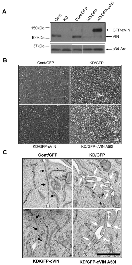
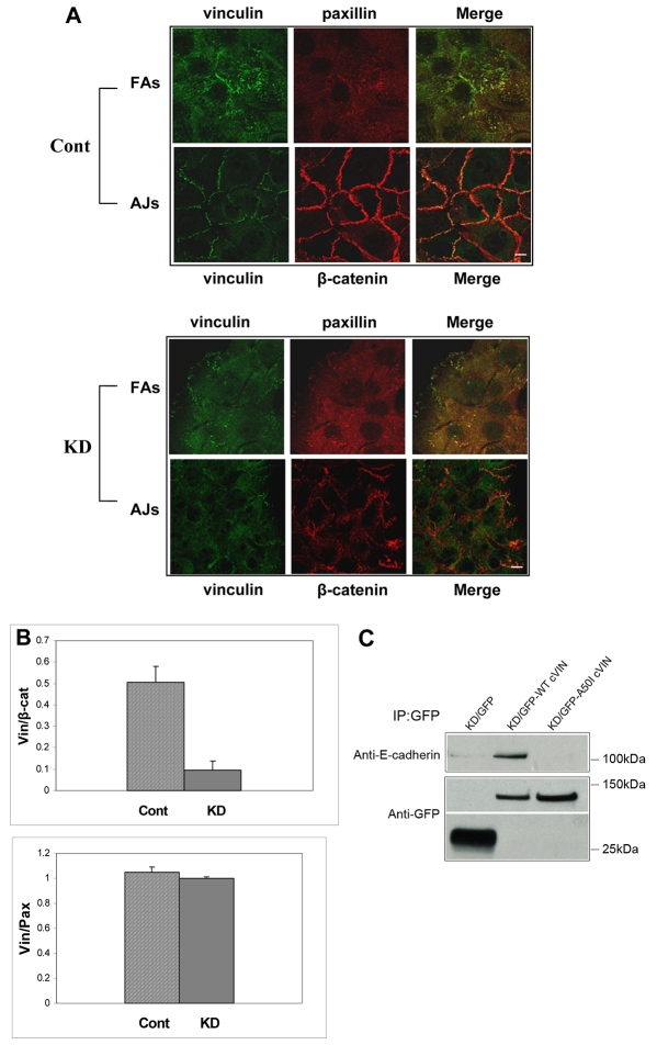
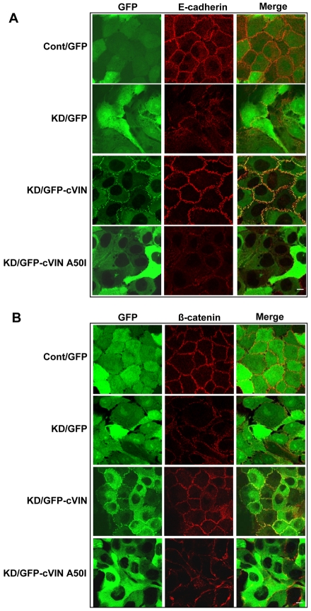
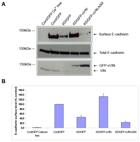
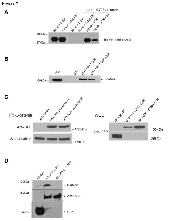
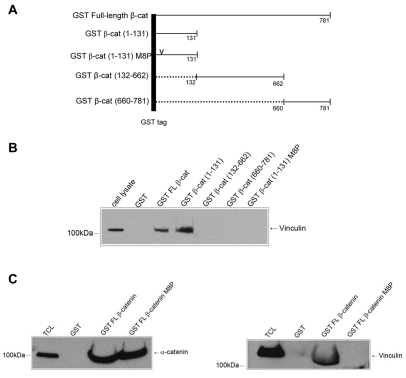
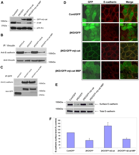
Vinculin was identified as a component of adherens junctions 30 years ago, yet its function there remains elusive. Deletion studies are consistent with the idea that vinculin is important for the organization of cell-cell junctions. However, this approach removes vinculin from both cell-matrix and cell-cell adhesions, making it impossible to distinguish its contribution at each site. To define the role of vinculin in cell-cell junctions, we established a powerful short hairpin-RNA-based knockdown/substitution model system that perturbs vinculin preferentially at sites of cell-cell adhesion. When this system was applied to epithelial cells, cell morphology was altered, and cadherin-dependent adhesion was reduced. These defects resulted from impaired E-cadherin cell-surface expression. We have investigated the mechanism for the effects of vinculin and found that the reduced surface E-cadherin expression could be rescued by introduction of vinculin, but not of a vinculin A50I substitution mutant that is defective for beta-catenin binding. These findings suggest that an interaction between beta-catenin and vinculin is crucial for stabilizing E-cadherin at the cell surface. This was confirmed by analyzing a beta-catenin mutant that fails to bind vinculin. Thus, our study identifies vinculin as a novel regulator of E-cadherin function and provides important new insight into the dynamic regulation of adherens junctions.


Figures

Fig. 1.
Fig. 1.
Stable inhibition of vinculin in breast epithelial cells by RNA interference. (A) MCF10a cells were infected with GFP, GFP-tagged chick vinculin (GFP-cVIN) or a mutant version of vinculin (GFP-cVIN A50I), and then infected a second time with either an empty vector (Cont) or a vector encoding a shRNA targeting human vinculin (KD). Lysates were harvested from cell lines stably expressing these plasmids, and western blot analysis was performed using antibodies against vinculin (VIN) or the p34-Arc subunit of the Arp2/3 complex as a loading control. All of the samples were prepared and analyzed in the same experiment; the white line indicates where the western blot was cut to remove some irrelevant samples. (B) Phase-contrast images of MCF10a cells stably expressing the indicated constructs. Scale bar: 100 μm. (C) Transmission EM micrographs of MCF10a cells stably expressing the indicated constructs. Scale bar: 1 μm. The black arrows indicate adherens junctions; the white arrows indicate regions between cells where adherens junctions are disrupted.
Fig. 2.
Fig. 2.
Inhibition of vinculin preferentially depleted vinculin from cell-cell adhesion but not cell-matrix adhesions. (A) MCF10a cells stably expressing the indicated constructs were stained with antibodies against the adherens junction (AJ) marker β-catenin or the focal adhesion (FA) marker (paxillin). Using confocal microscopy different focal planes were isolated and photographed. Scale bar: 10 μm. (B) Ratiometric analysis of vinculin, β-catenin (β-cat) and paxillin (Pax) fluorescence intensity at adherens junctions or focal adhesions. Fluorescence intensity of vinculin staining was expressed as a ratio of β-catenin or paxillin fluorescence intensity at the same individual cell—cell contacts. Data are means ± s.e.m. (n=30 and are representative of three independent experiments). (C) GFP, wild-type GFP-cVIN or GFP-cVIN A50I were immunoprecipitated, washed, fractionated by SDS-PAGE, and immunoblotted with an antibody against E-cadherin. The blot was stripped and re-probed for the precipitated levels of each GFP protein.
Fig. 3.
Fig. 3.
Phenotypes of vinculin knockdown cells are not due to altered cell-matrix adhesions. Cells were plated on coverslips coated with saturating concentrations of (A) fibronectin, (B) collagen, or (C) sub-saturating concentrations of fibronectin. The coverslips were washed, and the number of adherent cells per field of view (FOV) was counted. Thirty-five FOVs from three independent experiments were quantified and used to calculate the percent of adherent cells. Each bar represents the mean ± s.e.m.
Fig. 4.
Fig. 4.
Adhesion to cadherin extracellular domains is impaired in cells with reduced vinculin levels. For assessing homophilic ligation, dishes were coated with an anti-Fc antibody, washed, coated with human E-cadherin extracellular domains fused to Fc, and blocked with BSA. Cells expressing the indicated constructs were lifted, incubated in the coated dishes, and washed extensively with agitation. Cells were scored as adherent if they were phase gray. The mean number of cells that adhered per field of view ± s.e.m. was calculated by the means of three independent experiments. A sample of the knockdown cells rescued with wild-type vinculin was preincubated with the function-blocking antibody DECMA (+DECMA) prior to plating.
Fig. 5.
Fig. 5.
E-cadherin localization to adherens junctions is altered in cells with low levels of vinculin. MCF10a cells stably expressing the indicated constructs (green) were analyzed by immunofluorescence, following staining with antibodies against (A) E-cadherin (red) or (B) β-catenin (red). Representative images are shown. Scale bar: 10 μm.
Fig. 6.
Fig. 6.
Vinculin regulates surface expression of E-cadherin. (A) Surface E-cadherins were biotinylated. After the reaction was quenched, cells were lysed and the biotinylated proteins were recovered with streptavidin beads. Surface E-cadherin or whole-cell lysates were analyzed by SDS-PAGE and immunoblotted with E-cadherin antibody. In the control experiment (Cont/GFP Ca2+ free), cells were first incubated with EGTA to disassemble adherens junctions. (B) The amount of E-cadherin on the cell surface was quantified using densitometry. The mean level of E-cadherin on the cell surface in the control cells was set to 100%. Data presented are the mean ± s.e.m. from three independent experiments.
Fig. 7.
Fig. 7.
Substitution of A50I blocks β-catenin binding to vinculin, but not α-catenin. (A) In vitro binding analysis of α-catenin interaction with wild-type vinculin (His VIN 1-398) or mutant vinculin (His VIN 1-398 A50I) head sequence encompassing amino acids 1-398. Purified His-VIN or His-VIN A50I were incubated with purified GST or GST-tagged full-length α-catenin at room temperature for 30 minutes, and then recovered using glutathione beads. The beads were washed, after which the bound peptides were separated by SDS-PAGE and immunoblotted with His antibodies. Purified wild-type and mutant peptides were included to indicate the positions of the purified proteins. All of the samples were prepared and analyzed in the same experiment. The white line indicates where the western blot was cut to remove some irrelevant lanes. (B) Pull-down analysis of α-catenin interaction with wild-type vinculin (GST VIN 1-398) or mutant vinculin (GST VIN 1-398 A50I) head sequence. Purified GST-VIN or GST-VIN A50I attached to the glutathione beads were incubated with MDCK cell lysates. The beads were then washed, and subjected to western blotting analysis with an antibody against α-catenin. Total cell lysate (TCL) was also included. (C) Co-immunoprecipitation of α-catenin with GFP-cVIN or GFP-cVIN A50I. MCF10a cells expressing the indicated construct were lysed, α-catenin was immunoprecipitated, and the immunoprecipitates were recovered using protein A Sepharose. The bound proteins were fractionated by SDS-PAGE and immunoblotted with an antibody against GFP. The blot was stripped and re-probed for α-catenin. The expression levels of GFP-cVIN or GFP-cVIN A50I in the whole cell lysate (WCL) were also shown. (D) Co-immunoprecipitation of GFP-cVIN or GFP-cVIN A50I with β-catenin. MCF10a cells expressing the indicated construct were lysed and immunoprecipitated with a GFP antibody. The proteins were then recovered using protein G Sepharose. The beads were washed and fractionated by SDS-PAGE, and immunoblotted with an antibody against β-catenin. The blot was stripped and re-probed for GFP.
Fig. 8.
Fig. 8.
Substitution of M8P in β-catenin blocks vinculin binding. (A) Schematic of β-catenin truncations and the M8P mutation (v). (B) Pull-down analysis to map vinculin binding site on β-catenin. Purified GST protein or GST-tagged β-catenin (β-cat) fragments attached to the glutathione beads were incubated with MDCK cell lysates. The beads were washed and fractionated by SDS-PAGE and immunoblotted with vinculin. (C) Pull down analysis of α-catenin or vinculin interaction with wild-type β-catenin (GST FL β-catenin) or mutant β-catenin (GST FL β-catenin M8P). Purified GST FL β-catenin or GST FL β-catenin M8P attached to the glutathione beads were incubated with MDCK cell lysates. The beads were then washed and fractionated by SDS-PAGE, and immunoblotted with an antibody against α-catenin or vinculin. Total cell lysate (TCL) was also included.
Fig. 9.
Fig. 9.
Vinculin binding to β-catenin is required for cell surface E-cadherin expression. (A) Representative blots of MCF10a cells stably expressing the indicated constructs. Lysates were harvested from cell lines, and western blot analysis was performed using antibodies against vinculin (VIN) or the p34-Arc subunit of the Arp2/3 complex as a loading control (βKD, β-catenin knockdown; mβ-cat, mouse β-catenin). (B) Co-immunoprecipitation of vinculin with E-cadherin. MCF10a cells expressing the indicated construct were lysed and immunoprecipitated with a vinculin antibody. The proteins were then recovered using protein G Sepharose. The beads were washed and fractionated by SDS-PAGE, and immunoblotted with an antibody against E-cadherin. The blot was stripped and re-probed for vinculin. (C) Co-immunoprecipitation of GFP-mβ-catenin or GFP-mβ-catenin M8P with E-cadherin. MCF10a cells expressing the indicated construct were lysed and immunoprecipitated with a GFP antibody. The proteins were then recovered using protein G Sepharose. The beads were washed and fractionated by SDS-PAGE, and immunoblotted with an antibody against E-cadherin. The blot was stripped and re-probed for GFP. (D) MCF10a cells stably expressing the indicated GFP-proteins (green) were analyzed by carrying out immunofluorescence analysis with antibodies against E-cadherin (red). Representative images are shown. Scale bar: 10 μm. (E) The presence of E-cadherin at the surface in indicated cell lines was identified by surface biotinylation as described in Fig. 6. All of the samples were prepared and analyzed in the same experiment; the white line indicates where the western blot was cut to remove some irrelevant lanes. (F) Quantification of E-cadherin levels at the surface by comparison to the control cells. Data were obtained at described in Fig. 6 and are representative of three independent experiments.

Similar articles

Cited by

References

    1. Bakolitsa C., Cohen D. M., Bankston L. A., Bobkov A. A., Cadwell G. W., Jennings L., Critchley D. R., Craig S. W., Liddington R. C. (2004). Structural basis for vinculin activation at sites of cell adhesion. Nature 430, 583-586 - PubMed
    1. Barth A. I., Nathke I. S., Nelson W. J. (1997). Cadherins, catenins and APC protein: interplay between cytoskeletal complexes and signaling pathways. Curr. Opin. Cell Biol. 9, 683-690 - PubMed
    1. Bois P. R., Borgon R. A., Vonrhein C., Izard T. (2005). Structural dynamics of alpha-actinin-vinculin interactions. Mol. Cell. Biol. 25, 6112-6122 - PMC - PubMed
    1. Bryant D. M., Stow J. L. (2004). The ins and outs of E-cadherin trafficking. Trends Cell Biol. 14, 427-434 - PubMed
    1. Burridge K., Feramisco J. R. (1980). Microinjection and localization of a 130K protein in living fibroblasts: a relationship to actin and fibronectin. Cell 19, 587-595 - PubMed

Publication types

MeSH terms