Review
. 2010 Jan;90(1):179-206.
doi: 10.1152/physrev.00034.2009.

Lens gap junctions in growth, differentiation, and homeostasis

Affiliations
Review

Lens gap junctions in growth, differentiation, and homeostasis

Richard T Mathias et al. Physiol Rev. 2010 Jan.

Abstract

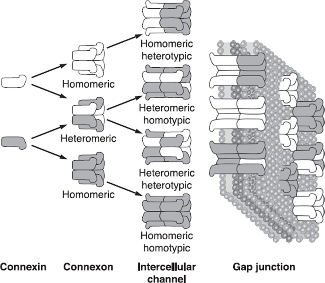
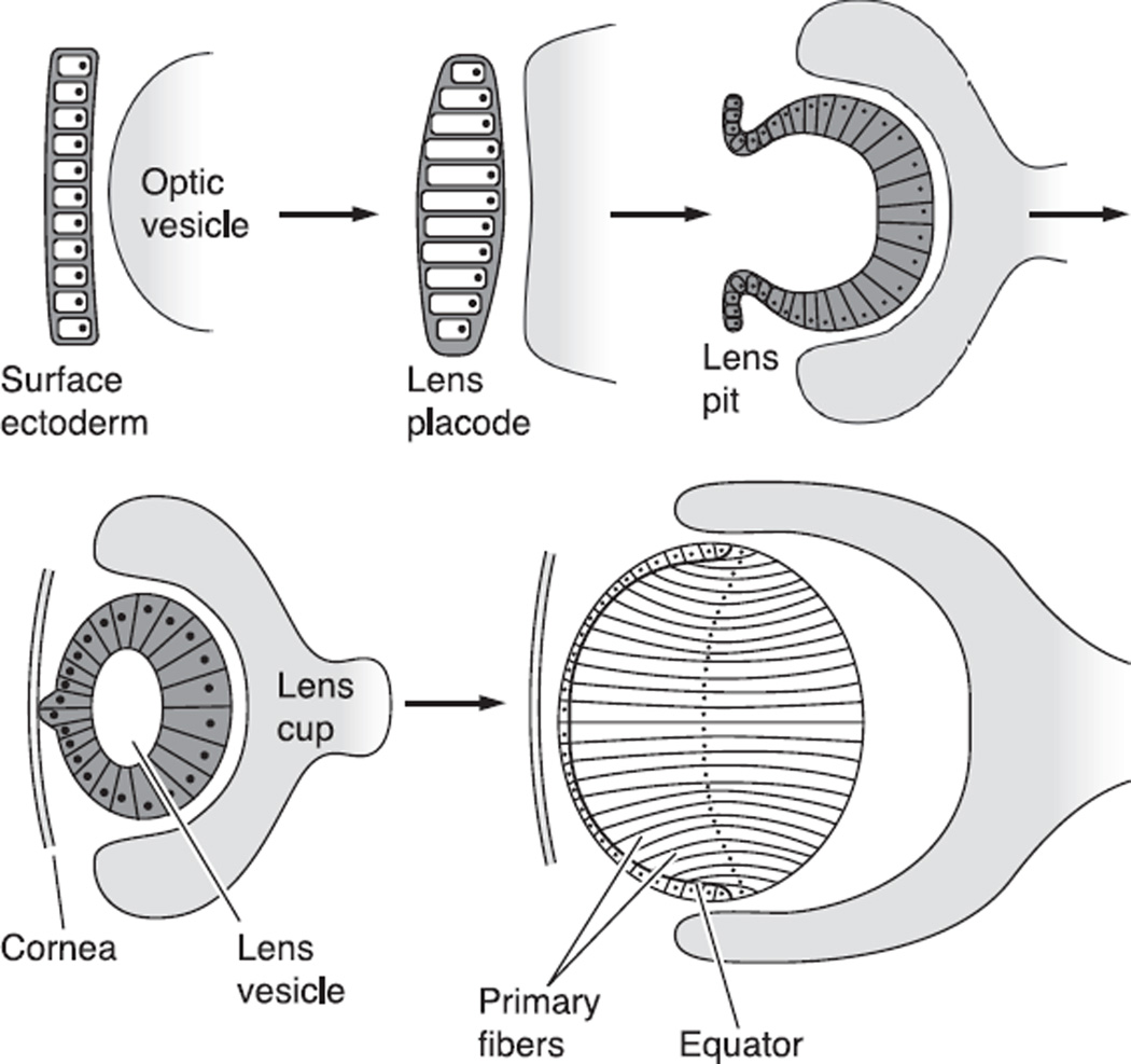
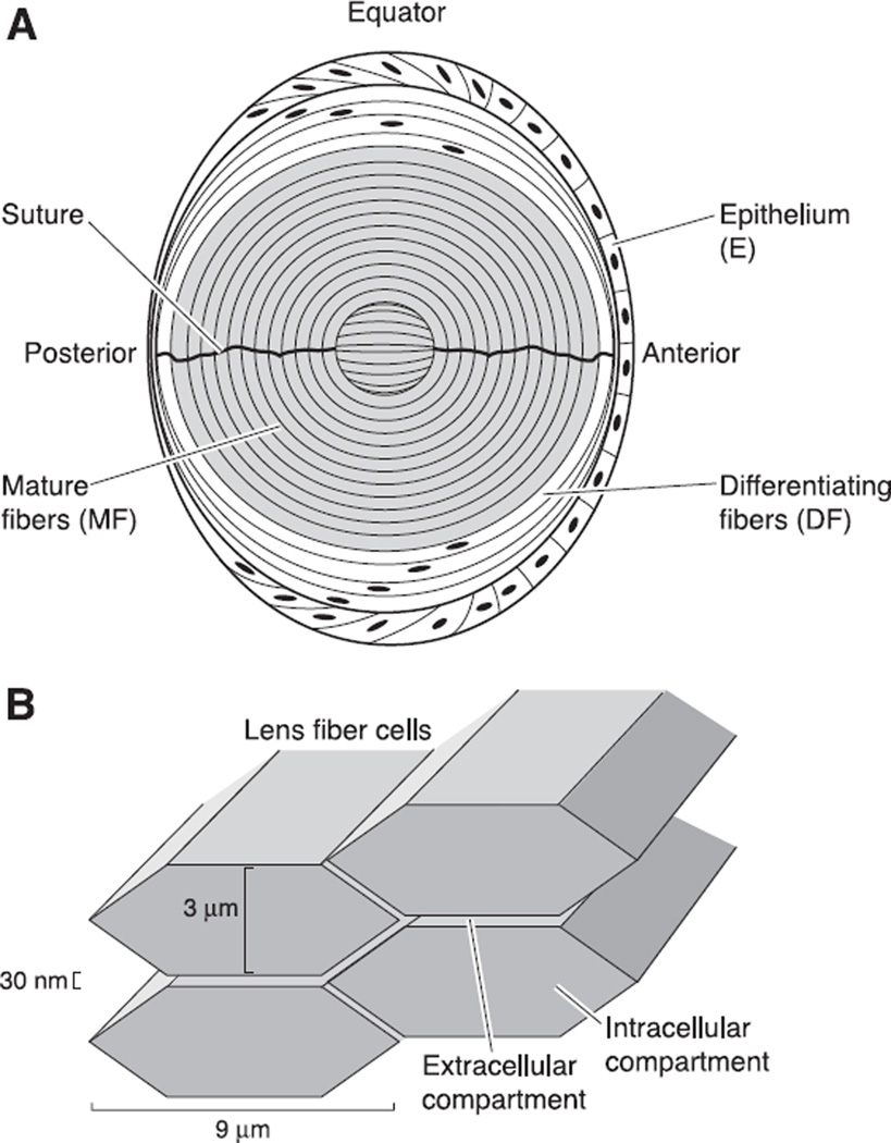
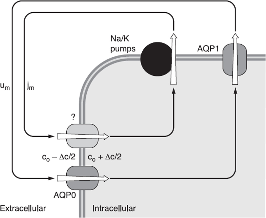
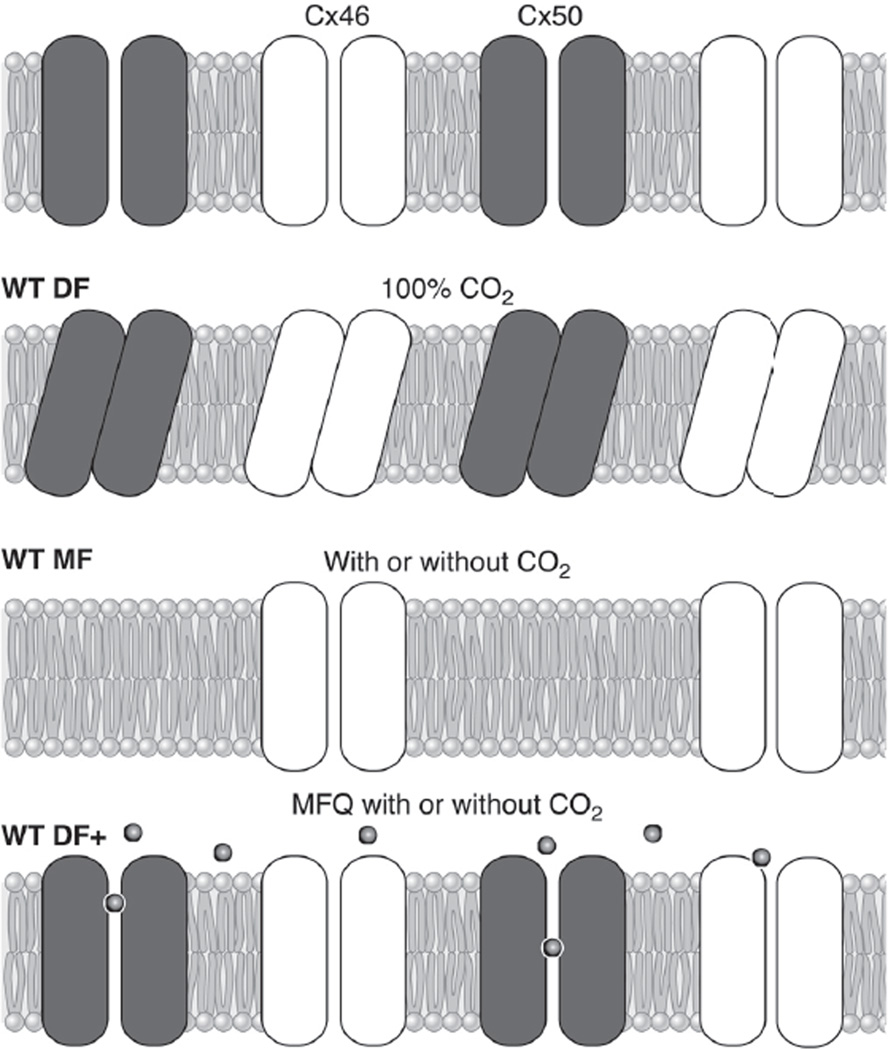
The cells of most mammalian organs are connected by groups of cell-to-cell channels called gap junctions. Gap junction channels are made from the connexin (Cx) family of proteins. There are at least 20 isoforms of connexins, and most tissues express more than 1 isoform. The lens is no exception, as it expresses three isoforms: Cx43, Cx46, and Cx50. A common role for all gap junctions, regardless of their Cx composition, is to provide a conduit for ion flow between cells, thus creating a syncytial tissue with regard to intracellular voltage and ion concentrations. Given this rather simple role of gap junctions, a persistent question has been: Why are there so many Cx isoforms and why do tissues express more than one isoform? Recent studies of lens Cx knockout (KO) and knock in (KI) lenses have begun to answer these questions. To understand these roles, one must first understand the physiological requirements of the lens. We therefore first review the development and structure of the lens, its numerous transport systems, how these systems are integrated to generate the lens circulation, the roles of the circulation in lens homeostasis, and finally the roles of lens connexins in growth, development, and the lens circulation.


Figures

Fig. 1
Fig. 1
The nomenclature for the different types of intercellular channels that can form a gap junction. The gap junction comprises many closely packed cell-to-cell channels connecting the cytoplasm of adjacent cells. Each channel is formed by the docking of two hemichannels, or connexons, one in each of the two cells. Each hemichannel is composed of six subunit connexins. The connexin family of proteins includes many isoforms that can intermix to form hemichannels and intact channels, as shown in the figure.
Fig. 2
Fig. 2
The stages of development of the embryonic lens starting from formation of the surface ectoderm and leading to formation of the initial identifiable lens, which has only primary fibers. [From Francis et al. (65), with permission from Elsevier Science.]
Fig. 3
Fig. 3
The structure of the mature lens. A: a sketch of the cellular structure of the lens. As described in the text, the transport properties of lens cells differ in three zones. The epithelium, which caps the front of the lens, expresses most of the active transport proteins in the lens. The mature fibers, which contain no organelles, fill the majority of the volume of the lens. They express membrane transporters for nutrients and antioxidants that are needed for homeostasis. Between the epithelium and mature fibers are the differentiating fibers, which still have organelles, but have different membrane transporters than the epithelium. B: the structure of the intracellular and extracellular compartments within the lens. When cut in cross-section (an equatorial section of the structure shown in A), the lens fibers are flattened hexagons of the approximate dimensions shown. The extracellular and intracellular compartments communicate through transporters in the fiber cell membranes.
Fig. 4
Fig. 4
The lens circulation. A: circulating currents enter the lens at both poles and exit at the equator. This panel indicates the overall pattern of current flow without regard to detail. B: a more detailed sketch of how the currents enter and exit the lens. The inward currents, which are carried by Na+, enter the lens along the extracellular spaces. The outward currents, which are also carried by Na+, are intracellular, flowing from cell to cell through gap junctions to reach the epithelium, where the Na+-K+ pumps transport the Na+ out of the lens to complete the current loops. The spatial separation of active Na+ extrusion from passive Na+ entry creates the circulation from surface to interior but does not explain the complex pattern. C: the factors responsible for the complex pattern of flow. Gap junction coupling conductance is concentrated in the equatorial region of differentiating fibers (DF), thus directing the outward intracellular flow to the equatorial epithelial cells, where Na+-K+ pump expression per unit area of lens surface is highly concentrated to transport the Na+ out at the equator. [From Mathias et al. (130), with permission from Springer Science + Business Media.]
Fig. 5
Fig. 5
The coupling through local osmosis of the circulating flow of solute (jm) to a circulation of fluid (um). In this model, circulating Na+ current creates a circulation of solute that drives fluid to move in the same pattern. The complex pattern ensures the fluid is well stirred and can carry fresh nutrients and antioxidants into the lens along the extracellular spaces to the central fiber cells, which are too far from the lens surface for diffusion to be effective. Thus the avascular lens generates its own internal microcirculatory system. [From Mathias et al. (130), with permission from Springer Science + Business Media.]
Fig. 6
Fig. 6
The methodology of whole lens impedance studies. A: the positioning of microelectrodes within the lens to inject current (I) and measure the induced voltage (V). The voltage microelectrode is moved to different locations to determine the spatial distribution of transport properties such as gap junction coupling conductance. B: typical records of the input impedance (Rin) as a function of the sinusoidal frequency of the injected current. The data were recorded at four depths into the lens, starting at 12% of the distance into the lens from the surface (r/a = 0.88), and culminating at 45% of the distance into the lens from the surface, where a is the radius and r is the distance from the center of the lens. Note the high frequencies, the input impedance asymptotes to a frequency-independent series resistance, RS. As described in the text, RS is proportional to the resistance of all the gap junctions between the point of voltage recording and the lens surface.
Fig. 7
Fig. 7
A summary of the properties of gap junction coupling in DF. A: the effects of knockout of Cx46 or Cx50 on coupling conductance of DF. The conductance has been normalized to the average value of wild-type (WT) coupling in DF, which is typically ~ 1 S/cm2 of cell-to-cell contact, so the normalized values are close to actual values in those units. The reductions in conductance in Cx46+/− and Cx46−/− lenses are from Gong et al. (78). The reductions in conductance in Cx50+/− and Cx50−/− lenses are from Baldo et al. (8). The conclusion is that both Cx46 and Cx50 contribute to coupling of DF, and their contribution to the coupling conductance is about equal. B: immunostaining of connexins. The green labeling in a is for the inner loop of Cx46. The red labeling in b is for the COOH terminus of Cx50. The yellow labeling in c is an overlay of the two top panels and indicates both connexins are in the same plaques. The inner loop of Cx46 is not cleaved at the DF to MF transition, whereas the COOH terminus of Cx50 is cleaved, hence the green labeling persists into MF in d, whereas red staining does not. [From Gong et al. (78), copyright 1998 National Academy of Sciences USA.]
Fig. 8
Fig. 8
A summary of the properties of gap junction coupling conductance in the mature fibers (MF). A: the effects of knockout of Cx46 and Cx50 on MF coupling conductance. The conductances are normalized to the average value in the DF, which is ~1 S/cm2 of cell-to-cell contact. The reductions in coupling conductance in the Cx46+/− and Cx46−/− lenses are from Gong et al. (78). The data from Cx50+/− lenses are from Baldo et al. (8). In the MF of WT lenses, the average coupling conductance is about half that of DF. Knocking out half of Cx46 (Cx46+/−) caused a 50% reduction in MF coupling conductance, whereas knocking out all of Cx46 (Cx46−/−) reduced coupling to zero. In contrast, knocking out half of Cx50 (Cx50+/−) had no effect on MF coupling. In the Cx50−/− lenses, coupling decreased, but so did all transport parameters, suggesting indirect effects. The Cx50+/− lenses were perfectly healthy, but there was no effect on coupling, suggesting Cx50 does not contribute to MF coupling, consistent with the results from the Cx46+/− and Cx46−/− lenses. B: the effects of knockout (KO) and knock in (KI) of Cx46 on MF coupling conductance. The KO data are from Gong et al. (78). The KI data are from Martinez-Wittinghan et al. (127). The normalized MF coupling conductance is almost linearly proportional to the number of copies of Cx46 being expressed, suggesting Cx46 is the only functional connexin in the MF.
Fig. 9
Fig. 9
Gating of lens gap junction channels. A: gating of gap junctions in WT lenses. When current is injected into a central fiber cell and voltage recorded in DF, the series resistance (RS in Fig. 6B) increases dramatically and reversibly when the lens is superfused with CO2 solution, indicating closure of DF gap junction channels. In contrast, when voltage is recorded in MF, the increase in series resistance is relatively small, indicating MF channels are not closing and the small increase is due to closure of DF channels. [From Martinez-Wittingham et al. (127), with permission from the Association for Research in Vision and Ophthalmology.] B: gating of DF gap junction channels in WT and Cx50−/− lenses. KO of Cx50 reduces DF coupling conductance to about half its normal value, but the remaining Cx46 channels have lost most of their pH sensitivity. Because the KO lenses were in such poor condition, we thought the lack of gating might be an indirect effect, but this was ruled out by the data in C. [Redrawn from Baldo et al. (8).] C: gating of DF gap junction channels in WT and mefloquine-treated lenses. Mefloquine (MFQ) blocks gap junction coupling conductance in channels made from Cx50 but not Cx46. DF gap junction coupling conductance drops to about half its normal value over a period of ~ 1 h in MFQ, suggesting blockade of all Cx50 channels, leaving Cx46 channels to provide coupling. When the MFQ lenses are superfused with CO2-containing solution, there is no closure of the remaining Cx46 channels, whereas in the untreated lens, both Cx50 and Cx50 channels close. The MFQ-treated lens was perfectly healthy, so the small residual gating seen in Cx50−/− lenses is actually due to their poor health, and in a healthy lens Cx46 channels are totally insensitive to pH. [From Martinez-Wittingham et al. (128), with permission from Springer Science + Business Media.]
Fig. 10
Fig. 10
A mode of cooperative gating to explain the data shown in Figure 9. [From Martinez-Wittingham et al. (128), with permission from Springer Science + Business Media.]
Fig. 11
Fig. 11
The effects of KO or KI of lens connexins on transparency. The Cx46+/− or Cx50+/− lenses are indistinguishable from WT, so a WT lens is not shown. Surprisingly, the Cx50(46/50) heterozygous KI lens gets a significant central cataract, even though its coupling conductance is at least as great as that of the Cx46+/− or Cx50+/− lenses. The Cx46−/− lens gets a dense central cataract. The Cx50−/− lens has a mild central cataract but is undersized and in poor physical shape. The Cx50(46/46) lens is as transparent as WT lenses, but it is undersized. It appears Cx50 is necessary for normal growth.
Fig. 12
Fig. 12
The radial variation in resting voltage in WT and Cx46−/− lenses. In WT lenses, there is about a 10-mV gradient between the central and surface fiber cells, as the voltage is about −60 mV in the center and drops to −70 mV at the surface. This is associated with the circulating ionic currents described in Figure 4. In the Cx46−/− lens, MF coupling is lost so the current cannot circulate through the MF and the voltage becomes flat. However, because the MF are also isolated from the epithelial K+channels, the cells depolarize to voltage dependent on their Cl~ and Na+ conductances. [From Gong et al. (78), copyright 1998 National Academy of Sciences USA.]
Fig. 13
Fig. 13
Calcium homeostasis in the lens and its relationship to gap junction coupling conductance. Ca2+ is expected to circulate between the surface epithelial cells where active transport takes place and the inner fiber cells where the passive leak takes place. Thus a center-to-surface concentration gradient should exist to drive diffusion, and that gradient should depend on fiber cell coupling conductance. [From Gao et al. (66), copyright 2004. Originally published in The Journal of General Physiology.]
Fig. 14
Fig. 14
Calcium concentration gradients measured with fura 2 in intact lenses from WT and Cx46 KO and KI lenses. In WT lenses, there is a center-to-surface Ca2+ gradient of ~400 nM, as the concentration goes from 700 nM at the center to 300 nM at the surface. In the Cx50(46/46) KI lenses, the MF coupling conductance is doubled and the concentration gradient is halved, now being 200 nM, as the concentration goes from 500 nM at the center to 300 nM at the surface. In the Cx46−/− KO lenses, the lack of MF coupling blocks the egress pathway for Ca2+, so it accumulates without regulation in the MF. [From Gao et al. (66), copyright 2004. Originally published in The Journal of General Physiology.]
Fig. 15
Fig. 15
The effect on intracellular homeostasis of knocking out GPX-1, an enzyme that protects the lens against H2O2-mediated oxidative damage. All lenses were ~2 mo old. The smooth curves are from a structurally based model of the lens circulation. The only detectable defect in the GPX-1 KO lenses was that MF coupling conductance was on average half the value in WT lenses, so the expected effect is that the same circulation of Na+ or Ca2+ will require twice the center-to-surface concentration gradient. A: intracellular Na+ concentration gradients measured with SBFI in WT and GPX-1 KO lenses. In WT lenses, the Na+ concentration gradient is ~10 mM, as the concentration goes from 17 mM at the center to 7 mM at the surface. In the GPX-1 KO lenses, the gradient is ~22 mM, as the concentration goes from 33 mM at the center to 11 mM at the surface. B: intracellular Ca2+ concentration gradients measured with fura 2 in WT and GPX-1 KO lenses. In the WT lens, the gradient is ~500 nM, as the concentration goes from 650 nM in the center to 150 nM at the surface. In the GPX-1 KO lenses, the gradient is ~850 nM, as the concentration goes from ~1,000 nM at the center to 150 nM at the surface. [From Wang et al. (204), with permission from Springer Science + Business Media.]

Similar articles

Cited by

References

    1. Addison PK, Berry V, Holden KR, Espinal D, Rivera B, Su H, Srivastava AK, Bhattacharya SS. A novel mutation in the connexin 46 gene (GJA3) causes autosomal dominant zonular pulverulent cataract in a Hispanic family. Mol Vis. 2006;12:791–795. - PubMed
    1. Al-Ghoul KJ, Kirk T, Kuszak AJ, Zoltoski RK, Shiels A, Kuszak JR. Lens structure in MIP-deficient mice. Anat Rec A Discov Mol Cell Evol Biol. 2003;273:714–730. - PubMed
    1. Alvarez LJ, Candia OA, Polikoff LA. Beta-adrenergic stimulation of Na(+)-K(+)-2Cl(−) cotransport activity in the rabbit lens. Exp Eye Res. 2003;76:61–70. - PubMed
    1. Alvarez LJ, Candia OA, Turner HC, Polikoff LA. Localization of a Na(+)-K(+)-2Cl(−) cotransporter in the rabbit lens. Exp Eye Res. 2001;73:669–680. - PubMed
    1. Alvarez LJ, Candia OA, Wolosin JM. Evidence for parallel Na(+)-H(+) and Na(+)-dependent Cl(−)-HCO3(−) exchangers in cultured bovine lens cells. Exp Eye Res. 1992;55:747–755. - PubMed

Publication types