. 2010 Mar;78(3):963-75.
doi: 10.1128/IAI.00925-09. Epub 2010 Jan 19.

Polysaccharide capsule and sialic acid-mediated regulation promote biofilm-like intracellular bacterial communities during cystitis

Affiliations

Polysaccharide capsule and sialic acid-mediated regulation promote biofilm-like intracellular bacterial communities during cystitis

Gregory G Anderson et al. Infect Immun. 2010 Mar.

Abstract

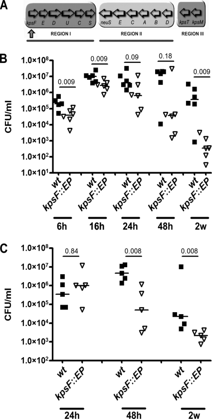
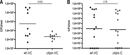
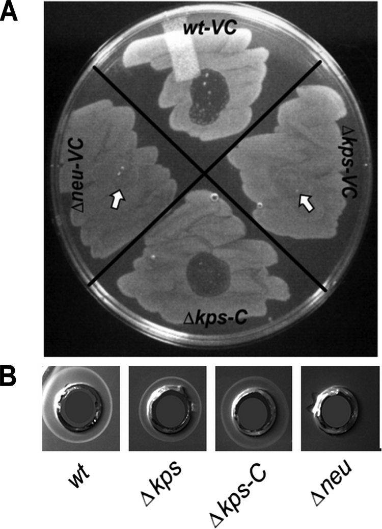
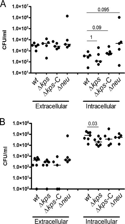
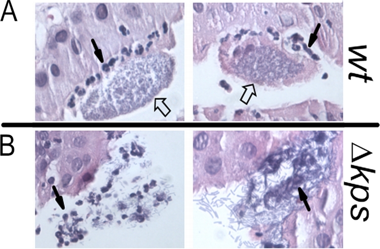
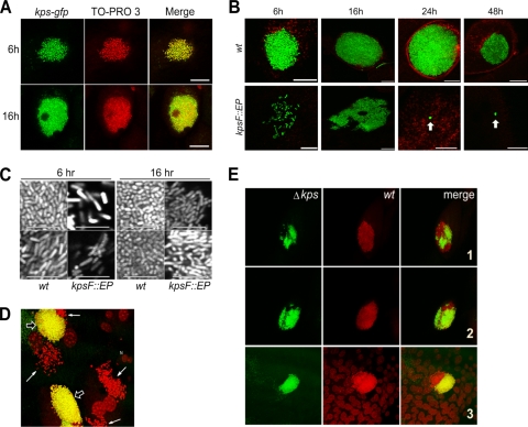
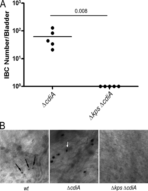
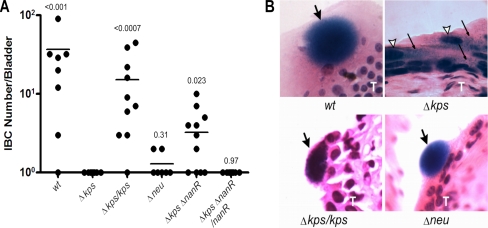
Uropathogenic Escherichia coli (UPEC) is the leading cause of urinary tract infections (UTIs). A murine UTI model has revealed an infection cascade whereby UPEC undergoes cycles of invasion of the bladder epithelium, intracellular proliferation in polysaccharide-containing biofilm-like masses called intracellular bacterial communities (IBC), and then dispersal into the bladder lumen to initiate further rounds of epithelial colonization and invasion. We predicted that the UPEC K1 polysaccharide capsule is a key constituent of the IBC matrix. Compared to prototypic E. coli K1 strain UTI89, a capsule assembly mutant had a fitness defect in functionally TLR4(+) and TLR4(-) mice, suggesting a protective role of capsule in inflamed and noninflamed hosts. K1 capsule assembly and synthesis mutants had dramatically reduced IBC formation, demonstrating the common requirement for K1 polysaccharide in IBC development. The capsule assembly mutant appeared dispersed in the cytoplasm of the bladder epithelial cells and failed to undergo high-density intracellular replication during later stages of infection, when the wild-type strain continued to form serial generations of IBC. Deletion of the sialic acid regulator gene nanR partially restored IBC formation in the capsule assembly mutant. These data suggest that capsule is necessary for efficient IBC formation and that aberrant sialic acid accumulation, resulting from disruption of K1 capsule assembly, produces a NanR-mediated defect in intracellular proliferation and IBC development. Together, these data demonstrate the complex but important roles of UPEC polysaccharide encapsulation and sialic acid signaling in multiple stages of UTI pathogenesis.


Figures

FIG. 1.
FIG. 1.
Unencapsulated UPEC has neutrophil-dependent and -independent fitness defects. (A) Organization of the capsule gene locus. Region I genes encode capsule assembly and export factors. Region II encodes synthesis factors. Region III products are involved in capsule export. Region I interruption (strain UTI89 kpsF::EP185) is designated by the upward-pointing arrow. Deletions of region I (Δkps) and region II (Δneu) used in later experiments are indicated by lines below the diagram. (B and C) CFU counts of isolates UTI89 (WT; closed squares) and UTI89 kpsF:: EP185 (open triangles) in infected bladders at time points between 6 h and 2 weeks in female C3H/HeN (TLR4+) and C3H/HeJ (TLR4) mice, respectively. Horizontal lines indicate the median values for the groups. P values were calculated using the Mann-Whitney nonparametric test.
FIG. 2.
FIG. 2.
Complementation of a region I kps deletion restores virulence. Competitive infections were performed with WT strain UTI89 attλ::PSSH10-1 (spectinomycin resistant) and the isogenic region I capsule deletion mutant UTI89 Δkps attHK::COM-GFP (kanamycin resistant; GFP expressing) for 48 h in C3H/HEN (TLR4+) mice. (A) Each strain carried the control vector pBAC-LacZ (designated VC). (B) UTI89 attλ::PSSH10-1 (WT) carries control vector pBAC-LacZ, and UTI89 Δkps attHK::COM-GFP carries complementing vector pBAC-LAC RegI kps, containing the entire region I capsule gene cluster under the control of its native promoter (designated C). A total of 10 mice were infected. Horizontal bars represent the geometric means. Statistical comparisons were performed using the Mann-Whitney nonparametric test.
FIG. 3.
FIG. 3.
Phage sensitivity and radial immunodiffusion of capsule synthesis and assembly mutants. (A) Lawns of each strain grown on L agar were exposed to a spot of fresh K1F phage lysate and incubated for ∼6 h at 37°C. Phage sensitivity, indicating fully surface-assembled K1 capsule, is evident by lytic zones. Arrows indicate where phage was spotted on nonlysed bacterial lawns. VC = vector control, pBR329. C = complement, pSX50. Identical results were obtained for VC = pBAC-LacZ and C = pBAC-LacZ RegI kps (data not shown). (B) Radial immunodiffusion plates containing K1-reactive polyclonal horse serum. Whole-cell lysates of each strain indicated were distributed into the respective wells. VC = vector control, pBR329. C = complement, pSX50.
FIG. 4.
FIG. 4.
Adherence, invasion, and early intracellular replication by K1 capsule mutants. Gentamicin protection assays were performed ex vivo on previously infected bladders of C3H/HeN mice at 1 and 6 h (panels A and B, respectively). Δkps-C designates UTI89 Δkps carrying the region I genes on pSX50. The remaining strains carried the control vector pBR329. Bars indicate the median values for the group, and P values were calculated by the Mann-Whitney nonparametric test. P > 0.1 for all comparisons at 6 h (panel B), except where indicated otherwise.
FIG. 5.
FIG. 5.
Neutrophil infiltration of IBC formed by WT and isogenic acapsular UPEC. Two representative sections of IBC formed by the WT or unencapsulated UTI89 Δkps at 12 h postinfection in C3H/HeN mice. Each is visualized under ×40 magnification and by hematoxylin-eosin staining. Closed arrows indicate representative neutrophils. Open arrows indicate characteristic UPEC-containing IBC.
FIG. 6.
FIG. 6.
Expression of capsule during IBC formation and contribution to IBC morphogenesis. (A) kps-gfp expression during IBC formation in WT UPEC. Infected bladders from female C3H/HEN mice infected for 6 h were counterstained with TO-PRO-3 (red) and imaged in whole mount by confocal microscopy. Representative images are shown. Scale bar = 50 μm. (B) IBC morphologies of UTI89 (WT) and UTI89 kpsF:: EP185 during bladder infection (C3H/HEN) from 6 h to 2 weeks. Bladders were counterstained with wheat germ agglutinin-Alexa 594. Arrows indicate microclusters of bacteria. Scale bar = 50 μm. (C) Magnified view of bacterial grouping (C3H/HEN mice) and morphology within intracellular collections of WT and kpsF:: EP185 mutant strains at 6 and 16 h p.i. Two representative images are shown for each strain at each time point. Scale bar = 10 μm. (D) Coinfection of bladders of female C3H/HEN mice with the GFP-expressing WT and the non-GFP expressing Δkps mutant at 6 h p.i. Representative images were taken in whole mount by confocal microscopy following counterstaining with TO-PRO-3. IBC formed by the WT is yellow (open arrows), and Δkps mutant bacteria are red (closed arrows). (E) Mixed IBC in the bladders during WT and Δkps mutant coinfection of female C3H/HEN mice at 6 h p.i. The Δkps mutant is green; the WT is red. Three representative examples are shown.
FIG. 7.
FIG. 7.
CdiA is not responsible for failure of capsule assembly mutants to form IBC. (A) Comparative IBC formation at 6 h postinfection (C3H/HEN mice) by capsule mutants and cdiA mutants in C3H/HeN mice. IBC were enumerated by Lac staining (see Materials and Methods). The horizontal bars represent the median values. P value was determined by the nonparametric Mann-Whitney test. (B) Representative images of Lac-stained IBC derived from UTI89 (WT) and the UTI89 ΔcdiA mutant at 6 h postinfection. Arrows indicate IBC present in the representative images. Each image was taken at the same magnification.
FIG. 8.
FIG. 8.
Mutation of nanR partially restores IBC formation in the capsule assembly mutant. (A) IBC formation was determined for each UTI89 derivative by whole-mount Lac staining at 6 h postinfection in female C3H/HeN mice. Horizontal bars indicate median values. P values are shown as calculated by the Mann-Whitney test in comparison to UTI89 Δkps. Complementation in trans was performed with pBAC-LAC RegI kps and pBAC-LAC nanR. (B) Hematoxylin-eosin-stained 8-μm sections of infected C3H/HEN bladders demonstrating IBC formation. Images were captured at ×400 magnification. Large filled arrows indicate IBC. Open arrows indicate apical epithelial cell nuclei. Thin black arrows indicate dispersed bacteria. A capital T indicates the transitional epithelium layer.

Similar articles

Cited by

References

    1. Alteri, C. J., S. N. Smith, and H. L. Mobley. 2009. Fitness of Escherichia coli during urinary tract infection requires gluconeogenesis and the TCA cycle. PLoS Pathog. 5:e1000448. - PMC - PubMed
    1. Anderson, G. G., J. J. Palermo, J. D. Schilling, R. Roth, J. Heuser, and S. J. Hultgren. 2003. Intracellular bacterial biofilm-like pods in urinary tract infections. Science 301:105-107. - PubMed
    1. Anderson, J. B., F. Parivar, G. Lee, T. B. Wallington, A. G. MacIver, R. A. Bradbrook, and J. C. Gingell. 1989. The enigma of interstitial cystitis—an autoimmune disease? Br. J. Urol. 63:58-63. - PubMed
    1. Aoki, S. K., R. Pamma, A. D. Hernday, J. E. Bickham, B. A. Braaten, and D. A. Low. 2005. Contact-dependent inhibition of growth in Escherichia coli. Science 309:1245-1248. - PubMed
    1. Baba, T., T. Ara, M. Hasegawa, Y. Takai, Y. Okumura, M. Baba, K. A. Datsenko, M. Tomita, B. L. Wanner, and H. Mori. 2006. Construction of Escherichia coli K-12 in-frame, single-gene knockout mutants: the Keio collection. Mol. Syst. Biol. 2:2006.0008. - PMC - PubMed

Publication types

Substances