. 2010 Apr;30(7):1689-702.
doi: 10.1128/MCB.01105-09. Epub 2010 Jan 19.

Characterization of PXK as a protein involved in epidermal growth factor receptor trafficking

Affiliations

Characterization of PXK as a protein involved in epidermal growth factor receptor trafficking

Hiroshi Takeuchi et al. Mol Cell Biol. 2010 Apr.

Abstract

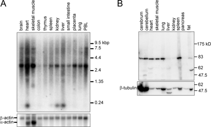
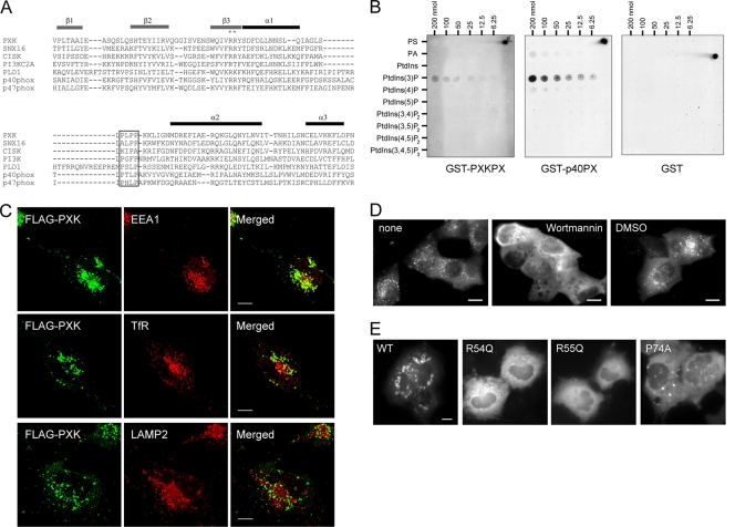
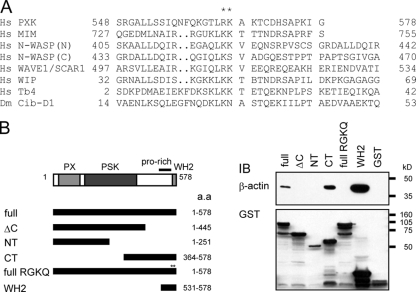
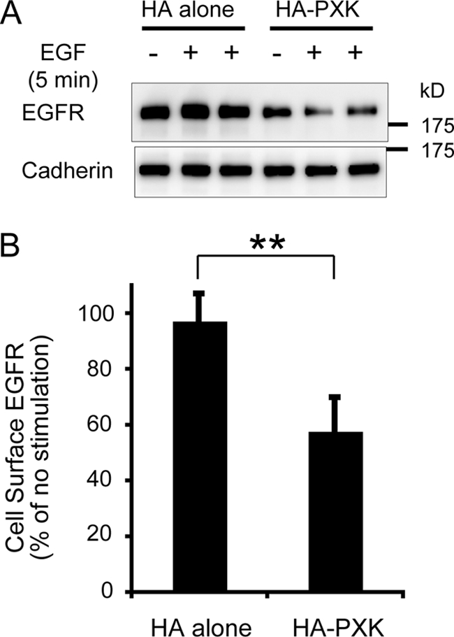
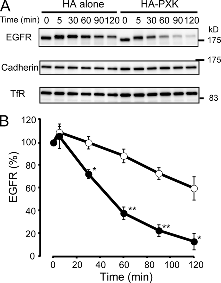
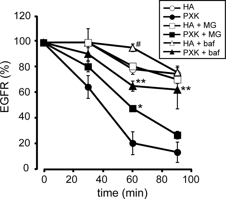
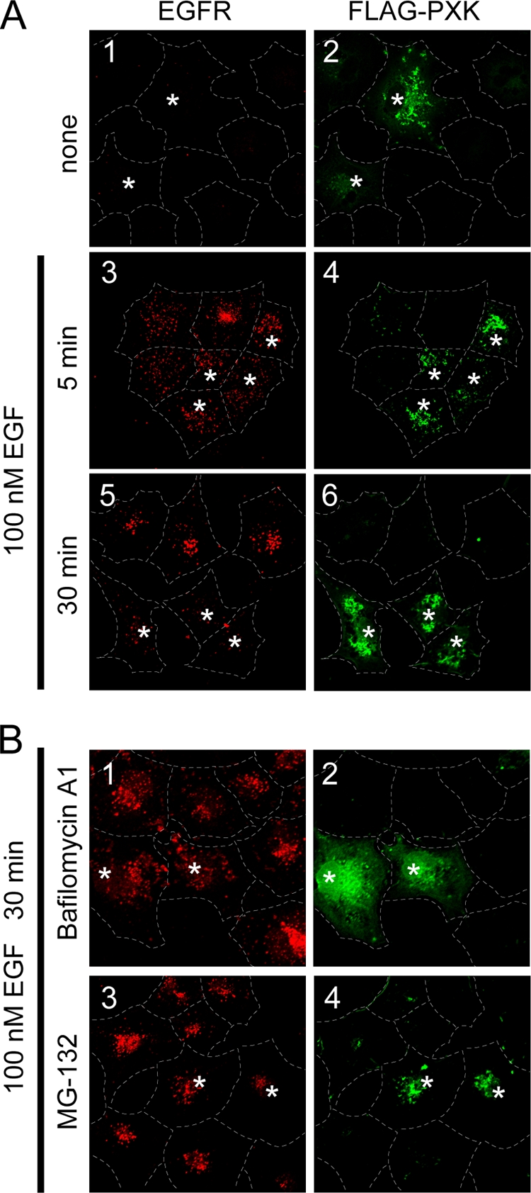
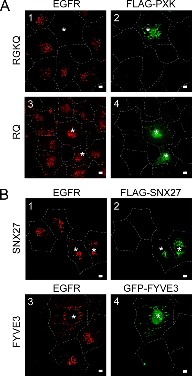
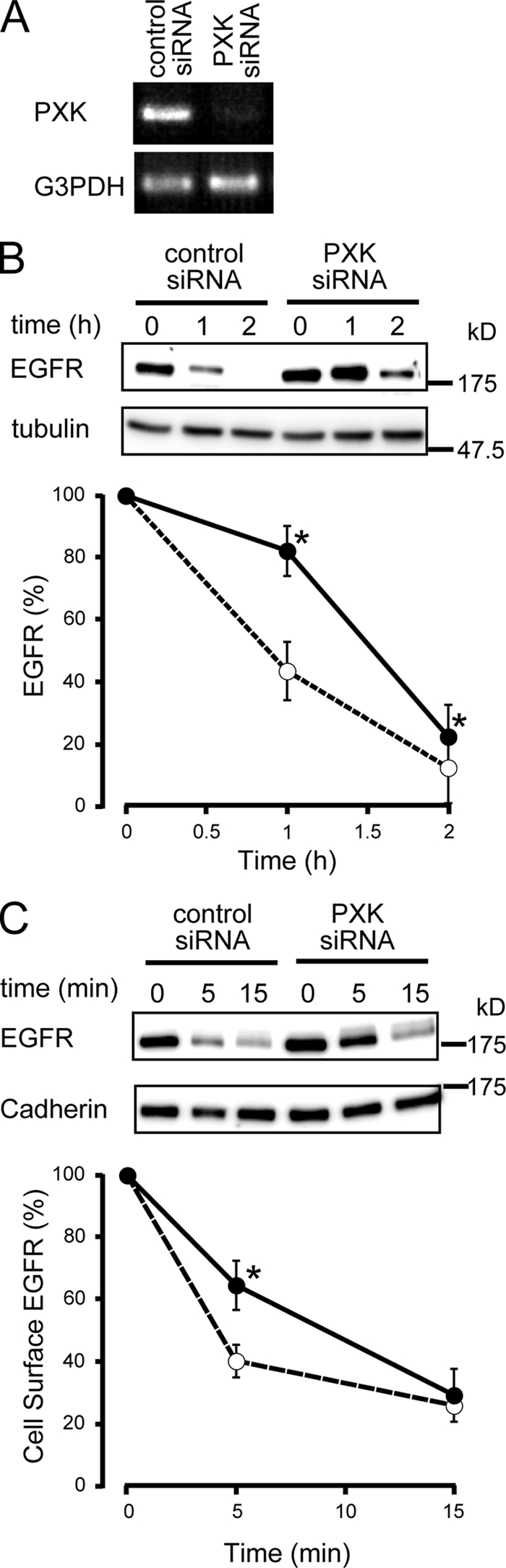
The phox homology (PX) domain is a phosphoinositide-binding module that typically binds phosphatidylinositol 3-phosphate. Out of 47 mammalian proteins containing PX domains, more than 30 are denoted sorting nexins and several of these have been implicated in internalization of cell surface proteins to the endosome, where phosphatidylinositol-3-phosphate is concentrated. Here we investigated a multimodular protein termed PXK, composed of a PX domain, a protein kinase-like domain, and a WASP homology 2 domain. We show that the PX domain of PXK localizes this protein to the endosomal membrane via binding to phosphatidylinositol 3-phosphate. PXK expression in COS7 cells accelerated the ligand-induced internalization and degradation of epidermal growth factor receptors by a mechanism requiring phosphatidylinositol 3-phosphate binding but not involving the WASP homology 2 domain. Conversely, depletion of PXK using RNA interference decreased the rate of epidermal growth factor receptor internalization and degradation. Ubiquitination of epidermal growth factor receptor by the ligand stimulation was enhanced in PXK-expressing cells. These results indicate that PXK plays a critical role in epidermal growth factor receptor trafficking through modulating ligand-induced ubiquitination of the receptor.


Figures

FIG. 1.
FIG. 1.
Tissue expression of PXK. (A) Tissue distribution of human PXK mRNA. The blot membrane containing 2 μg human mRNA in each lane was probed with a 662-bp fragment of EcoRI-digested PXK cDNA and reprobed with the control probe for β-actin. The sizes of the markers are shown on the right. (B) Tissue expression of PXK protein in rats. Rat tissue extract (40 μg protein) was subjected to immunoblot analysis of PXK by probing with rabbit anti-human PXK antiserum. A blot of β-tubulin as a loading control is also shown. The molecular sizes of the markers are shown at the right.
FIG. 2.
FIG. 2.
Endosomal localization of PXK depends on phosphoinositide binding of the PX domain. (A) Sequence alignment of several PX domains. Sequences were aligned using ClustalW and a Gonnet-weight matrix with a gap-opening penalty of 5 and extension penalty of 0.05 for multiple alignments, with some modification by hand to adjust the positions of proline-rich motifs. Highly conserved Arg residues are indicated by asterisks, and proline-rich regions are boxed. Secondary structure elements are aligned according to the PX domain of p40phox and are shown at the top. All PX domains in the alignment are from human homologs, and the GenBank accession numbers and corresponding amino acids are as follows: PXK, NP060241, 18 to 122; SNX16, NP690049, 109 to 214; CISK (cytokine-independent survival kinase), NP037389, 16 to 120; PI3K-C2α, NP002636, 1426 to 1534; PLD1, NP002653, 82 to 206; p40phox, NP038202, 23 to 136; and p47phox, NP000256, 8 to 121. (B) Phospholipid-binding property of the PX domain from human PXK. The ability of GST fusion proteins to bind a variety of phospholipids was analyzed using a protein-lipid overlay assay. Serial dilutions of the indicated phospholipids were spotted onto nitrocellulose membranes, which were then probed with GST fusion proteins, followed by detection using anti-GST antibody. Each GST fusion protein (1 pmol) was also spotted in the upper right corner of the membrane. PS, phosphatidylserine; PA, phosphatidic acid. (C) COS7 cells cultured on coverslips were transfected to express FLAG-PXK and visualized in green for FLAG-PXK and red for EEA1, TfR, or LAMP-2 by indirect immunofluorescence using a combination of rabbit antibody against FLAG and Alexa 594-conjugated rabbit IgG and mouse antibodies against EEA1, TfR, or LAMP-2 and Cy3-conjugated mouse IgG, respectively. The yellowish staining in the merged picture indicates colocalization with FLAG-PXK. For quantification, the number of the yellowish punctates and the total number of FLAG-PXK-positive, greenish punctuates were counted manually and the ratio was calculated. More than 10 cells were counted for each experiment. Scale bars, 10 μm. (D) Madin-Darby canine kidney (MDCK) cells cultured on coverslips were transfected to express PXK-EYFP, followed by treatment with 100 nM wortmannin or the vehicle, dimethyl sulfoxide (DMSO) for 20 min. Scale bars, 10 μm. (E) Subcellular localization of PXK-EGFP carrying point mutations in the PX domain in COS7 cells. WT, wild type. Scale bars, 10 μm.
FIG. 3.
FIG. 3.
The WH2 domain of PXK is a functional actin-binding domain. (A) Sequence alignment of WH2 motifs. Basic residues with asterisks are highly conserved. GenBank accession numbers of parental proteins are as follows: human PXK, NP060241; human MIM, NP055566; human N-WASP, NP003932; human WAVE1/Scar1, NP003922; human WIP, NP003378; human thymosin β4, NP066932; and Drosophila Ciboulot, NP726908. (B) Actin binding assay. Wild-type or mutant PXK fused to GST was expressed in COS7 cells and collected using glutathione beads from cell lysates. The intrinsic actin copurified with GST-PXK mutants was analyzed with anti-β-actin antibody. An immunoblot (IB) with anti-GST antibody is also shown. A schematic representation of PXK mutants is shown on the left. PX, PX domain; PSK, pseudo-kinase (kinase-like) domain; Pro-rich, proline-rich region before the WH2 domain (not the proline-rich region in the PX domain; see also Fig. 2A); WH2, WH2 domain.
FIG. 4.
FIG. 4.
PXK promotes ligand-induced EGFR internalization. (A) COS7 cells stably expressing HA or HA-PXK were starved for 12 h prior to stimulation with 10 nM EGF for 5 min. After EGF stimulation, cell surface proteins were biotinylated, followed by precipitation using streptavidin beads from the cell lysate and immunoblotting to detect the amounts of EGFR and pan-cadherin. (B) The density of each band, quantified using NIH Image software, is shown after normalization with cadherin. One hundred percent was obtained for each cell with no stimulation. Data are the means ± SEs from three experiments in duplicate. **, statistically different (P < 0.01) from the control.
FIG. 5.
FIG. 5.
Effect of PXK expression on ligand-induced EGFR degradation. (A) COS7 cells stably expressing HA alone and HA-PXK were serum starved for 12 h, followed by incubation with 20 μg/ml CHX and 100 nM EGF for the times indicated. Cells were lysed, and EGFR was analyzed by immunoblotting. The same blot was reprobed with anti pan-cadherin and TfR. (B) The density of EGFR, quantified using NIH Image software, was plotted after normalization with cadherin. Open and closed symbols indicate the values obtained with cells expressing HA alone and expressing HA-PXK, respectively. Data are the means ± SEs from three experiments. *, P < 0.05; **, P < 0.01 (different from cells expressing HA alone).
FIG. 6.
FIG. 6.
MG-132 and bafilomycin A1 block EGFR degradation accelerated by PXK. COS7 cells stably expressing HA alone and HA-PXK were starved for 12 h, followed by pretreatment with each inhibitor for 30 min in the presence of CHX. Cells were then stimulated with 100 nM EGF in medium containing inhibitors for the indicated times. The amount of EGFR remaining in cell lysates after EGF stimulation is shown as described for Fig. 5. Data are the means ± SEs from three experiments. #, P < 0.05 (different from HA cells [open circle]). *, P < 0.05; **, P < 0.01 (different from HA-PXK cells [closed circles]).
FIG. 7.
FIG. 7.
PXK expression accelerates EGFR degradation. COS7 cells cultured on coverslips were transfected with the gene for FLAG-PXK. Cells showing FLAG fluorescence are indicated by asterisks at the nucleus, and the cellular edges are traced by broken lines. (A) Thirty-six hours after transfection, cells were serum starved for 12 h, followed by preincubation with 20 μg/ml CHX and then left unstimulated (none) or stimulated with 100 nM EGF for 5 or 30 min. Cells were then fixed and treated with mouse anti-FLAG antibody and rabbit anti-EGFR antibody, followed by visualization with Alexa Fluor 488-conjugated anti-mouse antibody (green) and Alexa Fluor 594-conjugated anti-rabbit antibody (red), respectively. (B) Cells were preincubated with 10 μM MG-132 or 0.25 μM bafilomycin A1 in the presence of 20 μg/ml CHX for 30 min and then stimulated with 100 nM EGF for 30 min, followed by treatment as described above. Bars, 10 μm.
FIG. 8.
FIG. 8.
PXK expression accelerates EGFR degradation. Immunofluorescence analysis was performed as described for Fig. 7. (A) COS7 cells were transfected with genes for FLAG-tagged mutant PXK (RGKQ or RQ as indicated at the left), followed by visualization of EGFR and PXK. (B) COS7 cells were transfected with the gene for FLAG-SNX27 or EGFP-FYVE. Bars, 10 μm.
FIG. 9.
FIG. 9.
Effect of endogenous PXK depletion on ligand-induced internalization and degradation of EGFR. Endogenous PXK in HaCaT cells was depleted by transfecting with siRNA. (A) PXK silencing was confirmed by reverse transcription-PCR from total RNA of HaCaT cells transfected with siRNA specific against PXK or scrambled siRNA duplex as a control. Ethidium bromide staining is shown. (B) EGFR in HaCaT cell lysates stimulated with 200 nM EGF for the indicated times was analyzed as for Fig. 5. Open circles, control; closed circles, PXK-depleted cells. (C) EGFR remaining on the cell surface after 20 nM EGF stimulation for the indicated times was analyzed as for Fig. 4. Open circles, control; closed circles, PXK-depleted cells. Each point is the mean ± SE for three experiments. *, P < 0.05, different from control siRNA-treated cells (open circles).
FIG. 10.
FIG. 10.
Effect of PXK expression on uptake and recycling of transferrin. Biotinylated Tf in cells was monitored as an index of the receptors. (A) Control (open circles) or PXK-expressing (closed circles) cells were incubated in the presence of biotinylated Tf (10 μg/ml) for the times indicated. (B) Cells incubated with biotinylated Tf for 60 min were then incubated with 500 μg/ml nonlabeled Tf for 5 or 30 min. After fixation and permeabilization, biotinylated Tf was quantified with streptavidin-conjugated alkaline phosphatase. Each point is the mean ± SE from three experiments.
FIG. 11.
FIG. 11.
Effect of PXK expression on ubiquitination of EGFR. Cells expressing either HA alone (open symbols or bars) or HA-PXK (closed symbols or bars) were stimulated with 100 nM EGF for the indicated times. Cell extracts were then immunoprecipitated with 2 μg of anti-EGFR antibody and protein G-Sepharose, followed by Western blotting for ubiquitinated and total EGFR using antiubiquitin and anti-EGFR antibodies, respectively. Panels 1, typical blots; panels 2, density of EGFR quantified by NIH image software; panels 3, ratio of ubiquitination of EGFR. Data are the means ± SEs from three experiments. *, P < 0.05; **, P < 0.01 (different from cells expressing HA alone). Experiments were performed in the absence (A) or presence (B) of bafilomycin A1.

Similar articles

Cited by

References

    1. Ago, T., F. Kuribayashi, H. Hiroaki, R. Takeya, T. Ito, D. Kohda, and H. Sumimoto. 2003. Phosphorylation of p47phox directs phox homology domain from SH3 domain toward phosphoinositides, leading to phagocyte NADPH oxidase activation. Proc. Natl. Acad. Sci. U. S. A. 100:4474-4479. - PMC - PubMed
    1. Alwan, H. A., E. J. van Zoelen, and J. E. van Leeuwen. 2003. Ligand-induced lysosomal epidermal growth factor receptor (EGFR) degradation is preceded by proteasome-dependent EGFR de-ubiquitination. J. Biol. Chem. 278:35781-35790. - PubMed
    1. Arighi, C. N., L. M. Hartnell, R. C. Aguilar, C. R. Haft, and J. S. Bonifacino. 2004. Role of the mammalian retromer in sorting of the cation-independent mannose 6-phosphate receptor. J. Cell Biol. 165:123-133. - PMC - PubMed
    1. Badour, K., M. K. McGavin, J. Zhang, S. Freeman, C. Vieira, D. Filipp, M. Julius, G. B. Mills, and K. A. Siminovitch. 2007. Interaction of the Wiskott-Aldrich syndrome protein with sorting nexin 9 is required for CD28 endocytosis and cosignaling in T cells. Proc. Natl. Acad. Sci. U. S. A. 104:1593-1598. - PMC - PubMed
    1. Boudeau, J., D. Miranda-Saavedra, G. J. Barton, and D. R. Alessi. 2006. Emerging roles of pseudokinases. Trends Cell Biol. 16:443-452. - PubMed

Publication types

MeSH terms