. 2010 Mar;30(6):1457-66.
doi: 10.1128/MCB.01321-09. Epub 2010 Jan 19.

Yeast exonuclease 5 is essential for mitochondrial genome maintenance

Affiliations

Yeast exonuclease 5 is essential for mitochondrial genome maintenance

Peter M Burgers et al. Mol Cell Biol. 2010 Mar.

Abstract

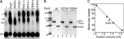
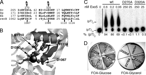
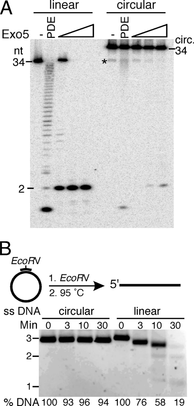
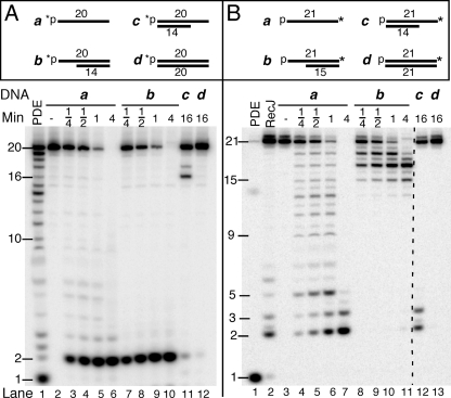
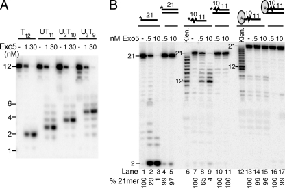
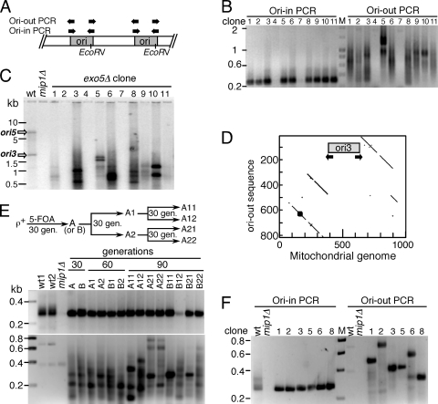
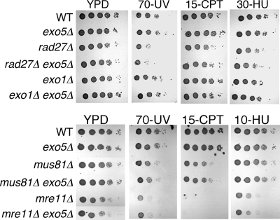
Yeast exonuclease 5 is encoded by the YBR163w (DEM1) gene, and this gene has been renamed EXO5. It is distantly related to the Escherichia coli RecB exonuclease class. Exo5 is localized to the mitochondria, and EXO5 deletions or nuclease-defective EXO5 mutants invariably yield petites, amplifying either the ori3 or ori5 region of the mitochondrial genome. These petites remain unstable and undergo continuous rearrangement. The mitochondrial phenotype of exo5Delta strains suggests an essential role for the enzyme in DNA replication and recombination. No nuclear phenotype associated with EXO5 deletions has been detected. Exo5 is a monomeric 5' exonuclease that releases dinucleotides as products. It is specific for single-stranded DNA and does not hydrolyze RNA. However, Exo5 has the capacity to slide across 5' double-stranded DNA or 5' RNA sequences and resumes cutting two nucleotides downstream of the double-stranded-to-single-stranded junction or RNA-to-DNA junction, respectively.


Figures

FIG. 1.
FIG. 1.
Cloning of EXO5 and overproduction of Exo5 nuclease. (A) Extracts from the indicated deletion mutants of strain BY4741 were subjected to partial purification. Standard assay mixtures contained 2 μl of the fraction that flowed through the mixed-bed ion exchanger (see Materials and Methods). Controls were no extract (−) or partially purified Exo5. Migration positions of phosphate (Pi) and mono- and dinucleotide (1 and 2, respectively) are indicated. (B) Overexpression and purification of Exo5. Lane 1, GST-Exo5 after glutathione column chromatography; lane 2, after cutting with 3C protease and heparin-Sepharose chromatography; lanes 3 and 4, the same for the Exo5-D270A mutant; lanes 5 and 6, the same for the Exo5-D320A mutant. Each lane contained ∼2 μg of protein. wt, wild type. The values on the left are molecular sizes in kilodaltons. (C) Superose 12 gel filtration analysis of Exo5 in buffer A200. Marker proteins are bovine serum albumin dimer and monomer, ovalbumin, and carbonic anhydrase.
FIG. 2.
FIG. 2.
Mutational analysis of EXO5. (A) Threading analysis of Exo5. Motifs conserved with E. coli RecB nuclease are shown. Sc, S. cerevisiae; Sp, S. pombe; Hs, Homo sapiens. (B) Active site of the RecB nuclease domain from the crystal structure of E. coli RecBCD (31). The active-site divalent metal ion (black) is coordinated by Asp1067 and Asp1080 (dark gray), which are part of the two gray beta strands, 1057 to 1072 and 1078 to 1082, respectively. The active site is closed off by the α helix at 1107 to 1125 (light gray) with the conserved Gln1110 and Tyr1114 residues (dark gray). (C) Standard nuclease assay mixtures contained the indicated concentrations of wild-type (wt) or mutant Exo5 for 10 min. Analysis was by TLC. (D) Strain PY209 (exo5Δ::HIS3 but [rho+] because it contains pBL253 [EXO5 URA3]) was transformed with the empty vector (vector) or pBL256 (EXO5), pBL256-270 (exo5-D270A), or pBL256-320 (exo5-D320A). Transformants were streaked onto FOA plates to select for loss of plasmid pBL253, with either glucose or glycerol as the carbon source. Plates were photographed after 2 days (glucose plates) or 4 days (glycerol plates) of growth at 30°C.
FIG. 3.
FIG. 3.
Exo5 lacks endonuclease activity. (A) Standard nuclease assay mixtures contained either 10 nM linear or circular 34-mer oligonucleotide 32P-c81 (see Materials and Methods) and 1 nM, 10 nM, or 100 nM Exo5. Analysis was on a 7 M urea-20% polyacrylamide gel. Note that the circular (circ.) oligonucleotide migrates slower than the linear form and is contaminated with 5% linear form (asterisk). This contaminating linear form is converted into the dinucleotide by Exo5. PDE, snake venom phosphodiesterase ladder. (B) Standard assay mixtures contained 5 nM circular or linear bluescript SK2 ssDNA (linearized by hybridizing a 12-mer oligonucleotide across the EcoRV site, followed by cutting with EcoRV and heating to restore complete single strandedness) and 200 nM Exo5. Aliquots taken after the indicated times were analyzed on a 1.2% agarose gel. Note that under these conditions the linear form migrates slightly slower that the circular form.
FIG. 4.
FIG. 4.
Substrate specificity of Exo5. (A) 5′-labeled substrates. 5′-32P-labeled v31 was either single stranded (a) or hybridized to a twofold excess of oligonucleotide c50 (b), oligonucleotide c49 (c), or oligonucleotide c41 (d). (B) 3′-labeled substrates. 3′-P-v31-32P-dA was either single stranded (a) or hybridized to a twofold excess of oligonucleotide cT50 (b), oligonucleotide c49 (c), or oligonucleotide cT41 (d). An asterisk indicates the position of the label. All of the assay mixtures contained 10 nM labeled substrate, 200 mM NaCl, and 10 nM Exo5 for the indicated times at 30°C. Assay mixtures were analyzed on 7 M urea-20% polyacrylamide gels. PDE, partial digestion with snake venom phosphodiesterase, a 3′ exonuclease; RecJ, partial digestion with E. coli RecJ, a 5′ exonuclease. A dotted guide line is added in panel B. The values on the left of each panel are oligomer sizes.
FIG. 5.
FIG. 5.
Exo5 slides across 5′ RNA sequences. (A) Standard assay mixtures contained the indicated 5′-labeled DNA or RNA-DNA chimera at 10 nM/either 1 or 30 nM Exo5 for 5 min at 30°C. Assay mixtures were analyzed on a 7 M urea-20% polyacrylamide gel. (B) Standard assay mixtures contained the indicated 5′-labeled DNA or RNA-DNA chimera at 10 nM (in order from the left) v71, v71/c72, vR73, vR73/c72, Bio-vR74 plus streptavidin, or Bio-vR74/c72, plus streptavidin. Assay mixtures contained either 0.5 or 10 nM Exo5 for 5 min at 30°C and were analyzed on 7 M urea-20% polyacrylamide gels. Klen., sequence ladder generated by partial degradation with the DNA polymerase I Klenow fragment. The values beside the lanes are oligomer sizes.
FIG. 6.
FIG. 6.
Analysis of mitochondrial DNA in exo5Δ petite mutants. (A) Head-to-tail arrangement of amplified ORI regions. A unique EcoRV restriction site localized asymmetrically in both ori3 and ori5 is shown. The ori-in and ori-out primers were used for PCR analysis of either the ORI sequences or the inter-ORI sequence regions, respectively. (B) PCR analysis of DNA from 11 individual exo5Δ clones after 90 generations of growth. (C) Southern analysis of total yeast DNA from the clones in panel B, digested with EcoRV, and probed with a mixture of ori3 and ori5 probes. The probes were generated by PCR labeling of mitochondrial DNA obtained from ori3- and ori5-containing petites. Analysis of DNA from the wild type (lane 1) shows the expected sizes for the wild-type (wt) ori3 and ori5 EcoRV fragments, marked with arrows. (D) The ori-out PCR products shown in panel B were sequenced in both directions. The presence of different overlapping sequence runs allowed only approximate sequence determination. For one representative clone, a matrix analysis of the ori-out sequence with that of the mitochondrial genome is shown (coordinates 54168 to 55167), identifying a 23-nt identity per 25-nt sliding window. Regions directly to the left and right of ori3 were identified; two internal repeats are apparent. (E) Instability of [rho] genomes during serial propagation. Two independent clones (clone A with ori3 amplified and clone B with ori5 amplified) were obtained by eviction of EXO5 from PY209 and grown for 30 generations (gen.) by serial dilution. They were plated on YPD, and two colonies were picked (A1 and A2 for the experiment with clone A) and grown for another 30 generations. Again, two colonies were picked per culture, yielding clones A11, A12, A21, and A22, and grown. The scheme for clone A is shown; the clone B scheme was the same. DNA from each isolate was prepared and subjected to PCR analysis with ori-in primers (top) and ori-out primers (bottom). (F) Clones 1, 2, 3, 5, 6, and 8 (as shown in panel B) were transformed with pBL253 (URA3 EXO5), and DNA from one transformant of each clone was subjected to PCR analysis.
FIG. 7.
FIG. 7.
exo5Δ mutant strains are not damage sensitive. All strains were mip1Δ ([rho0]) and, in addition, had the indicated genotype. Serial 10-fold dilutions of late-log-phase cells, from 105 to 10 cells per spot, were spotted onto YPD plates or YPD plates containing the indicated concentrations of hydroxyurea (HU, mM) or camptothecin (CPT, μg/ml). Some YPD plates were irradiated with the indicated fluency of UV254. Plates were grown for 3 days at 30°C and photographed. WT, wild type.

References

    1. Bernardi, G. 2005. Lessons from a small, dispensable genome: the mitochondrial genome of yeast. Gene 354:189-200. - PubMed
    1. Burgers, P. M. 2009. Polymerase dynamics at the eukaryotic DNA replication fork. J. Biol. Chem. 284:4041-4045. - PMC - PubMed
    1. Burgers, P. M. J., G. A. Bauer, and L. Tam. 1988. Exonuclease V from Saccharomyces cerevisiae. A 5′-3′-deoxyribonuclease that produces dinucleotides in a sequential fashion. J. Biol. Chem. 263:8099-8105. - PubMed
    1. Büttner, S., T. Eisenberg, D. Carmona-Gutierrez, D. Ruli, H. Knauer, C. Ruckenstuhl, C. Sigrist, S. Wissing, M. Kollroser, K. U. Frohlich, S. Sigrist, and F. Madeo. 2007. Endonuclease G regulates budding yeast life and death. Mol. Cell 25:233-246. - PubMed
    1. Bylund, G. O., J. Majka, and P. M. Burgers. 2006. Overproduction and purification of RFC-related clamp loaders and PCNA-related clamps from Saccharomyces cerevisiae. Methods Enzymol. 409:1-11. - PubMed

Publication types

MeSH terms